J Korean Ophthalmol Soc.
2008 Jan;49(1):57-64. 10.3341/jkos.2008.49.1.57.
The Changes in Macular Thickness after Phacoemulsification in Patients with Non-diabetes and Nonproliferative Diabetic Retinopathy
- Affiliations
-
- 1Department of Ophthalmology, Chungnam National University College of Medicine, Daejeon, Korea. shchoi@cnu.ac.kr
- KMID: 2211183
- DOI: http://doi.org/10.3341/jkos.2008.49.1.57
Abstract
-
PURPOSE: To compare the changes in macular thickeness between non-diabetic group and a mild or moderate nonproliferative diabetic retinopathy group after phacoemulsification.
METHODS
This study consisted of 32 eyes of 22 patients who underwent phacoemulsification. The non-diabetic group included 20 eyes of 15 patients; the diabetic group (mild or moderate nonproliferative diabetic retinopathy) included 12 eyes of 7 patients. Macular thickness using optical coherence tomography (OCT) and corrected visual acuity were measured before surgery and 1 day, 1 week, 1 month, 2 months, 6 months and 12 months after surgery.
RESULTS
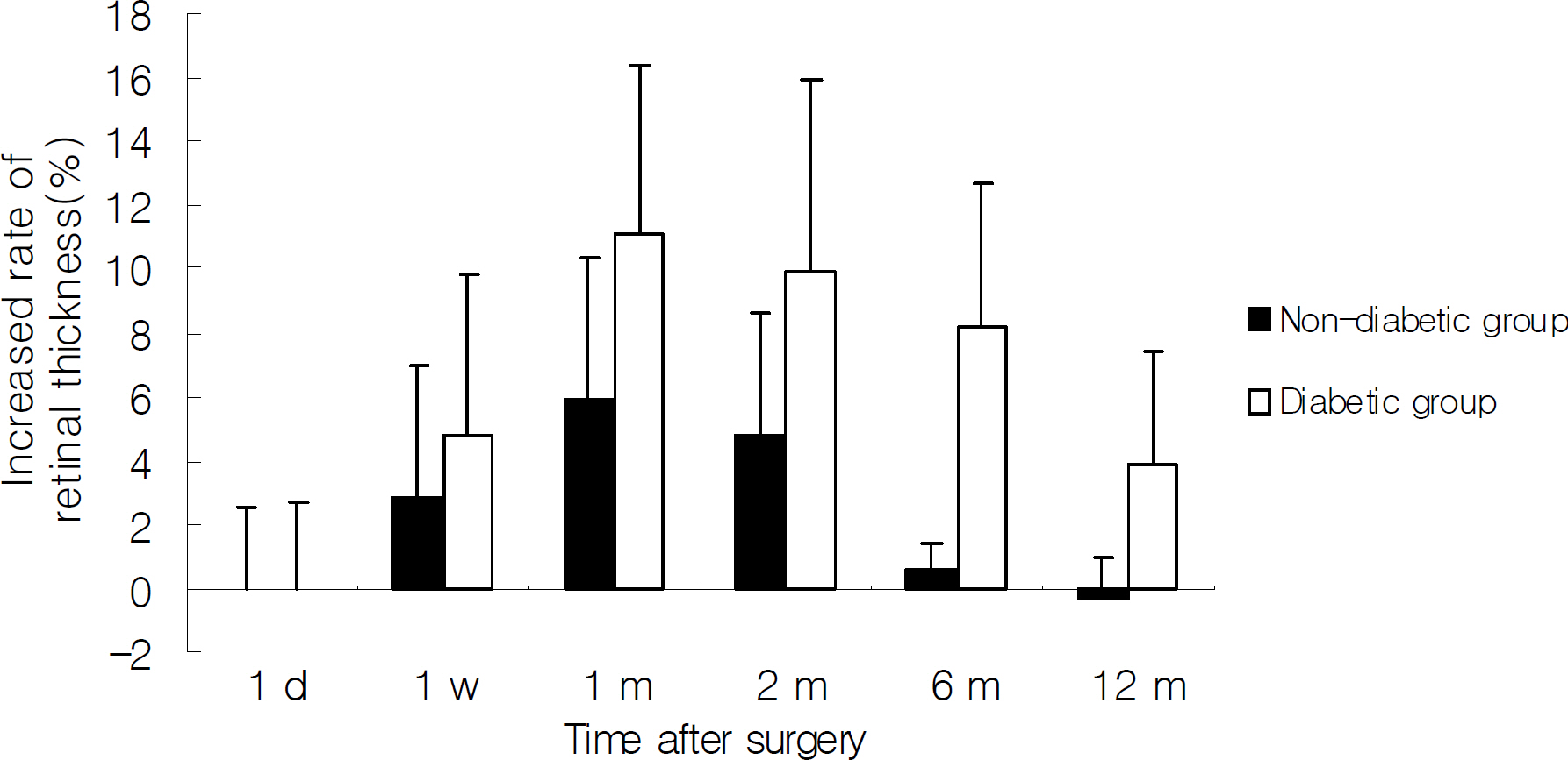
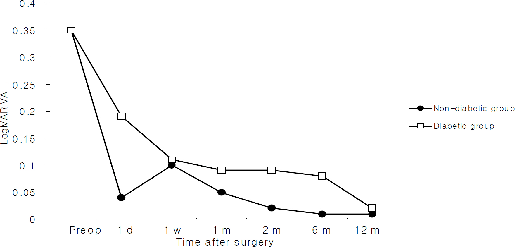
In the non-diabetic group, the macular thickness increased by 2.8+/-4.2% at 1 week, 5.9+/-4.5% at 1 month, 4.8+/-3.8% at 2 months, 0.6+/-0.8% at 6 months, and -0.3+/-1.2% at 12 months after surgery, while it increased by 4.8+/-5.0% at 1 week, 11.1+/-5.2% at one month, 9.9+/-6.0% at two months, 8.1+/-4.6% at 6 months, 3.9+/-3.5% at 12 months in the diabetic group. The increased amount of macular thickness was significantly higher in the diabetic group than in the non-diabetic group at 1 month, 2 months, 6 months, and 12 months. Visual acuity was not significantly different between the diabetic and non-diabetic groups. In the non-diabetic group, 2 months after the operation, LogMAR below 0.02 (Snellen 0.95) were remained with best corrected visual acuity. Similarly to non-diabetic patients, diabetic patients needed 12 months to reach best corrected visual acuity.
CONCLUSIONS
Macular thickness increased in both diabetic and non-diabetic groups after phacoemulsification, and the increased amount of macular thickness was significantly greater and lasted longer in the diabetic group compared with the non-diabetic group. In cases of mild or moderate nonproliferative diabetic retinopathy, macular thickness change due to cataract surgery did not influence visual acuity.
Keyword
MeSH Terms
Figure
Cited by 1 articles
-
The Results of a Combination of Cataract Surgery and Intravitreal Bevacizumab Injection for Diabetic Macular Edema
Bu Ki Kim, Eui Yong Kweon, Dong Wook Lee, Min Ahn, Nam Chun Cho
J Korean Ophthalmol Soc. 2010;51(7):954-960. doi: 10.3341/jkos.2010.51.7.954.
Reference
-
References
1. Rossetti L, Autelitano A. Cystoid macular edema following cataract surgery. Curr Opin Ophthalmol. 2000; 11:65–72.
Article2. Powe NR, Schein OD, Gieser SC, et al. Synthesis of the literature on visual acuity and complications following cataract extraction and intraocular lens implantation. Arch Ophthalmol. 1994; 112:239–52.3. Riley AF, Malik TY, Grupcheva CN, et al. The Auckland cataract study: comorbidity, surgical techniques and clinical outcomes in a public hospital service. Br J Ophthalmol. 2002; 86:185–90.
Article4. Ursell PG, Spalton DJ, Whitcup SM. Cystoid macular oedema after phacoemulsification: relationship to blood aqueous barrier damage and visual acuity. J Cataract Refract Surg. 1999; 25:1492–7.5. Simon N, Andrew R, Hussain P, et al. Correlations between optical coherence tomography measurement of macular thickness and visual acuity after cataract extraction. Clin Experiment Ophthalmol. 2006; 34:124–9.6. Ching HY, Wong AC, Wong CC, et al. Cystoid macular oedema and changes in retinal thickness after phacoemulsifi cation with optical coherence tomography. Eye. 2006; 20:297–303.7. Ah-Fat FG, Sharma MK, Majid MA, Yang YC. Vitreous loss during conversion from conventional extracapsular cataract extraction to phacoemulsification. J Cataract Refract Surg. 1998; 24:801–5.
Article8. Henry MM, Henry LM. A possible cause of chronic cystic maculopathy. Ann Ophthalmol. 1977; 9:455–7.9. Irvine SR. A newly defined vitreous syndrome following cataract surgery interpreted according to recent concepts of the structure of the vitreous. Am J Ophthalmol. 1953; 36:599–619.10. Jampol LM, Sanders DR, Kraff MC. Prophylaxis and therapy of aphakic cystoid macular edema. Surv Ophthalmol. 1984; 28:535–9.
Article11. Dowler JG, Hykin PG, Lightman SL, Hamilton AM. Visual acuity following extracapsular cataract extraction in diabetes: a meta-analysis. Eye. 1995; 9:313–7.
Article12. Schatz H, Atienza D, McDonald HR, Johnson RN. Severe diabetic retinopathy after cataract surgery. Am J Ophthalmol. 1994; 117:314–21.
Article13. Pollack A, Leiba H, Bukelman A, Oliver M. Cystoid macular oedema following cataract extraction in patients with diabetes. Br J Ophthalmol. 1992; 76:221–4.
Article14. Dowler JG, Sehmi KS, Hykin PG, Hamilton AM. The natural history of macular edema after cataract surgery in diabetes. Ophthalmology. 1999; 106:663–8.
Article15. Smith RE, Godfrey WA, Kimura SJ. Complications of chronic cyclitis. Am J Ophthalmol. 1976; 82:277–82.
Article16. Ursell PG, Spalton DJ, Whitcup SM, Nussenblatt RB. Cystoid macular edema after phacoemulsification: Relationship to blood ?aqueous barrier damage and visual acuity. J Cataract Refract Surg. 1999; 25:1492–7.17. Benson WE, Brown GC, Tasman W, et al. Extracapsular cataract extraction with placement of a posterior chamber lens in patients with diabetic retinopathy. Ophthalmology. 1993; 100:730–8.
Article18. Cheng H, Franklin SL. Treatment of cataracts in diabetics with and without retinopathy. Eye. 1988; 2:607–14.19. Cunliffe IA, Flanagan DW, George ND, et al. Extracapsular cataract surgery in patients with lens implantation in patients with and without proliferative retinopathy. Br J Ophthalmol. 1991; 75:9–12.20. Hykin PG, Gregson RM, Stevens JD, Hamilton AM. Extracapsular cataract extraction in proliferative diabetic retinopathy. Ophthalmology. 1993; 100:394–9.
Article21. Jaffe GJ, Burton TJ. Progression of nonproliferative diabetic retinopathy following cataract extraction. Arch Ophthalmol. 1988; 106:745–9.
Article22. Jaffe GJ, Burton TC, Kuhn E, et al. Progression of nonproliferative daibetic retinopathy and visual outcome after extracapsular cataract extraction and intraocular lens implantation. Am J Ophthalmol. 1992; 114:448–56.23. Pollack A, Dotan S, Oliver M. Course of diabetic retinopathy following cataract surgery. Br J Ophthalmol. 1991; 75:2–8.
Article24. Pollack A, Dotan S, Oliver M. Progression of diabetic retinopathy after cataract extraction. Br J Ophthalmol. 1991; 75:547–51.
Article25. Miyak K. Indomethacin in the treatment of postoperative cystoid macular edema. Surv Ophthalmol. 1984; 28:554–68.26. Yannauzzi LA, Landau AN, Turtz AI. Incidence of aphakic cystoid macular edema with the use of topical indomethacin. Ophthalmology. 1981; 88:947–54.
Article27. Jung HJ, Hyun JH, Kim YI, et al. Normal Macular Thickness Measured Macular Mapping of OCT3. J Korean Ophthalmol Soc. 2004; 45:962–968.28. Neubauer AS, Pringlinger S, Ullrich S, et al. Comparison of foveal thickness measured with the retinal thickness analyzer and optical coherence tomography. Retina. 2001; 21:596–601.
Article29. Massin P, Vicat E, Haouchine B, et al. Reproducibility of retinal mapping using optical coherence tomography. Arch Ophthalmol. 2001; 119:135–42.
Article30. Matthias B, Ronald CG, Jeffrey ML, Robert R. Reproducibility of retinal thickness measurements in normal eyes using optical coherence tomography. Ophthalmic Surg Lasers. 1998; 29:280–5.31. Lee HS, Shin HH, Byun YJ. Retinal thickness after cataract surgery measured by optical coherence tomography. J Korean Ophthalmol Soc. 2004; 45:203–8.32. Kent D, Vinores SA, Campochiaro PA. Macular oedema: the role of soluble mediators. Br J Ophthalmol. 2000; 84:542–5.
Article33. Bazan NG. Metabolism of arachidonic acid in the retina and retinal pigment epithelium: biological effects of oxygenated metabolites of arachidonic acid. Prog Clin Biol Res. 1989; 312:15–37.34. Kim SJ, Equi R, Bressler NM. Analysis of macular edema after cataract surgery in patients with diabetes using optical coherence tomography. Ophthalmology. 2007; 114:881–9.
Article35. Diabetic Retinopathy Clinical Research Network. Browning DJ, Glassman AR, Aiello LP, et al. Relationship between optical coherence tomography-measured central thickness and visual acuity in diabetic macular edema. Ophthalmology. 2007; 114:525–36.
- Full Text Links
- Actions
-
Cited
- CITED
-
- Close
- Share
- Similar articles
-
- Quantitative Analysis of Macular Thickness with OCT Map
- Retinal Thickness After Cataract Surgery Measured by Optical Coherence Tomography
- Diurnal Variation of Macular Thickness in Diabetic Macular Edema
- Changes in Macular Thickness after Cataract Surgery According to Optical Coherence Tomography
- Influence of Phacoemulsification on the Progression of Diabetic Retinopathy


