Korean J Gastroenterol.
2010 Sep;56(3):196-200. 10.4166/kjg.2010.56.3.196.
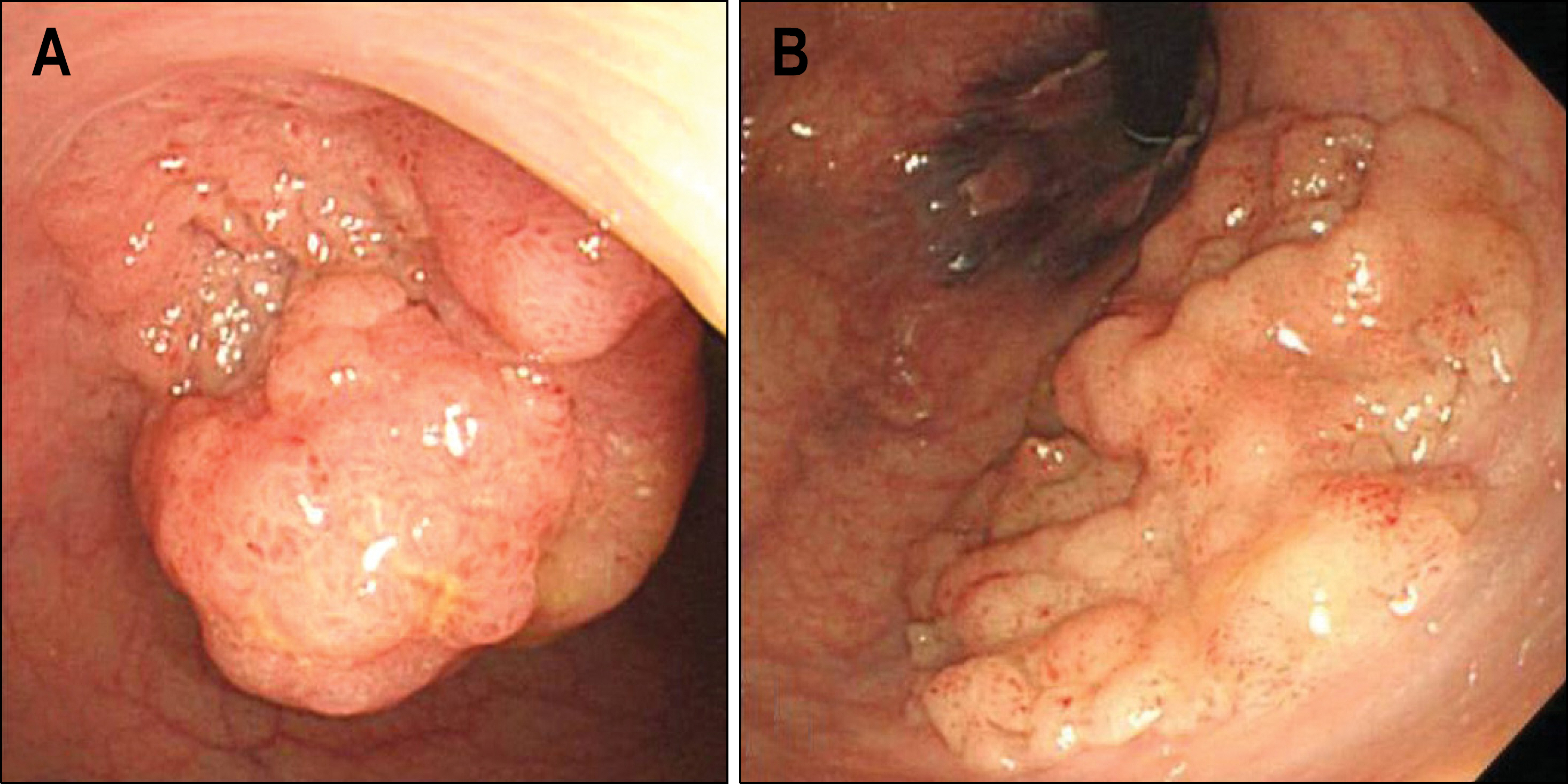
A Case of Synchronous Colonic Laterally Spreading Tumors Treated by Sequential Endoscopic Submucosal Dissection Performed on Two Consecutive Days
- Affiliations
-
- 1Department of Internal Medicine, Keimyung University School of Medicine, Daegu, Korea. Seenae99@Dsmc.Or.Kr
- KMID: 948782
- DOI: http://doi.org/10.4166/kjg.2010.56.3.196
Abstract
- Endoscopic submucosal dissection (ESD) is an useful therapeutic technique for large gastrointestinal epithelial tumors that it provides an en bloc resection. Although there is some controversy about the role of ESD for colorectal lesions, for large lesions in the distal rectum, ESD has the advantage of preserving anal function. However, the large amount of insufflating gas used during the procedure can cause severe abdominal pain and discomfort. Moreover, high intra-luminal pressure caused by a by large amount of gas can cause a micro-perforation. There is no consensus as to whether ESD is the optimal treatment for synchronous large colorectal laterally spreading tumors (LSTs) that cannot be removed en-bloc by conventional endoscopic mucosal resection. Here, a case with two neighboring synchronous large LSTs, one located in the rectum and the other in the distal sigmoid colon, were sequentially removed by separate ESD procedures performed on two consecutive days in a patient who could not tolerate a long procedure.
MeSH Terms
Figure
Reference
-
1. Kudo S. Endoscopic mucosal resection of flat and depressed types of early colorectal cancer. Endoscopy. 1993; 25:455–461.
Article2. Saito Y, Fujii T, Kondo H, et al. Endoscopic treatment for laterally spreading tumors in the colon. Endoscopy. 2001; 33:682–686.
Article3. Tanaka S, Haruma K, Oka S, et al. Clinicopathologic features and endoscopic treatment of superficially spreading colorectal neoplasms larger than 20 mm. Gastrointest Endosc. 2001; 54:62–66.
Article4. Gotoda T, Kondo H, Ono H, et al. A new endoscopic mucosal resection procedure using an insulation-tipped electro-surgical knife for rectal flat lesions: report of two cases. Gastrointest Endosc. 1999; 50:560–563.
Article5. Yamamoto H, Kawata H, Sunada K, et al. Successful en-bloc resection of large superficial tumors in the stomach and colon using sodium hyaluronate and small-caliber-tip transparent hood. Endoscopy. 2003; 35:690–694.
Article6. Fujishiro M, Yahagi N, Nakamura M, et al. Successful outcomes of a novel endoscopic treatment for GI tumors: endoscopic submucosal dissection with a mixture of high-molec-ular-weight hyaluronic acid, glycerin, and sugar. Gastrointest Endosc. 2006; 63:243–249.
Article7. Kim JJ, Lee JH, Jung HY, et al. EMR for early gastric cancer in Korea: a multicenter retrospective study. Gastrointest Endosc. 2007; 66:693–700.8. Min BH, Lee JH, Kim JJ, et al. Clinical outcomes of endoscopic submucosal dissection (ESD) for treating early gastric cancer: comparison with endoscopic mucosal resection after circumferential precutting (EMR-P). Dig Liver Dis. 2009; 41:201–209.
Article9. Tanaka S, Oka S, Chayama K. Colorectal endoscopic submucosal dissection: present status and future perspective, in-cluding its differentiation from endoscopic mucosal resection. J Gastroenterol. 2008; 43:641–651.
Article10. Leung FW. Methods of reducing discomfort during colonoscopy. Dig Dis Sci. 2008; 53:1462–1467.
Article11. Dellon ES, Hawk JS, Grimm IS, Shaheen NJ. The use of carbon dioxide for insufflation during GI endoscopy: a systematic review. Gastrointest Endosc. 2009; 69:843–849.
Article12. Uraoka T, Saito Y, Matsuda T, et al. Endoscopic indications for endoscopic mucosal resection of laterally spreading tu-mours in the colorectum. Gut. 2006; 55:1592–1597.
Article13. Tanaka S, Oka S, Kaneko I, et al. Endoscopic submucosal dissection for colorectal neoplasia: possibility of standardization. Gastrointest Endosc. 2007; 66:100–107.
Article14. Antillon MR, Bartalos CR, Miller ML, Diaz-Arias AA, Ibdah JA, Marshall JB. En bloc endoscopic submucosal dissection of a 14-cm laterally spreading adenoma of the rectum with involvement to the anal canal: expanding the frontiers of endoscopic surgery (with video). Gastrointest Endosc. 2008; 67:332–337.
Article
- Full Text Links
- Actions
-
Cited
- CITED
-
- Close
- Share
- Similar articles
-
- Unexpected Delayed Colon Perforation after the Endoscopic Submucosal Dissection with Snaring of a Laterally Spreading Tumor
- Submerging Endoscopic Submucosal Dissection Leads to Successful En Bloc Resection of Colonic Laterally Spreading Tumor with Submucosal Fat
- Current Status of Colorectal Endoscopic Submucosal Dissection in Korea
- Successful Endoscopic Resection of Residual Colonic Mucosa-Associated Lymphoid Tissue Lymphoma after Polypectomy
- Endoscopic Submucosal Dissection of a Colonic Calcifying Fibrous Tumor


