Clin Endosc.
2020 Jul;53(4):487-490. 10.5946/ce.2019.138.
Endoscopic Submucosal Dissection of a Colonic Calcifying Fibrous Tumor
- Affiliations
-
- 1Department of Internal Medicine, St. Vincent’s Hospital, College of Medicine, The Catholic University of Korea, Suwon, Korea
- KMID: 2504954
- DOI: http://doi.org/10.5946/ce.2019.138
Abstract
- A 49-year-old woman was referred to our hospital for further treatment due to the suspicion of a submucosal tumor in a routine screening colonoscopy. On colonoscopy, a 1-cm sized subepithelial mass with normal overlying mucosa in the hepatic flexure was found. Endoscopic ultrasonography (EUS) showed a homogenous hypoechoic lesion arising from the second and third layer. We were unable to make a final diagnosis because the lesion showed a small tumor with atypical macroscopic morphology including EUS findings. Therefore, endoscopic submucosal dissection was performed for the diagnostic treatment of the tumor. Submucosal dissection was performed just above the muscle layer, and the tumor was removed completely and reliably without any acute complications such as perforation. Based on histopathological findings, we diagnosed a benign, calcifying fibrous tumor (CFT). The present case is the first report of successful endoscopic diagnosis and treatment of colonic CFT mimicking a submucosal tumor.
Keyword
Figure
-
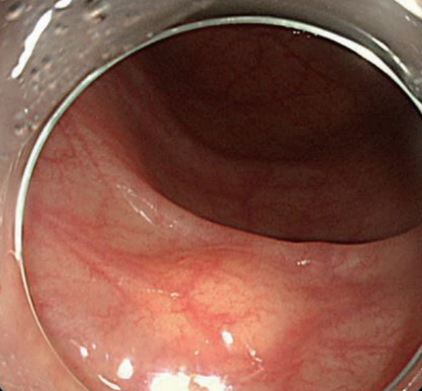
Fig. 1. Endoscopic view of colonic subepithelial tumor.
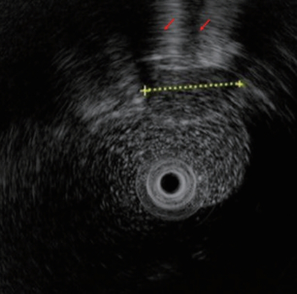
Fig. 2. Endoscopic ultrasonographic evaluation of colonic subepithelial tumor.

Fig. 3. (A) Injection by conventional needle. (B) Circumferential incision. (C) After completion of the whole procedure.

Fig. 4. Pathologic findings of the tumor. (A) Hematoxylin and eosin stained specimens showing hypocellular spindle cell proliferation, abundant hyalinized collagen, and prominent lymphoplasmacytic inflammatory infiltration. (B) Immunohistochemical staining showing tumor cells that were negative for S100.
Reference
-
1. Fetsch JF, Montgomery EA, Meis JM. Calcifying fibrous pseudotumor. Am J Surg Pathol. 1993; 17:502–508.
Article2. Chorti A, Papavramidis TS, Michalopoulos A. Calcifying fibrous tumor: review of 157 patients reported in international literature. Medicine (Baltimore). 2016; 95:e3690.3. Li BJ, Yang XD, Chen WX, Shi YH, Nie ZH, Wu J. Calcifying fibrous tumor of stomach: a case report. Medicine (Baltimore). 2017; 96:e8882.4. Rosenthal NS, Abdul-Karim FW. Childhood fibrous tumor with psammoma bodies. Clinicopathologic features in two cases. Arch Pathol Lab Med. 1988; 112:798–800.5. Pezhouh MK, Rezaei MK, Shabihkhani M, et al. Clinicopathologic study of calcifying fibrous tumor of the gastrointestinal tract: a case series. Hum Pathol. 2017; 62:199–205.
Article6. Kwon JG, Kim EY, Kim YS, et al. [Accuracy of endoscopic ultrasonographic impression compared with pathologic diagnosis in gastrointestinal submucosal tumors]. Korean J Gastroenterol. 2005; 45:88–96.7. Hwang JH, Saunders MD, Rulyak SJ, Shaw S, Nietsch H, Kimmey MB. A prospective study comparing endoscopy and EUS in the evaluation of GI subepithelial masses. Gastrointest Endosc. 2005; 62:202–208.
Article8. Quaedvlieg PF, Visser O, Lamers CB, Janssen-Heijen ML, Taal BG. Epidemiology and survival in patients with carcinoid disease in The Netherlands. An epidemiological study with 2391 patients. Ann Oncol. 2001; 12:1295–1300.
Article9. Ponsaing LG, Kiss K, Loft A, Jensen LI, Hansen MB. Diagnostic procedures for submucosal tumors in the gastrointestinal tract. World J Gastroenterol. 2007; 13:3301–3310.
Article10. Ogasawara N, Izawa S, Mizuno M, et al. Gastric calcifying fibrous tumor removed by endoscopic submucosal dissection. World J Gastrointest Endosc. 2013; 5:457–460.
Article
- Full Text Links
-
- Actions
-
Cited
- CITED
-
- Close
- Share
-
- Similar articles
-
- Calcifying Fibrous Pseudotumor of the Stomach That Was Diagnosed by Endoscopic Submucosal Dissection
- Successful Endoscopic Resection of Residual Colonic Mucosa-Associated Lymphoid Tissue Lymphoma after Polypectomy
- History and Development of Accessories for Endoscopic Submucosal Dissection
- Submerging Endoscopic Submucosal Dissection Leads to Successful En Bloc Resection of Colonic Laterally Spreading Tumor with Submucosal Fat
- Unexpected Delayed Colon Perforation after the Endoscopic Submucosal Dissection with Snaring of a Laterally Spreading Tumor
