Disc Hemorrhages in Patients with both Normal Tension Glaucoma and Branch Retinal Vein Occlusion in Different Eyes
- Affiliations
-
- 1Department of Ophthalmology, Hallym University College of Medicine, Seoul, Korea.
- 2Department of Ophthalmology, Seoul National University College of Medicine, Seoul, Korea. kihopark@snu.ac.kr
- KMID: 754640
- DOI: http://doi.org/10.3341/kjo.2007.21.4.222
Abstract
- PURPOSE: To document the clinical features of disc hemorrhage in patients with branch retinal vein occlusion (BRVO) and normal tension glaucoma (NTG), and to evaluate the relationship between BRVO and NTG with disc hemorrhages. METHODS: From July 2001 to May 2006, sixteen patients with both NTG and BRVO in different eyes were successively collected from outpatient population of Seoul National University Hospital in this observational case series. The frequency and location of disc hemorrhages, history of associated systemic diseases, and the order of the time of diagnosis between NTG and BRVO were studied. RESULTS: All patients had unilateral BRVO, and their mean age was 63.3+/-10.6 years. Disc hemorrhages were detected in eight patients (50%) during the mean follow-up of 26.8 months (range, 3-96 months). Six patients (75%) had disc hemorrhages in the non-BRVO eyes and two patients (25%) in BRVO eyes. Five hemorrhages (62.5%) were located at inferior-temporal quadrant of the optic disc. History of systemic hypertension was identified in 12 patients (75.0%). In 11 patients (68.8%), NTG was diagnosed at the same time as BRVO. CONCLUSIONS: A higher frequency of disc hemorrhages was identified in patients with both BRVO and NTG. Therefore, some cases of NTG, especially with disc hemorrhages, may share a common vascular pathophysiology with BRVO.
MeSH Terms
-
Adult
Aged
Eye Hemorrhage/*etiology/pathology/physiopathology
Female
Fluorescein Angiography
Follow-Up Studies
Fundus Oculi
Glaucoma, Open-Angle/*complications/pathology/physiopathology
Humans
Intraocular Pressure
Male
Middle Aged
Optic Disk/*pathology
Optic Nerve Diseases/*etiology/pathology/physiopathology
Retinal Vein Occlusion/*complications/pathology
Retrospective Studies
Severity of Illness Index
Visual Acuity
Figure
-
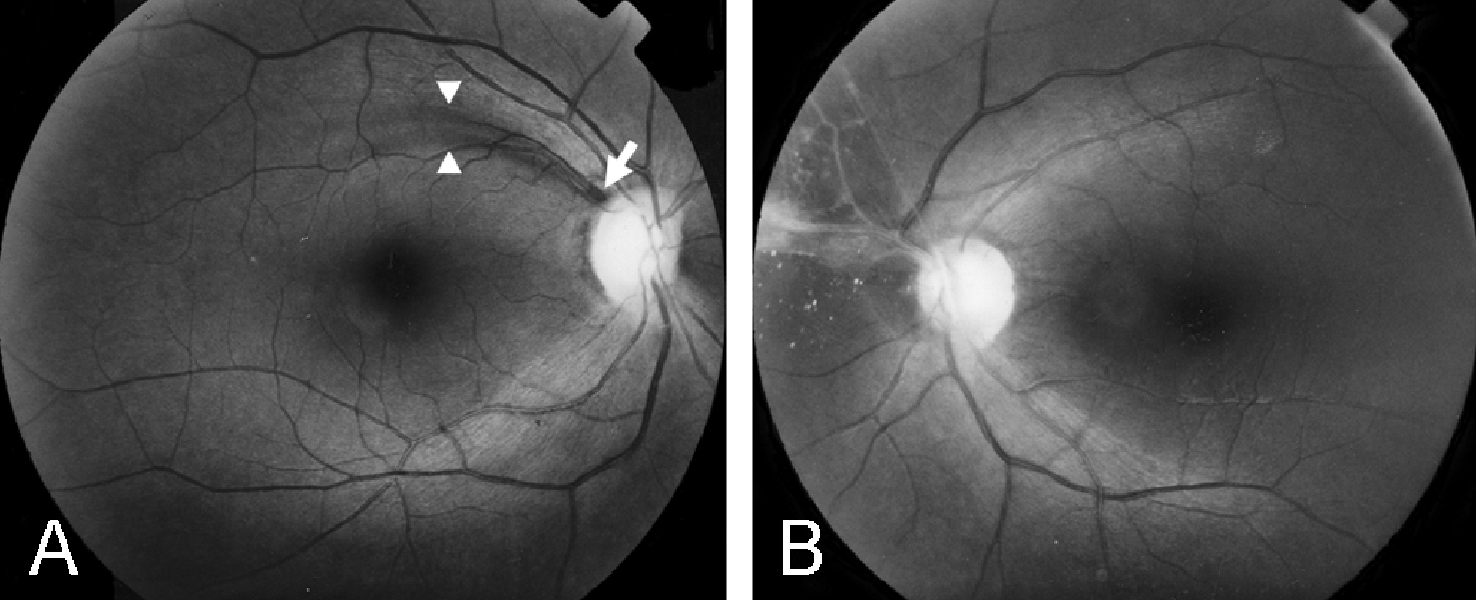
Fig. 1 Retinal nerve fiber layer (RNFL) photographs of a 67 year-old female patient (Case 10). (A) Right eye shows a small wedge shape RNFL defect (arrowheads) with a disc hemorrhage (white arrow) at 11 o'clock of disc. (B) Left eye shows features of chronic phase branch retinal vein occlusion (BRVO), i.e., vascular sheathing, epiretinal membrane, and collateral vessels that note a previous event of BRVO at superior nasal quadrant of retina.
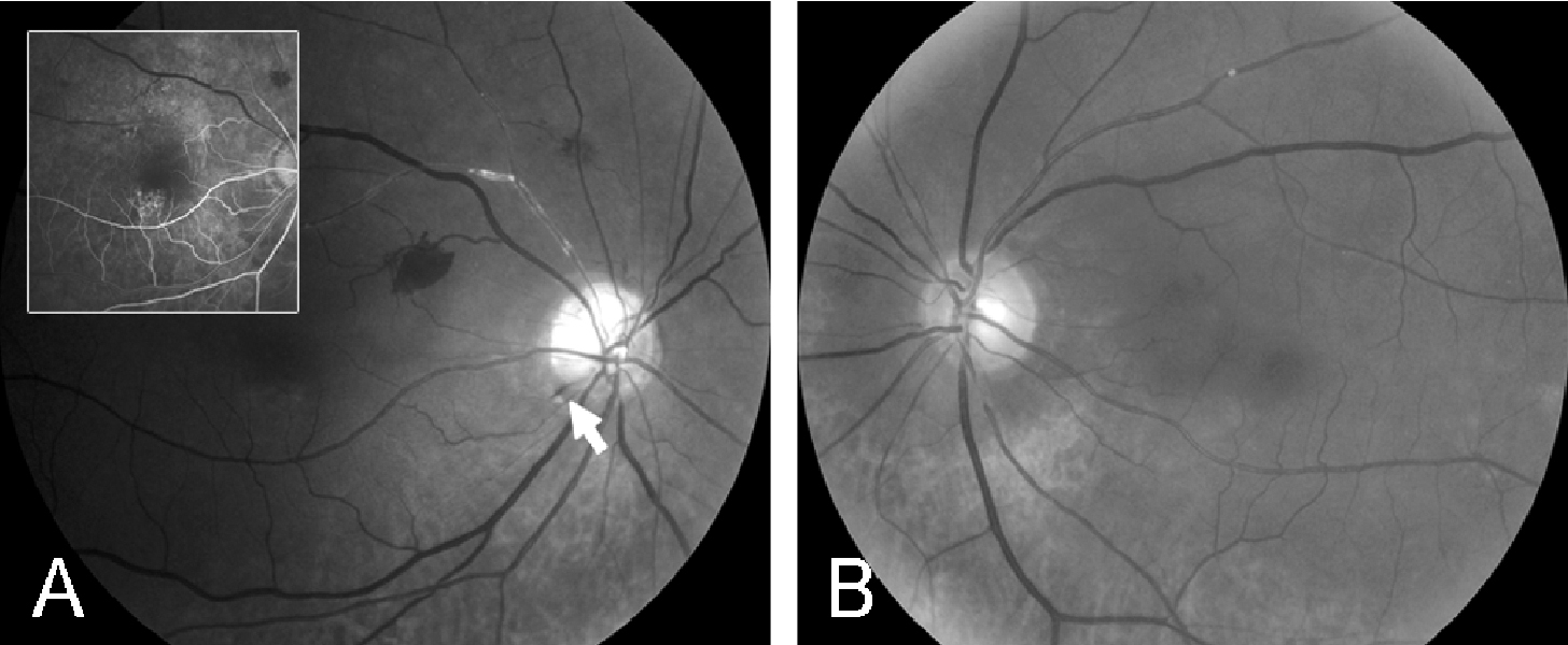
Fig. 2 Retinal nerve fiber layer (RNFL) photographs and fluorescein angiogram of a 73 year-old male patient (Case 1). (A) Right eye shows a small, splint hemorrhage at 7 o'clock of disc (white arrow), and vascular sheathing and retinal hemorrhages at superior temporal quadrant. Fluorescein angiography of right eye (inset picture), taken 3 months prior to RNFL photographies, shows a venous filling delay at superior temporal quadrant. (B) Left eye shows diffuse thinning of RNFL.
Cited by 2 articles
-
Lamina Cribrosa Thickness in the Fellow Eyes of Patients with Unilateral Retinal Vein Occlusion
Yong Il Kim, Tea Yoon Lee, Kyoo Won Lee, Jin Seon Kim
J Korean Ophthalmol Soc. 2015;56(11):1736-1741. doi: 10.3341/jkos.2015.56.11.1736.Comparison of Inner Retinal Thickness between the Fellow Eyes of Unilateral Branch Retinal Vein Occlusion and Normal Control
Gyu Chul Chung, Dong Eun Lee, Chang Ki Yoon, Hyun Woong Kim, Jung Lim Kim
J Korean Ophthalmol Soc. 2017;58(2):165-170. doi: 10.3341/jkos.2017.58.2.165.
Reference
-
1. Susanna R, Drance SM, Douglas GR. Disc hemorrhage in patients with elevated intraocular pressure. Occurrence with and without field changes. Arch Ophthalmol. 1979. 97:284–285.2. Bengtsson B, Holmin C, Krakau CE. Disc haemorrhage and glaucoma. Acta Ophthalmol (Copenh). 1981. 59:1–14.3. Kitazawa Y, Shirato S, Yamamoto T. Optic disc hemorrhage in low-tension glaucoma. Ohthalmology. 1986. 93:853–857.4. Diehl DLC, Quigley HA, Miller NR, et al. Prevalence and significance of disc hemorrhage in a longitudinal study of glaucoma. Arch Ophthalmol. 1990. 108:545–550.5. Ishida K, Yamamoto T, Sugiyama K, et al. Disk hemorrhage is a significantly negative prognostic factor in normal-tension glaucoma. Am J Ophthalmol. 2000. 129:707–714.6. Allingham RR. Allingham RR, Damji K, Freedman S, editors. Chronic open-angle glaucoma and normaltension glaucoma. Shield's textbook of glaucoma. 2005. 5th ed. Philadelphia: Lippincott Williams & Wilkins;chap. 11.7. Sonnsjo B, Krakau CE. Arguments for a vascular glaucoma etiology. Acta Ophthalmol (Copenh). 1993. 71:433–444.8. Soni KG, Woodhouse DF. Retinal vascular occlusion as a presenting feature of glaucoma simplex. Br J Ophthalmol. 1971. 55:192–195.9. Frucht J, Shapiro A, Merin S. Intraocular pressure in retinal vein occlusion. Br J Ophthalmol. 1984. 68:26–28.10. Beaumont PE, Kang HK. Cup-to-disc ratio, intraocular pressure, and primary open-angle glaucoma in retinal venous occlusion. Ophthalmology. 2002. 109:282–286.11. Hayreh SS, Zimmerman MB, Beri M, et al. Intraocular pressure abnormalities associated with central and hemicentral retinal vein occlusion. Ophthalmology. 2004. 111:133–141.12. Luntz MH, Scjenker HI. Retinal vascular accidents in glaucoma and ocular hypertension. Surv Ophthalmol. 1980. 25:163–167.13. Risk factors for branch retinal vein occlusion. Am J Ophthalmol. 1993. 116:286–296.14. Klein BE, Meuer SM, Knudtson MD, et al. The relationship of optic disc cupping to retinal vein occlusion: The beaver dam eye study. Am J Ophthalmol. 2006. 141:859–862.15. Kim SJ, Park KH. Four cases of normal-tension glaucoma with disk hemorrhage combined with branch retinal vein occlusion in the contralateral eye. Am J Ophthalmol. 2004. 137:357–359.16. Heijl A. Frequent disk photography and computerized perimetry in eyes with optic disk haemorrhage. Acta Ophthalmol. 1986. 64:274–281.17. Klein BE, Klein R, Sponsel WE, et al. Prevalence of glaucoma. The Beaver Dam Eye Study. Ophthalmology. 1992. 99:1499–1504.18. Healey PR, Mitchell P, Smith W, et al. Optic disc hemorrhages in a population with and without signs of glaucoma. Ophthalmology. 1998. 105:216–223.19. Yamamoto T, Iwase A, Kawase K, et al. Optic disc hemorrhages detected in a large-scale eye screening project. J Glaucoma. 2004. 13:356–360.20. Hendrickx KH, van den Enden A, Rasker MT, Hoyng PF. Cumulative incidence of patients with disc hemorrhages in glaucoma and the effect of therapy. Ophthalmology. 1994. 101:1165–1172.21. Rubinstein K, Jones EB. Retinal vein occlusion: long-term prospects, 10 years follow-up on 143 patients. Br J Ophthalmol. 1976. 60:148–150.22. Sonnsjo B. Similarities between disc haemorrhages and thrombosis of the retinal veins. Int Ophthalmol. 1992. 16:235–238.23. Lee JB, Cho YS, Choe YJ, Hong YJ. The prevalence of glaucoma in Korean adults. J Korean Ophthalmol Soc. 1993. 34:65–69.24. Alterman M, Henkind P. Radial peripapillary capillaries of the retina:II. Possible role in Bjerrum scotoma. Br J Ophthalmol. 1968. 52:26–31.25. Duker JS, Brown GC. Anterior location of the crossing artery in branch retinal vein occlusion. Arch Ophthalmol. 1989. 107:998–1000.26. Zhao J, Sastry SM, Sperduto RD, et al. Arteriovenous crossing patterns in branch retinal vein occlusion. The Eye Disease Case-Control Study Group. Ophthalmology. 1993. 100:423–428.27. Lindblom B. Open angle glaucoma and non-central retinal vein occlusion--the chicken or the egg? Acta Ophthalmol Scand. 1998. 76:329–333.
- Full Text Links
-
- Actions
-
Cited
- CITED
-
- Close
- Share
-
- Similar articles
-
- Vitrectomy for Vitreous Hemorrhage Associated with Branch Retinal Vein Occlusion
- Bevacizumab Therapy for Branch Retinal Vein Occlusion Associated with Normal Tension Glaucoma
- Clinical observation of the bilateral branch vein occlusion
- Cup-to-Disc Ratio, Intraocular Pressure, and Occlusion Site in Branch Retinal Vein Occlusion
- Clinical Analysis of the Hemispheric Retinal Vein Occlusion
