Unenhanced Spiral CT in Acute Ureteral Colic: A Replacement for Excretory Urography?
- Affiliations
-
- 1Sung Kyun Kwan Univ, Sch Med Dept Radiol Samsung Med Ctr, Kangnam Gu, 50 Ilwon Dong, Seoul 135710, South Korea
- KMID: 754118
- DOI: http://doi.org/10.3348/kjr.2001.2.1.14
Abstract
OBJECTIVE
To compare the usefulness of unenhanced spiral CT (UCT) with that of excretory urography (EU) in patients with acute flank pain. MATERIALS AND METHODS: Thirty patients presenting with acute flank pain under-went both UCT and EU. Both techniques were used to determine the presence, size, and location of urinary stone, and the presence or absence of secondary signs was also evaluated. The existence of ureteral stone was confirmed by its removal or spontaneous passage during follow-up. The absence of a stone was determined on the basis of the clinical and radiological evidence. RESULTS: Twenty-one of the 30 patients had one or more ureteral stones and nine had no stone. CT depicted 22 of 23 calculi in the 21 patients with a stone, and no calculus in all nine without a stone. The sensitivity and specificity of UCT were 96% and 100%, respectively. EU disclosed 14 calculi in the 21 patients with a stone and no calculus in eight of the nine without a stone. UCT and EU demon-strated secondary signs of ureterolithiasis in 15 and 17 patients, respectively. CONCLUSION: For the evaluation of patients with acute flank pain, UCT is an excellent modality with high sensitivity and specificity. In near future it may replace EU.
Keyword
MeSH Terms
Figure
-

Fig. 1 A 43-year-old patient with acute flank pain. A, B. Transaxial scans through the kidneys and bladder, respectively. Dilatation of the left renal pelvis (arrow in A) and a left distal ureter stone (arrow in B) are seen. Arrowhead: right renal calyceal stone.
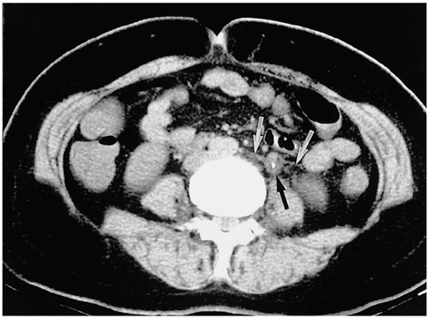
Fig. 2 A 46-year-old man with acute flank pain. A transaxial CT scan through the lower poles of the kidneys demonstrates soft tissue rim sign around the ureter stone (black arrow) and periureteral strand in adjacent fat tissue (white arrows).
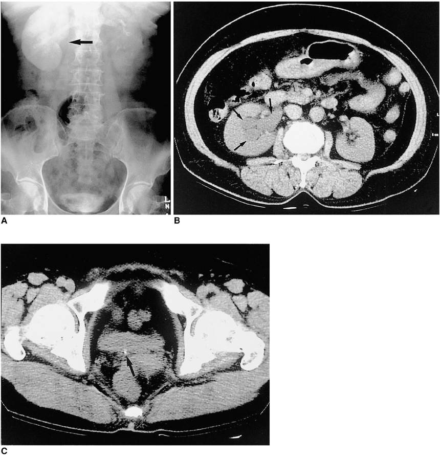
Fig. 3 A 57-year-old woman with acute right flank pain. A: EU; B, C: Transaxial UCT scans through the kidneys and bladder. A dense delayed nephrogram is seen in A, but the entire EU study revealed no stone shadow. On UCT a tiny stone at the right ureterovesical junction (arrow in C) is seen along with dilatation of the renal pelvocalyceal system (arrows in B).

Fig. 4 A 45-year-old male patient with acute flank pain. A: EU; B, C: UCT. Two separate stones (arrows) are demonstrated in the left proximal ureter in A, but on UCT (B, C) they are seen as one elongated stone. UCT (C), however, demonstrates small renal stones (arrowheads), not seen in A, at the lower pole of the left kidney.
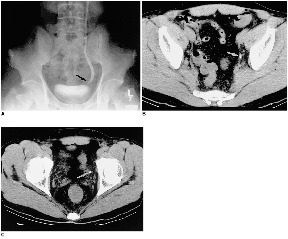
Fig. 5 A 53-year-old man with acute flank pain. A: EU; B, C: Two consecutive CT scans of the pelvis. A single stone is seen in the left distal ureter in A; UCT, however, clearly demonstrates two separate distal ureter stones (white arrows) in B and C. The smaller stone in the more distal ureter, seen in C, is obscured in A.
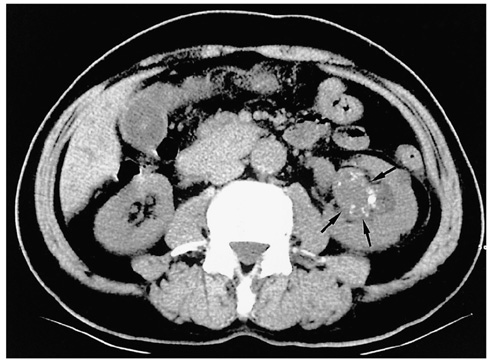
Fig. 6 Incidentally discovered transitional cell carcinoma of the left kidney (arrows) in a 72-year-old man who presented with acute flank pain and hematuria.
Reference
-
1. Sierakowski R, Finlayson B, Landes RR, et al. The frequency of urolithiasis in hospital discharge diagnoses in the United States. Invest Urol. 1978. 15:438–441.2. Miller OF, Rineer SK, Reichard SR, et al. Prospective comparison of unenhanced spiral computed tomography and intravenous urogram in the evaluation of acute flank pain. Urology. 1998. 52:982–987.3. Levine JA, Neitlich J, Verga M, Dalrymple N, Smith RC. Ureteral calculi in patients with flank pain: correlation of plain radiography with unenhanced helical CT. Radiology. 1997. 204:27–31.4. Smith RC, Ronsenfield AT, Choe KA, et al. Acute flank pain: comparison of non-contrast-enhanced CT and intravenous urography. Radiology. 1995. 194:789–794.5. Miller OF, Kane CJ. Unenhanced helical computed tomography in the evaluation of acute flank pain. Curr Opin Urol. 2000. 10:123–129.6. Dalymple NC, Verga M, Anderson KR, et al. The value of unenhanced helical computed tomography in the management of acute flank pain. J Urol. 1998. 159:735–774.7. Smith RC, Verga M, McCarthy S, Rosenfield AT. Acute ureteral obstruction: value of secondary signs of helical unenhanced CT. AJR. 1996. 167:1109–1113.8. Heneghan JP, Dalrymple NC, Verga M, Rosenfield AT, Smith RC. Soft tissue "rim" sign in the diagnosis of ureteral calculi with use of unenhanced helical CT. Radiology. 1997. 202:709–711.9. Kawashima A, Sandler CM, Boridy IC, et al. Unenhanced helical CT of ureterolithiasis: value of the tissue rim sign. AJR. 1997. 168:997–1000.10. Boridy IC, Nikloaidis P, Kawashima A, Sandler CM, Goldman SM. Noncontrast helical CT for ureteral stones. World J Urol. 1998. 16:18–21.11. Boulay I, Holtz P, Foley WD, White B, Begun FP. Ureteral calculi: diagnostic efficacy of helical CT and implications for treatment of patients. AJR. 1999. 172:1485–1490.12. Niall O, Russel J, McGregor R, Duncan H, Mullins J. A comparison of noncontrast computed tomography with excretory urography in the assessment of acute flank pain. J Urol. 1999. 161:534–537.13. Fielding JR, Steele G, Fox LA, Heller H, Loughlin KR. Spiral computed tomography in the evaluation of acute flank pain: a replacement for excretory urography. J Urol. 1997. 157:2071–2073.14. Chen MYM, Zagoria RJ. Can noncontrast helical computed tomography replace intravenous urography for evaluation of patients with acute urinary tract colic? J Emerg Med. 1999. 17:299–303.15. Yilmaz S, Sindel T, Arslan G, et al. Renal colic: comparison of spiral CT, US and IVU in the detection of ureteral calculi. Eur Radiol. 1998. 8:212–217.16. Sommer FG, Jeffrey RB Jr, Rubin GD, et al. Detection of ureteral calculi in patients with suspected renal colic: value of reformatted noncontrast helical CT. AJR. 1995. 165:509–513.17. Denton ERE, Mackenzie A, Greenwell T, Popert R, Rankin SC. Unenhanced helical CT for renal colic: is the radiation dose justifiable? Clin Radiol. 1999. 54:444–447.18. Vieweg J, Teh C, Freed K, et al. Unenhanced helical computed tomography for the evaluation of patients with acute flank pain. J Urol. 1998. 160:679–684.19. Svedstrom E, Alanen A, Nurmi M. Radiologic diagnosis of renal colic: the role of plain films, excretory urography and sonography. Eur J Radiol. 1990. 11:180–183.20. Mutgy A, Williams JW, Nettleman M. Renal colic: utility of the plain abdominal roentgenogram. Arch Intern Med. 1991. 151:1589–1591.21. Saita H, Matsukawa M, Fukushima H, Ohyama C, Nagata Y. Ultrasound diagnosis of ureteral stones: its usefulness with subsequent excretory urography. J Urol. 1987. 140:28–31.22. Berland LL, Smith JK. Multidetector-array CT: Once again, technology creates new opportunities. Radiology. 1998. 209:327–329.23. Hu H, He HD, Foley WD, et al. Four multidetector-row helical CT: image quality and volume coverage speed. Radiology. 2000. 215:55–62.24. Kawashima A, Sandler CM, West O, et al. Detection of ureterolithiasis with unenhanced multidetector-array helical CT: comparison of 2.5-mm and 5.0-mm slice thickness. Radiology. 2000. 217(P):223.
- Full Text Links
-
- Actions
-
Cited
- CITED
-
- Close
- Share
-
- Similar articles
-
- Is excretory urography necessary in the staging work-up of uterine cervical carcinoma?
- Comparision of Unenhanced Helical Computerized Tomography and Intravenous Urography in the Radiologic Evaluation of Acute Flank Pain
- An Aspergilloma Mistaken for a Pelviureteral Stone on Nonenhanced CT: A Fungal Bezoar Causing Ureteral Obstruction
- The Value of Excretory Urography in Staging Bladder Cancer
- The Usefulness of Unenhanced Helical Computerized Tomography in Patients with Urinary Calculi
