Korean J Radiol.
2003 Sep;4(3):191-193. 10.3348/kjr.2003.4.3.191.
Gossypiboma of the Leg: MR Imaging Characteristics: A Case Report
- Affiliations
-
- 1Department of Radiology, Tri-Service General Hospital, National Defense Medical Center, Taipei, Taiwan.
- KMID: 754024
- DOI: http://doi.org/10.3348/kjr.2003.4.3.191
Abstract
- We report a 22-year-old man with a solid mass in the right proximal leg, which was furned out to be a gossypiboma. MR imaging revealed a well-defined mass lesion that showed intermediate signal intensity at T1-weighted imaging (T1WI) and slightly high signal intensity at T2-weighted imaging (T2WI). Wavy, low-signal-intensity stripes were visible within the fluid-filled central cavity. At surgical exploration, a sponge, retained after previous knee surgery, was discovered, and it was found that a granuloma had developed. Pathologic examination revealed granulomatous inflammation, with lymphocyte and giant cell infiltration. The presence of wavy, low-signal-intensity gauze fibers at T2WI may be a characteristic MR appearance of gossypiboma.
Keyword
Figure
-
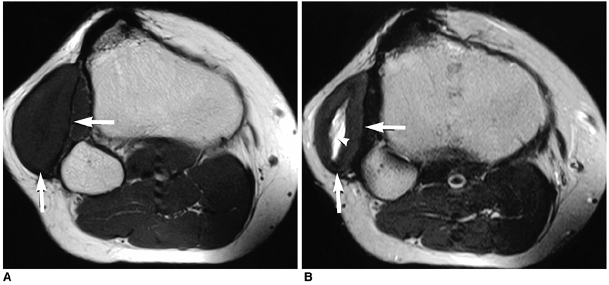
Fig. 1 MR imaging of the right proximal leg. Axial T1-weighted (A) and T2-weighted (B) MR images depict a well-circumscribed, elliptical mass (arrows) with a central, fusiform, fluid-filled cavity in the deep subcutaneous layer of the right proximal leg. Note the wavy, low-signal-intensity structures (arrowhead) in the central cavity, representing the gauze fibers.
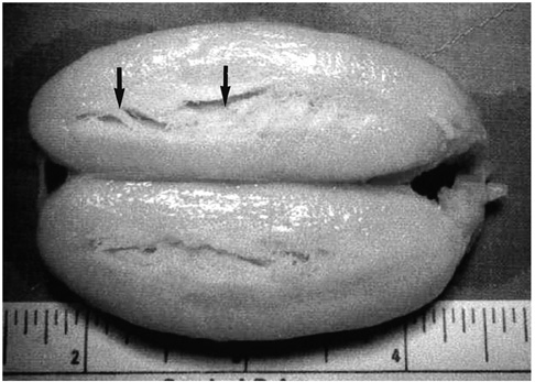
Fig. 2 Gross pathologic specimen shows a well-encapsulated mass with a fusiform central cavity. The gauze fibers (arrows) are clearly depicted.
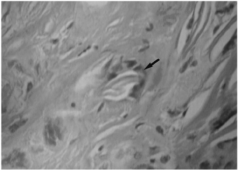
Fig. 3 Microscopic examination of the tumor revealed fibrosis, and lymphocyte and giant cell infiltration had occurred. Note the presence of giant cells, which phagocytosed the gauze fibers (arrow).
Reference
-
1. O'Connor AR, Coakley FV, Meng MV, Eberhardt S. Imaging of retained surgical sponges in the abdomen and pelvis. AJR Am J Roentgenol. 2003. 180:481–489.2. Choi BI, Kim SH, Yu ES, Chung HS, Han MC, Kim CW. Retained surgical sponge: diagnosis with CT and sonography. AJR Am J Roentgenol. 1988. 150:1047–1050.3. Kuwashima S, Yamato M, Fujioka M, Ishibashi M, Kogure H, Tajima Y. MR findings of surgical sponges and towels: report of two cases. Radiation Med. 1993. 11:98–101.4. Sugimura H, Tamura S, Kakitsubata Y, et al. Magnetic resonance imaging of retained surgical sponges. Clin Imaging. 1992. 16:259–262.5. Nabors MW, McCrary ME, Clemente RJ, et al. Identification of a retained surgical sponge using magnetic resonance imaging. Neurosurgery. 1986. 18:496–498.6. Roumen RMH, Weerdenburg HPG. MR features of a 24-year-old gossypiboma. Acta Radiol. 1998. 29:176–178.7. Olnick HM, Weens SH, Rogers JV Jr. Radiologic diagnosis of retained surgical sponges. J Am Med Assoc. 1955. 159:1525–1527.8. Resnick D. Resnick Donald, editor. Tumor and tumor-like lesions of soft tissues. Diagnosis of bone and joint disorders. 2002. 4th ed. Philadelphia: Saunders;4129–4273.
- Full Text Links
-
- Actions
-
Cited
- CITED
-
- Close
- Share
-
- Similar articles
-
- Intracranial Gossypiboma Mimicking a Recurrent Low Grade Astrocytoma: Case Report
- Pathologic Fracture of Femoral Neck due to Mass suspicious of Gossypiboma in Proximal Thigh: Case Report
- Retroperitoneal Gossypiboma
- Gossypiboma Mimicking a Soft Tissue Tumor
- A Case Report of Neglected Gossypiboma Causing Abdominal Pain for 20 Years Post-Cesarean Section
