Hyaline Vascular-Type Castleman Disease Presenting as an Esophageal Submucosal Tumor: Case Report
- Affiliations
-
- 1Department of Diagnostic Radiology, College of Medicine, Dong-A University, Pusan, Korea. gnlee@daunet.donga.ac.kr
- 2Department of Pathology, College of Medicine, Dong-A University, Pusan, Korea.
- 3Department of Thoracic and Cardiovascular Surgery, College of Medicine, Dong-A University, Pusan, Korea.
- 4Department of Internal Medicine, College of Medicine, Dong-A University, Pusan, Korea.
- KMID: 753919
- DOI: http://doi.org/10.3348/kjr.2006.7.1.73
Abstract
- Castleman disease is a relatively rare disorder of lymphoid tissue that involves the gastrointestinal tract in a variety of clinical and pathologic manifestations. A submucosal location has never been described in the medical literature. We report a case of esophageal Castleman disease involving thesubmucosal layer in a 62-year-old man, which was confirmed on pathology. Esophagography and CT demonstrated an intramural tumor, and a leiomyoma or leiomyosarcoma was suspected based on the known incidence of such tumors.
Keyword
MeSH Terms
Figure
-
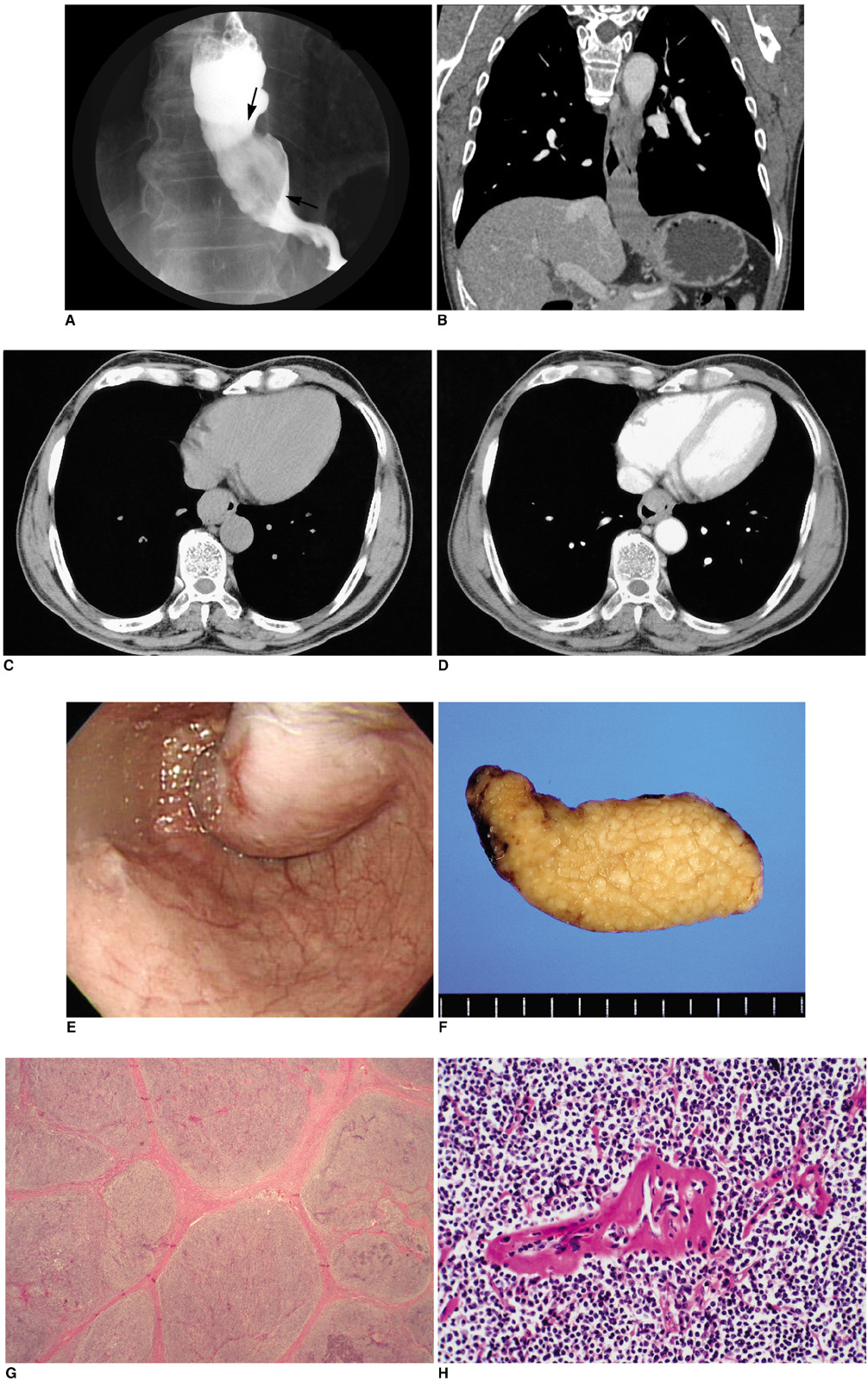
Fig. 1 A 62-year-old man with esophageal Castleman disease. A. Spot radiograph from a single-contrast esophagogram reveals a 6-cm filling defect (arrows) arising from the lateral esophageal wall. B. Coronal reformation CT image shows a definite extent of the disease. C, D. Axial CT images, before (C) and after contrast injection (D), display focal thickening in the anterior wall of the lower esophagus, with circumferential luminal air, and the intramural mass shows homogeneous enhancement. E. Endoscopy reveals a smoothly bulging mass with smooth surface and focal reddish coloration on the distal esophagus. F. Macroscopy of the excised tumor demonstrates a multilobulated mass with whitish color and smooth margin without hemorrhage or necrosis. G, H. Microscopically, the esophageal mass reveals nodular lymphoid areas, a marked expansion of the mantle zone, and small, relatively inconspicuous germinal centers (H & E staining, ×10). The follicles show marked vascular proliferation with hyalinization (H & E staining, ×200).
Reference
-
1. Zamir A, Prasher G, Moukarzel AA, Zeien L, Feldman F. Castleman's disease: a rare cause of hematemesis. J Pediatr Gastroenterol Nutr. 1999. 28:112–115.2. Wengrower D, Libson E, Okon E, Goldin E. Gastrointestinal manifestation in Castleman's disease. Am J Gastroenterol. 1990. 85:1179–1182.3. Kaneko T, Takahashi S, Takeuchi T, Goto T, Kitamura T. Castleman's disease in the retroperitoneal space. J Urol. 2003. 169:265–266.4. Kojima M, Nakamura S, Iijima M, Murayama K, Sakata N, Masawa N. Lymphoid variant of hyaline vascular Castleman's disease containing numerous mantle zone lymphocytes with clear cytoplasm. APMIS. 2005. 113:75–80.5. Seo BK, Oh YW, Cho KR, Lee NJ, Kim JH, Kim IS, et al. Imaging findings of Castleman's disease localized in the axilla: a case report. Korean J Radiol. 2002. 3:136–139.6. Park KS, Choi YJ, Song KS. Hyaline-vascular type Castleman's disease involving both orbits. Acta Ophthalmol Scand. 2002. 80:537–539.7. Gaunt GA, Gostout BS, Remstein E, Cliby WA. Pelvic Castleman disease presenting as vaginal occlusion. Obstet Gynecol. 2002. 100:1082–1085.8. Sotrel A, Castellano-Sanchez AA, Prusmack C, Birchansky S, Brathwaite C, Ragheb J. Castleman's disease in a child presenting with a partly mineralized solitary meningeal mass. Pediatr Neurosurg. 2003. 38:232–237.9. Serin E, Ozer B, Gumurdulu Y, Yildirim T, Barutcu O, Boyacioglu S. A case of castleman's disease with "downhill" varices in the absence of superior vena cava obstruction. Endoscopy. 2002. 34:160–162.10. Shirakusa T, Iwsaki A, Okazaki M. Downhill esophageal varices caused by benign giant lymphoma. Scand J Thorac Cardiovasc Surg. 1988. 22:135–138.
- Full Text Links
-
- Actions
-
Cited
- CITED
-
- Close
- Share
-
- Similar articles
-
- Hyaline-vascular Variant of Castleman's Disease in Retroperitoneum
- A Case of Retroperitoneal Castleman`s Disease
- Roentgenogram of the Issue : A Case of Castleman`s Disease(Hyaline Vascular type)
- A Case of Castleman's Disease Presenting as an Isolated Hypervascular Tumor in Neck
- A Case of Localized Castleman's Disease in a Patient with Rheumatoid Arthritis
