Evaluating anchorage loss in upper incisors during distalization of maxillary posterior teeth using clear aligners in adult patients: A prospective randomized study
- Affiliations
-
- 1Faculty of Dentistry, Karadeniz Technical University, Trabzon, Türkiye
- KMID: 2554368
- DOI: http://doi.org/10.4041/kjod23.150
Abstract
Objective
To evaluate the effect of clear aligner treatment and differential sequence distalization of maxillary posterior teeth on anchorage loss in the upper incisors (U1s).
Methods
This study used lateral cephalometries and digital models of 12 patients treated with 33% sequential distalization (group 1, mean age: 22.9 ± 0.7 years, five males, seven females) and 12 treated with 50% sequential distalization (group 2, mean age: 25.83 ± 0.5 years, three males, nine females) acquired before and after distalization of upper second premolars (U5) and upper first molars (U6) and upper second molars (U7). The amount of distalization was determined as 2.5 mm in both the groups. Independent Samples t test was used to compare normally distributed parameters. Mann– Whitney U and Wilcoxon tests were used to compare parameters that were not normally distributed.
Results
In both groups, the posterior teeth significantly moved by tipping distally and the U1s were displaced anteriorly. Increase in maxillary posterior transverse width (P < 0.001) and distopalatal rotation were observed in U5, U6, and U7 after distalization. It was also observed that U1 was significantly more proclined (1.82°; P < 0.001) and protruded (0.62 mm; P < 0.001), and the overjet (0.45 mm; P < 0.001) increased more in group 1 than in group 2.
Conclusions
After sequential distalization of maxillary posterior teeth, more anchorage loss was observed in the anterior region in group 1 than in group 2.
Keyword
Figure
-
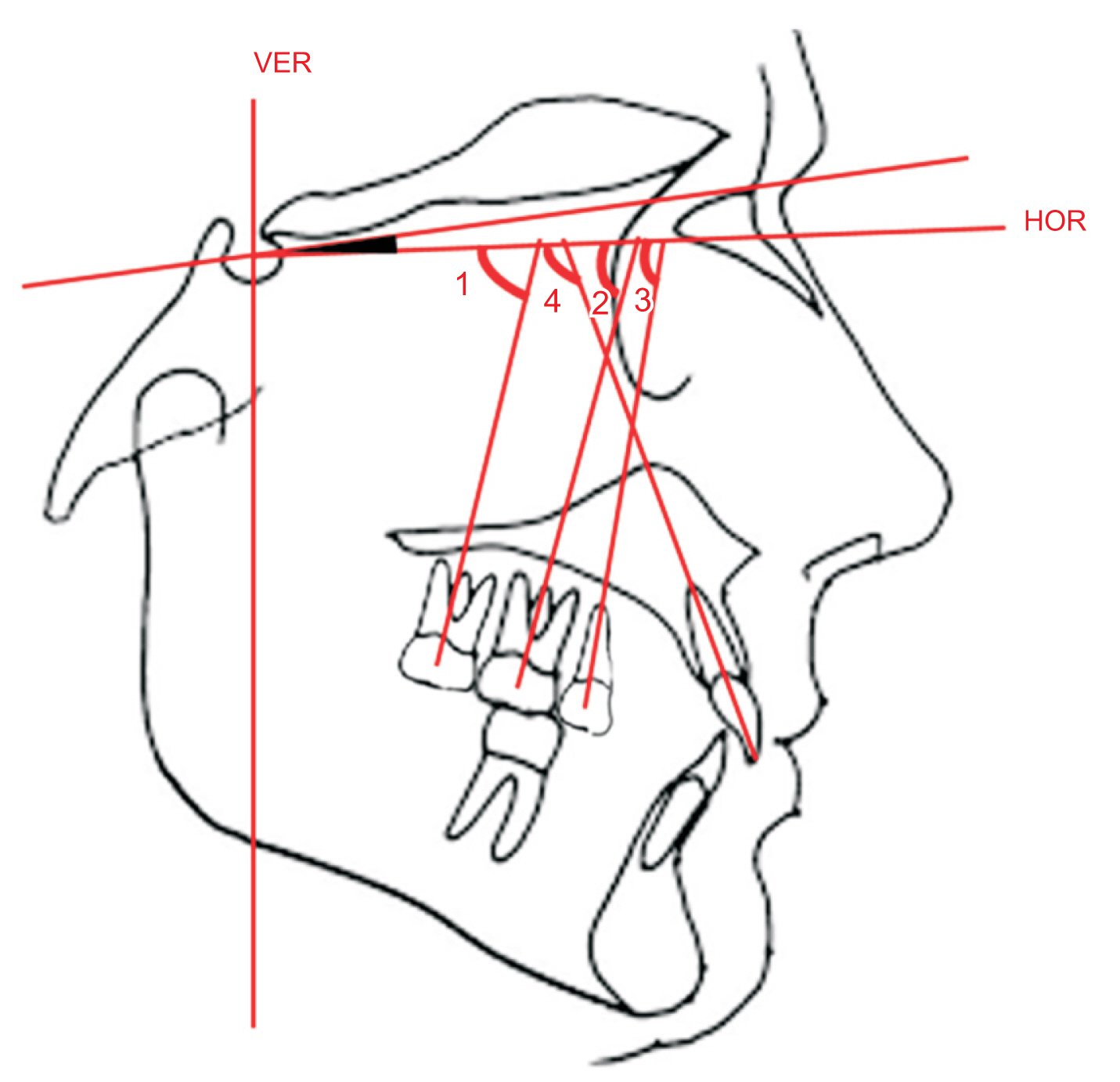
Figure 1 Dental angular measurements on cephalometric radiography. (1) U7/HR, (2) U6/HR, (3) U5/HR, (4) U1/HR. Determination of the long-axis of the teeth: crown tip of U1 and root apex point were marked, crown centroid point of U5 and root apex point were marked, crown centroid point of U6 and U7 and trifurcation point were marked. Centroid point of crown: the midpoint of the mesial and distal convexity of the crown of the molar tooth was accepted as the centroid point of crown. U7, upper second molars; U6, upper first molars; U5, upper second premolars; U1, upper incisor; HR, horizontal plane; VER, vertical plane; HOR, horizontal plane.
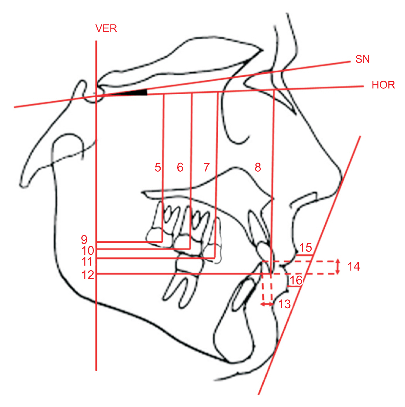
Figure 2 Dental linear measurements on cephalometric radiography. (5) The distance from crown centroid point of U7 to the horizontal plane (U7-HR), (6) the distance from crown centroid point of U6-HR, (7) the distance from crown centroid point of U5-HR, (8) the distance from crown tip point of U1-HR, (9) the distance from crown centroid point of U7 to the vertical plane (U7-VR), (10) the distance from crown centroid point of U6-VR, (11) the distance from crown centroid point of U5-VR, (12) the distance from crown tip point of U1-VR, (13) overjet, (14) overbite, (15) Ls-E, (16) Li-E. U7, upper second molars; U6, upper first molars; U5, upper second premolars; U1, upper incisor; Ls, labrale superius; Li, labrale inferius; E, the line between the soft tissue pogonion and tip of the nose; VER, vertical plane; SN, sella-nasion plane; HOR, horizontal plane.
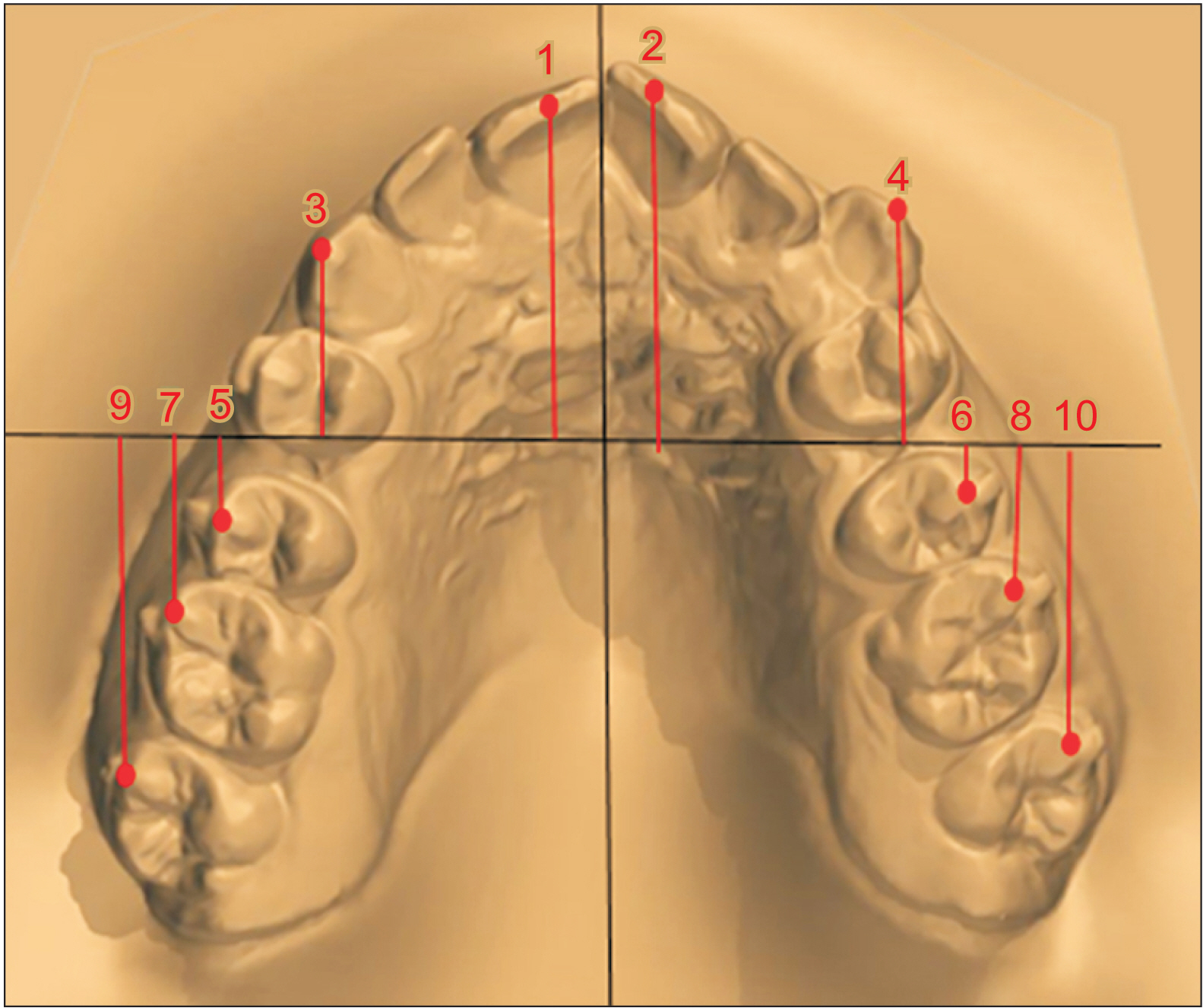
Figure 3 Sagittal linear measurements on the digital model. Line G: line tangent to inferior border of palatal rugae and perpendicular to midpalatal raphe. (1) The distance from incisor tip of 11 to G line, (2) the distance from incisor tip of 21 to G line, (3) the distance from incisor tip of 13 to G line, (4) the distance from crown tip of 23 to G line, (5) the distance from buccal cusp tip of 15 to G line, (6) the distance from buccal cusp tip of 25 to G line, (7) the distance from mesiobuccal cusp tip of 16 to G line, (8) the distance from mesiobuccal cusp tip of 26 to G line, (9) the distance from mesiobuccal cusp tip of 17 to G line, (10) the distance from mesiobuccal cusp tip of 27 to G line.
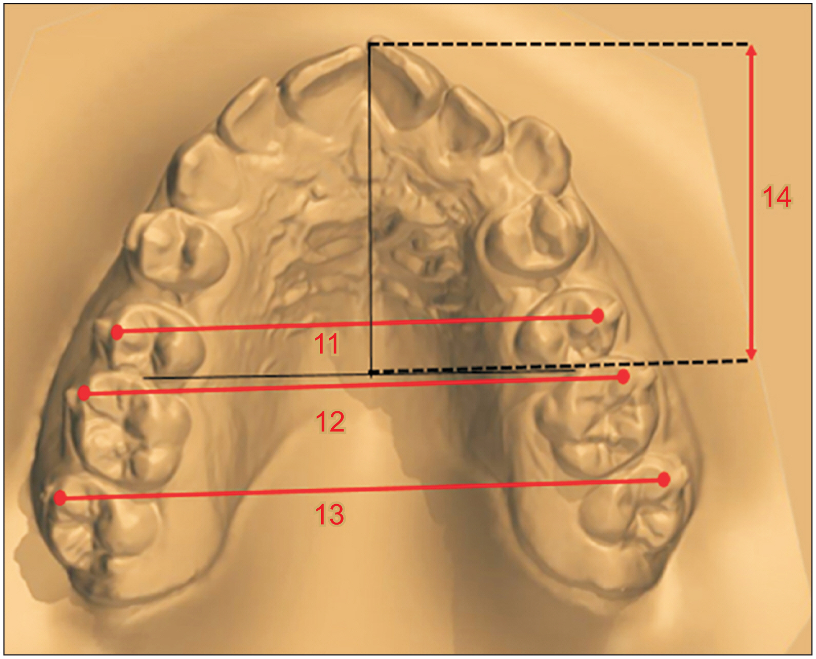
Figure 4 Transverse measurements and arc length (mm) on the digital model. (11) Transverse width between 15–25, (12) transverse width between 16–26, (13) transverse width between 17–27, (14) arch length: the distance between the contact point of the upper incisors and the mesial contact points of the upper first molars.
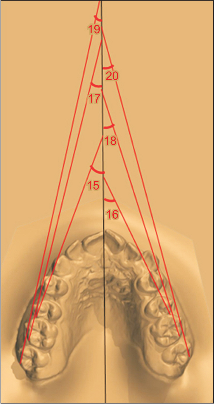
Figure 5 Rotation measurements on the digital model. Upper right second premolar: the angle between the mid-palatal raphe and the line passing through the mesial and distal convex ridges of right maxillary second premolar. Upper left second premolar: the angle between the mid-palatal raphe and the line passing through the mesial and distal convex ridges of left maxillary second premolar. Upper right first molar: the angle between the mid-palatal raphe and the line passing through the mesiobuccal and distobuccal cusps of the right maxillary first molar. Upper left first molar: the angle between the mid-palatal raphe and the line passing through the mesiobuccal and distobuccal cusps of the left maxillary first molar. Rotation measurement of upper right second molar: the angle between the mid-palatal raphe and the line passing through the mesiobuccal and distobuccal cusps of the right maxillary second molar. Upper left second molar: the angle between the mid-palatal raphe and the line passing through the mesiobuccal and distobuccal cusps of the left maxillary second molar.
Reference
-
1. Lione R, Franchi L, Laganà G, Cozza P. 2015; Effects of cervical headgear and pendulum appliance on vertical dimension in growing subjects: a retrospective controlled clinical trial. Eur J Orthod. 37:338–44. https://doi.org/10.1093/ejo/cju061. DOI: 10.1093/ejo/cju061. PMID: 25316493.
Article2. Park CO, Sa'aed NL, Bayome M, Park JH, Kook YA, Park YS, et al. 2017; Comparison of treatment effects between the modified C-palatal plate and cervical pull headgear for total arch distalization in adults. Korean J Orthod. 47:375–83. https://doi.org/10.4041/kjod.2017.47.6.375. DOI: 10.4041/kjod.2017.47.6.375. PMID: 29090125. PMCID: PMC5653686.
Article3. Clemmer EJ, Hayes EW. 1979; Patient cooperation in wearing orthodontic headgear. Am J Orthod. 75:517–24. https://doi.org/10.1016/0002-9416(79)90070-8. DOI: 10.1016/0002-9416(79)90070-8. PMID: 286553.
Article4. Ravera S, Castroflorio T, Garino F, Daher S, Cugliari G, Deregibus A. 2016; Maxillary molar distalization with aligners in adult patients: a multicenter retrospective study. Prog Orthod. 17:12. https://doi.org/10.1186/s40510-016-0126-0. DOI: 10.1186/s40510-016-0126-0. PMID: 27041551. PMCID: PMC4834290. PMID: 970deba192ee417f921a2b9ccfcdac13.
Article5. Bolla E, Muratore F, Carano A, Bowman SJ. 2002; Evaluation of maxillary molar distalization with the distal jet: a comparison with other contemporary methods. Angle Orthod. 72:481–94. https://pubmed.ncbi.nlm.nih.gov/12401059/.6. Fontana M, Cozzani M, Caprioglio A. 2012; Non-compliance maxillary molar distalizing appliances: an overview of the last decade. Prog Orthod. 13:173–84. https://doi.org/10.1016/j.pio.2011.10.002. DOI: 10.1016/j.pio.2011.10.002. PMID: 23021121.
Article7. Yu IJ, Kook YA, Sung SJ, Lee KJ, Chun YS, Mo SS. 2014; Comparison of tooth displacement between buccal mini-implants and palatal plate anchorage for molar distalization: a finite element study. Eur J Orthod. 36:394–402. https://doi.org/10.1093/ejo/cjr130. DOI: 10.1093/ejo/cjr130. PMID: 22051536.
Article8. Fuziy A, Rodrigues de Almeida R, Janson G, Angelieri F, Pinzan A. 2006; Sagittal, vertical, and transverse changes consequent to maxillary molar distalization with the pendulum appliance. Am J Orthod Dentofacial Orthop. 130:502–10. https://doi.org/10.1016/j.ajodo.2004.12.031. DOI: 10.1016/j.ajodo.2004.12.031. PMID: 17045150.
Article9. Fontana M, Cozzani M, Caprioglio A. 2012; Soft tissue, skeletal and dentoalveolar changes following conventional anchorage molar distalization therapy in class II non-growing subjects: a multicentric retrospective study. Prog Orthod. 13:30–41. https://doi.org/10.1016/j.pio.2011.07.002. DOI: 10.1016/j.pio.2011.07.002. PMID: 22583585.
Article10. Mariani L, Maino G, Caprioglio A. 2014; Skeletal versus conventional intraoral anchorage for the treatment of class II malocclusion: dentoalveolar and skeletal effects. Prog Orthod. 15:43. https://doi.org/10.1186/s40510-014-0043-z. DOI: 10.1186/s40510-014-0043-z. PMID: 25138818. PMCID: PMC4138549.
Article11. Melsen B. 2011; Northcroft lecture: how has the spectrum of orthodontics changed over the past decades? J Orthod. 38:134–43. quiz 145https://doi.org/10.1179/14653121141362. DOI: 10.1179/14653121141362. PMID: 21677105.
Article12. Schupp W, Haubrich J, Neumann I. 2010; Treatment of anterior open bite with the Invisalign system. J Clin Orthod. 44:501–7. https://pubmed.ncbi.nlm.nih.gov/21105589/.13. Joffe L. 2003; Invisalign: early experiences. J Orthod. 30:348–52. https://doi.org/10.1093/ortho/30.4.348. DOI: 10.1093/ortho/30.4.348. PMID: 14634176.
Article14. Caruso S, Nota A, Ehsani S, Maddalone E, Ojima K, Tecco S. 2019; Impact of molar teeth distalization with clear aligners on occlusal vertical dimension: a retrospective study. BMC Oral Health. 19:182. https://doi.org/10.1186/s12903-019-0880-8. DOI: 10.1186/s12903-019-0880-8. PMID: 31409348. PMCID: PMC6692944. PMID: a52f54f493354496bfddc508889c341b.
Article15. Samoto H, Vlaskalic V. 2014; A customized staging procedure to improve the predictability of space closure with sequential aligners. J Clin Orthod. 48:359–67. https://pubmed.ncbi.nlm.nih.gov/25083756/.16. Saif BS, Pan F, Mou Q, Han M, Bu W, Zhao J, et al. 2022; Efficiency evaluation of maxillary molar distalization using Invisalign based on palatal rugae registration. Am J Orthod Dentofacial Orthop. 161:e372–9. https://doi.org/10.1016/j.ajodo.2021.11.012. DOI: 10.1016/j.ajodo.2021.11.012. PMID: 34974928.
Article17. Papadopoulos MA. 2003; Meta-analysis in evidence-based orthodontics. Orthod Craniofac Res. 6:112–26. https://pubmed.ncbi.nlm.nih.gov/12809274/. DOI: 10.1034/j.1600-0854.2003.3r275.x. PMID: 12809274.
Article18. Simon M, Keilig L, Schwarze J, Jung BA, Bourauel C. 2014; Treatment outcome and efficacy of an aligner technique--regarding incisor torque, premolar derotation and molar distalization. BMC Oral Health. 14:68. https://doi.org/10.1186/1472-6831-14-68. DOI: 10.1186/1472-6831-14-68. PMID: 24923279. PMCID: PMC4068978.
Article19. Angelieri F, de Almeida RR, Janson G, Castanha Henriques JF, Pinzan A. 2008; Comparison of the effects produced by headgear and pendulum appliances followed by fixed orthodontic treatment. Eur J Orthod. 30:572–9. https://doi.org/10.1093/ejo/cjn060. DOI: 10.1093/ejo/cjn060. PMID: 19054813.
Article20. Kirjavainen M, Kirjavainen T, Hurmerinta K, Haavikko K. 2000; Orthopedic cervical headgear with an expanded inner bow in class II correction. Angle Orthod. 70:317–25. https://pubmed.ncbi.nlm.nih.gov/10961782/.21. Mossaz CF, Byloff FK, Kiliaridis S. 2007; Cervical headgear vs pendulum appliance for the treatment of moderate skeletal class II malocclusion. Am J Orthod Dentofacial Orthop. 132:616–23. https://doi.org/10.1016/j.ajodo.2005.11.043. DOI: 10.1016/j.ajodo.2005.11.043. PMID: 18005835.
Article22. Caprioglio A, Fontana M, Longoni E, Cozzani M. 2013; Long-term evaluation of the molar movements following Pendulum and fixed appliances. Angle Orthod. 83:447–54. https://doi.org/10.2319/050812-378.1. DOI: 10.2319/050812-378.1. PMID: 23075060. PMCID: PMC8763082.
Article23. Al-Thomali Y, Basha S, Mohamed RN. 2017; Pendulum and modified pendulum appliances for maxillary molar distalization in class II malocclusion - a systematic review. Acta Odontol Scand. 75:394–401. https://doi.org/10.1080/00016357.2017.1324636. DOI: 10.1080/00016357.2017.1324636. PMID: 28502196.
Article24. Schütze SF, Gedrange T, Zellmann MR, Harzer W. 2007; Effects of unilateral molar distalization with a modified pendulum appliance. Am J Orthod Dentofacial Orthop. 131:600–8. https://doi.org/10.1016/j.ajodo.2005.09.031. DOI: 10.1016/j.ajodo.2005.09.031. PMID: 17482078.
Article25. Acar AG, Gürsoy S, Dinçer M. 2010; Molar distalization with a pendulum appliance K-loop combination. Eur J Orthod. 32:459–65. https://doi.org/10.1093/ejo/cjp136. DOI: 10.1093/ejo/cjp136. PMID: 20231213.
Article26. Chiu PP, McNamara JA Jr, Franchi L. 2005; A comparison of two intraoral molar distalization appliances: distal jet versus pendulum. Am J Orthod Dentofacial Orthop. 128:353–65. https://doi.org/10.1016/j.ajodo.2004.04.031. DOI: 10.1016/j.ajodo.2004.04.031. PMID: 16168332.
Article27. Ngantung V, Nanda RS, Bowman SJ. 2001; Posttreatment evaluation of the distal jet appliance. Am J Orthod Dentofacial Orthop. 120:178–85. https://doi.org/10.1067/mod.2001.114645. DOI: 10.1067/mod.2001.114645. PMID: 11500660.
Article28. Brickman CD, Sinha PK, Nanda RS. 2000; Evaluation of the Jones jig appliance for distal molar movement. Am J Orthod Dentofacial Orthop. 118:526–34. https://doi.org/10.1067/mod.2000.110332. DOI: 10.1067/mod.2000.110332. PMID: 11094366.
Article29. Patterson BD, Foley PF, Ueno H, Mason SA, Schneider PP, Kim KB. 2021; Class II malocclusion correction with Invisalign: is it possible? Am J Orthod Dentofacial Orthop. 159:e41–8. https://doi.org/10.1016/j.ajodo.2020.08.016. DOI: 10.1016/j.ajodo.2020.08.016. PMID: 33223374.
Article30. Cui JY, Ting L, Cao YX, Sun DX, Bing L, Wu XP. 2022; Morphology changes of maxillary molar distalization by clear aligner therapy. Int J Morphol. 40:920–6. https://doi.org/10.4067/S0717-95022022000400920. DOI: 10.4067/S0717-95022022000400920.
Article31. Hu YR, Song BL, Li B, Shi RY, Liu SY, Gu ZX. 2023; Three-dimensional analysis of maxillary dentition during molar distalization with clear aligners under different movement designs: an in vitro experiment. Zhonghua Kou Qiang Yi Xue Za Zhi. 58:265–70. Chinese. https://doi.org/10.3760/cma.j.cn112144-20220731-00431.32. Gulati S, Kharbanda OP, Parkash H. 1998; Dental and skeletal changes after intraoral molar distalization with sectional jig assembly. Am J Orthod Dentofacial Orthop. 114:319–27. https://doi.org/10.1016/s0889-5406(98)70215-x. DOI: 10.1016/S0889-5406(98)70215-X. PMID: 9743138.
Article33. Wilmes B, Drescher D. 2010; Application and effectiveness of the Beneslider: a device to move molars distally. World J Orthod. 11:331–40. https://pubmed.ncbi.nlm.nih.gov/21490998/.34. Henry RG. 1956; Relationship of the maxillary first permanent molar in normal occlusion and malocclusion: an intraoral study. Am J Orthod. 42:288–306. https://doi.org/10.1016/0002-9416(56)90128-2. DOI: 10.1016/0002-9416(56)90128-2.
Article35. Lione R, Paoloni V, De Razza FC, Pavoni C, Cozza P. 2022; The efficacy and predictability of maxillary first molar derotation with Invisalign: a prospective clinical study in growing subjects. Appl Sci. 12:2670. https://doi.org/10.3390/app12052670. DOI: 10.3390/app12052670. PMID: ff1dd15a801a40029372e2330d21c639.
Article36. Ghosh J, Nanda RS. 1996; Evaluation of an intraoral maxillary molar distalization technique. Am J Orthod Dentofacial Orthop. 110:639–46. https://doi.org/10.1016/s0889-5406(96)80041-2. DOI: 10.1016/S0889-5406(96)80041-2. PMID: 8972811.
Article
- Full Text Links
-
- Actions
-
Cited
- CITED
-
- Close
- Share
-
- Similar articles
-
- Nonextraction treatment of Class II division 2 in an adult patient using microimplant anchorage (MIA)
- Zygoma-gear appliance for intraoral upper molar distalization
- Noncompliance screw supported maxillary molar distalization in a parallel manner
- Comparison of the predicted and achieved labiolingual inclinations of the maxillary central incisors in adult Class II division 2 malocclusions treated with clear aligners
- Maxillary alveolar bone evaluation following dentoalveolar expansion with clear aligners in adults: A cone-beam computed tomography study
