Lymph Node Stations of Pancreas Which Are Identified in Real Color Sectioned Images of a Cadaver With Pancreatic Cancer
- Affiliations
-
- 1Department of Anatomy, Dongguk University School of Medicine, Gyeongju, Korea
- KMID: 2548543
- DOI: http://doi.org/10.3346/jkms.2023.38.e392
Abstract
- Background
In pancreatic cancer surgery, anatomical understanding of lymph node metastases is required. Distinguishing lymph nodes in computed tomography or magnetic resonance imaging is challenging for novice doctors and medical students because of their small size and similar color to surrounding tissues. This study aimed to enhance our understanding of the clinical anatomy of lymph node stations relevant to pancreatic cancer using newly sectioned images of a cadaver with true color and high resolution and their three-dimensional (3D) models.
Methods
An 88-year-old female cadaver who died of pancreatic cancer was serially sectioned. Among the sectioned images of the whole body (0.05 mm-sized pixel, 48 bits color), images of the abdomen were selected, and examined to identify lymph nodes and nearby structures. 34 structures (9 in digestive system; 1 in urinary system; 2 in cardiovascular system; 22 in lymphatic system) were segmented on the sectioned images. Based on the sectioned and segmented images, volume and surface models were produced.
Results
Among the known 28 lymph node stations, 21 stations were identified through location, size, and color of normal and abnormal structures in the sectioned images and 3D models. Two near the splenic artery could not be separated from the cancer tissue, and the remaining five were not clearly identified. In the surface models, the shape and location of lymph node stations could be confirmed with nearby structures.
Conclusion
The lymph node stations relevant to pancreatic cancer can be anatomically understood by using the sectioned images and 3D models which contain true color and high resolution.
Keyword
Figure
-
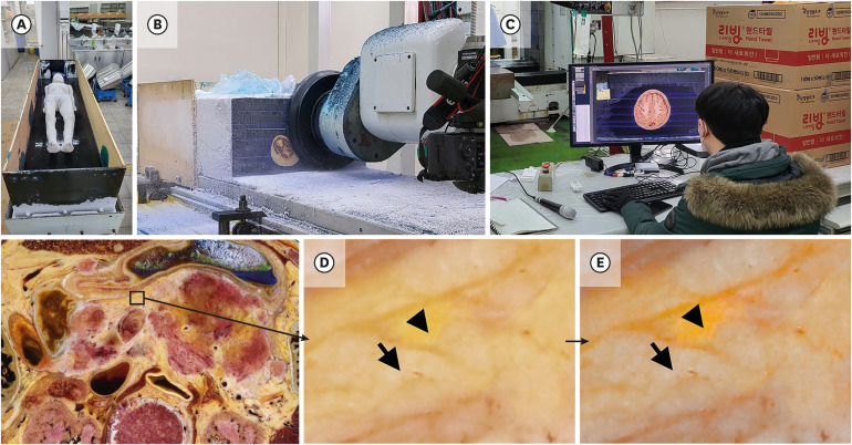
Fig. 1 Making processes of the sectioned images. (A) A cadaver is fixed by gelatin embedding agent in the embedding box. (B) Embedding box is milled by milling disc to make sectioned surface. (C) The sectioned surface is photographed and checked by anatomist to make sectioned image. Abdominal region in sectioned images of (D) original color and (E) a refined color after adjusting color purity in the image by Vibrance and Brightness/Contrast tools in Adobe Photoshop (Arrow = adipose tissue, Arrow head = parenchymal tissue of pancreas).
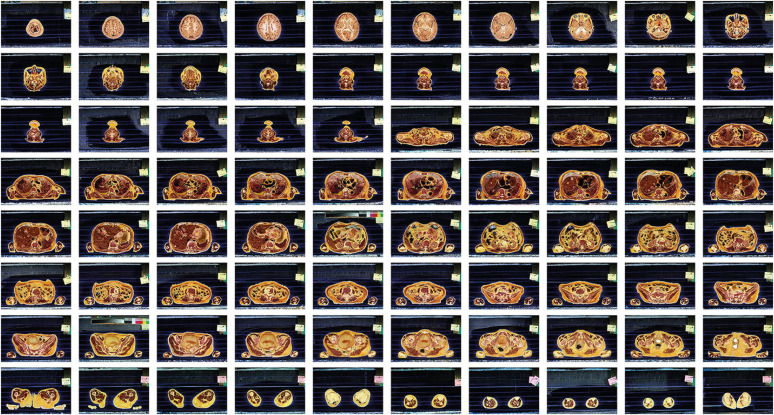
Fig. 2 Specimen sectioned images from head to toe of female whole body.
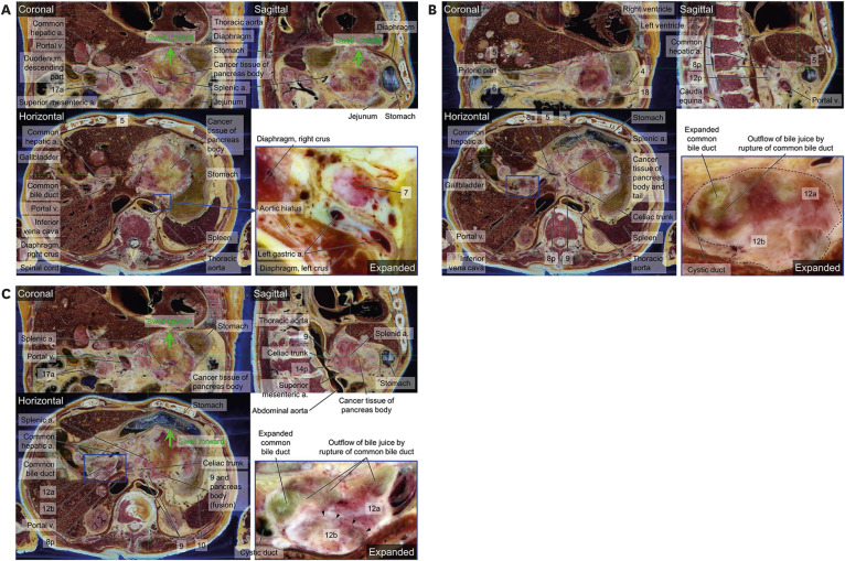
Fig. 3 Horizontal, coronal, and sagittal sectioned images from abdomen volume model on the MRIcroGL. Only horizontal images are aligned from top (A) to bottom (C) of the pancreas. (A) Both S-5 and S-7 are isolated from cancer tissue of pancreas. A part of cancer tissue of body of pancreas is touched in diaphragm, not stomach (Green arrow). Common bile duct is expanded. (B) S-3, S-5, S-6, S-8a, S-8p, and S-18 are separated whereas S-4, S-9, 12a, and 12b are fused with neighboring structures. There is serious cancer tissue in body of pancreas. (C) S-8p is isolated whereas S-9, S-12a, S-12b, S-14p, and S-17a are fused with cancer tissue of pancreas. S-12a and S-12b are fused together, but they can be divided (Arrow head). The stomach is squashed, due to swelling of the pancreas (Green arrow).‘S-’ is omitted in from S-1 to S-18.a. = artery, v. = vein.
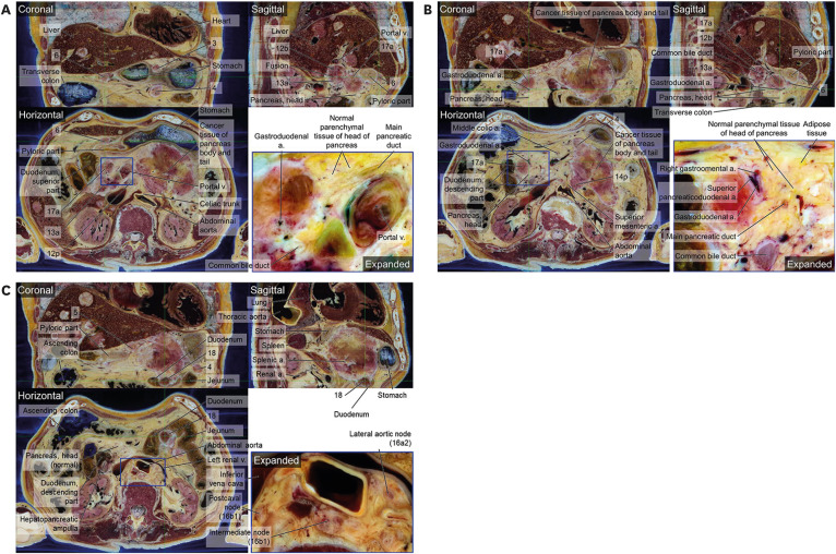
Fig. 4 Horizontal, coronal, and sagittal sectioned images from abdomen volume model on the MRIcroGL. Only horizontal images are aligned from top (A) to bottom (C) of the pancreas. (A) S-3 and S-6 are isolated respectively whereas 4, 12b, 12p, 13a, and 17a are fused with one another or pancreas. In the inferior portion of head of pancreas, normal parenchymal tissue is observed. (B) In the inferior portion of head of pancreas, normal tissue is larger than abnormal tissue. S-6 is isolated and S-4, S-12b, S-13a, S-14p, and S-17a are fused. (C) S-4, S-5, and 18 are observed. Duodenum is pressed by S-18. In the case of S-16a2 and 16b1, there were left lumbar nodes (Lateral aortic nodes), intermediate lumbar nodes, right lumbar nodes (postcaval nodes) as anatomical terminology. ‘S-’ is omitted in from S-1 to S-18.a. = artery, v. = vein.
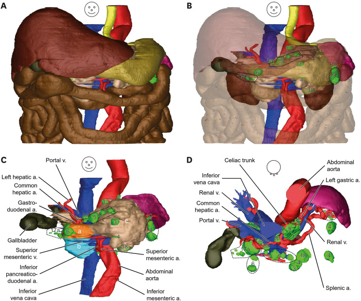
Fig. 5 Surface models of abdominal region including pancreatic cancer and its lymph node metastases. (A) Shape and location of structures of the abdominal region are observed opaquely. (B) In anterior view of translucent liver, stomach, small intestine, and large intestine, lymph node stations can be observed. (C) In the anterior view of structures except liver, stomach, small intestine, large intestine, and kidney, lymph node stations around the pancreas are identified in green. (D) In the superior view, lymph node stations around branches of the celiac trunk are identified.a. = artery, v. = vein.aSuperior and bInferior portions of head of pancreas, cBody and tail of pancreas.
Reference
-
1. Kim KS, Kwon J, Kim K, Chie EK. Impact of resection margin distance on survival of pancreatic cancer: a systematic review and meta-analysis. Cancer Res Treat. 2017; 49(3):824–833. PMID: 27561314.
Article2. Rhee H, Park MS. The role of imaging in current treatment strategies for pancreatic adenocarcinoma. Korean J Radiol. 2021; 22(1):23–40. PMID: 32901458.
Article3. Zhang J, Zhang L, Li C, Yang C, Li L, Song S, et al. LOX-1 is a poor prognostic indicator and induces epithelial-mesenchymal transition and metastasis in pancreatic cancer patients. Cell Oncol (Dordr). 2018; 41(1):73–84. PMID: 29168159.
Article4. Isaji S, Murata Y, Kishiwada M. New Japanese Classification of Pancreatic Cancer. Neoptolemos JP, Urrutia R, Abbruzzese JL, Büchler MW, editors. Pancreatic Cancer. New York, NY, USA: Springer New York;2018. p. 1021–1037.5. Sobin LH, Gospodarowicz MK, Wittekind C. TNM Classification of Malignant Tumours. Hoboken, NJ, USA: John Wiley & Sons;2011.6. Park JS, Chung MS, Hwang SB, Lee YS, Har DH, Park HS. Visible Korean human: improved serially sectioned images of the entire body. IEEE Trans Med Imaging. 2005; 24(3):352–360. PMID: 15754985.
Article7. Park HS, Choi DH, Park JS. Improved sectioned images and surface models of the whole female body. Int J Morphol. 2015; 33(4):1323–1332.
Article8. Chung BS, Han M, Har D, Park JS. Advanced sectioned images of a cadaver head with voxel size of 0.04 mm. J Korean Med Sci. 2019; 34(34):e218. PMID: 31456382.
Article9. Shin DS, Jang HG, Hwang SB, Har DH, Moon YL, Chung MS. Two-dimensional sectioned images and three-dimensional surface models for learning the anatomy of the female pelvis. Anat Sci Educ. 2013; 6(5):316–323. PMID: 23463707.
Article10. Park JS, Chung MS, Shin DS, Har DH, Cho ZH, Kim YB, et al. Sectioned images of the cadaver head including the brain and correspondences with ultrahigh field 7.0 T MRIs. Proc IEEE Inst Electr Electron Eng. 2009; 97(12):1988–1996.
Article11. You Y, Kim CY, Kim SK, Chung BS, Har D, Choi J, et al. Advanced-sectioned images obtained by microsectioning of the entire male body. Clin Anat. 2022; 35(1):79–86. PMID: 34591338.
Article12. You Y, Park JS. A novel human brainstem map based on true-color sectioned images. J Korean Med Sci. 2023; 38(10):e76. PMID: 36918030.
Article13. Kim SK, Hur MS, Park JS. Real color sectioned images and correspondence with ultrasound images of the palmar wrist. Appl Sci. 2022; 12(1):299.
Article14. Park JS, Jung YW. Peeled images and sectioned images from real-color volume models of foot. Surg Radiol Anat. 2021; 43(1):37–43. PMID: 32676743.
Article15. Kim CY, Park JS, Chung BS. Real color model of a cadaver for deep brain stimulation of the subthalamic nucleus. Appl Sci. 2021; 11(11):4999.
Article16. Chung BS, Park HS, Park JS, Hwang SB, Chung MS. Sectioned and segmented images of the male whole body, female whole body, male head, and female pelvis from the Visible Korean. Anat Sci Int. 2021; 96(1):168–173. PMID: 32803722.
Article17. Park JS. 2D browsing software and 3D PDF of canine ear based on real color sectioned images. Int J Morphol. 2020; 38(1):147–152.
Article18. Kwon K, Park JS, Shin BS. Virtual anatomical and endoscopic exploration method of internal human body for training simulator. J Korean Med Sci. 2020; 35(12):e90. PMID: 32233159.
Article19. Park JS. Cross-Sectional Atlas of the Human Head: With 0.1-mm Pixel Size Color Images. Berlin, Germany: Springer;2018.20. Park JS, You Y. Cross-Sectional Atlas of Human Brainstem: With 0.06-mm Pixel Size Color Images. Singapore: Springer Nature Singapore;2023.21. Harisinghani MG. Atlas of Lymph Node Anatomy. 2nd ed. Cham, Switzerland: Springer Nature Switzerland AG;2013. p. 59–88.22. Chung BS, Park JS. Real-color volume models made from real-color sectioned images of Visible Korean. J Korean Med Sci. 2019; 34(10):e86. PMID: 30886552.
Article23. Moore KL, Dalley AF, Agur AM. Clinically Oriented Anatomy. 8th ed. Philadelphia, PA, USA: Wolters Kluwer;2018. p. 488–491.24. Park JS, Chung MS, Hwang SB, Lee YS, Har DH. Technical report on semiautomatic segmentation using the Adobe Photoshop. J Digit Imaging. 2005; 18(4):333–343. PMID: 16003588.
Article25. FCAT. Terminologia Anatomica. Book & CD-ROM edition ed. New York, NY, USA: Thieme Medical Publishers;2002.26. Ertürk MA, Raaijmakers AJ, Adriany G, Uğurbil K, Metzger GJ. A 16-channel combined loop-dipole transceiver array for 7 Tesla body MRI. Magn Reson Med. 2017; 77(2):884–894. PMID: 26887533.
- Full Text Links
-
- Actions
-
Cited
- CITED
-
- Close
- Share
-
- Similar articles
-
- Identification of cranial nerve ganglia using sectioned images and three-dimensional models of a cadaver
- Registration of Cadaver's Sectioned Images to Patient's Head MRIs
- Automated Techniques for the Sectioned Images of Visible Korean
- Real-Color Volume Models Made from Real-Color Sectioned Images of Visible Korean
- Three types of the serial segmented images suitable for surface reconstruction
