Restor Dent Endod.
2022 May;47(2):e19. 10.5395/rde.2022.47.e19.
Morphotypes of the apical constriction of maxillary molars: a micro-computed tomographic evaluation
- Affiliations
-
- 1Faculty of Dentistry, The University of Hong Kong, Sai Ying Pun, Hong Kong
- 2Department of Conservative Dentistry and Endodontics, Sri Ramachandra Faculty of Dental Sciences, Sri Ramachandra Institute of Higher Education and Research (Deemed to be University), Chennai, TN, India
- 3Department of Conservative Dentistry and Endodontics, SRM Dental College, SRM University (Deemed to be University) Ramapuram, Chennai, TN, India
- 4Adhish Multispeciality Dental Clinic, Chennai, TN, India
- 5Department of Conservative Dentistry and Endodontics, Thai Moogambigai Dental College and Hospital, Dr. MGR Educational and Research Institute (Deemed to be University), Chennai, TN, India
- KMID: 2548123
- DOI: http://doi.org/10.5395/rde.2022.47.e19
Abstract
Objectives
The aim of this study was to evaluate and compare the apical constriction (AC) and apical canal morphology of maxillary first and second molars, using micro-computed tomography (micro-CT).
Materials and Methods
The anatomical features of 313 root canals from 41 maxillary first molars and 57 maxillary second molars of patients with known age and sex were evaluated using micro-CT, with a resolution of 26.7 µm. The factors evaluated were the presence or absence of AC, the morphotypes, bucco-lingual dimension, mesio-distal dimension, and the profile (shape) of AC and the apical root canal. The apical root canal dimensions, location of the apical foramen (AF), AC to AF distance, and presence of accessory canals in the apical 5 mm were also assessed. Descriptive and analytical statistics were used for data evaluation.
Results
AC was present in all 313 root canals. Patients’ age and sex did not significantly impact either AC or the apical canal dimensions. The most common AC morphotype detected was the traditional (single) constriction (52%), followed by the parallel (29%) morphotype. The mean AC dimensions in maxillary first molars were not significantly different from those in maxillary second molars. Sixty percent of AF were located within 0.5 mm from the anatomic apex.
Conclusions
The most common morphotype of AC detected was the traditional constriction. Neither patients’ age nor sex had a significant impact on the dimensions of the AC or the apical root canal. The majority of AF (60%) were located within 0.5 mm from the anatomic apex.
Figure
-
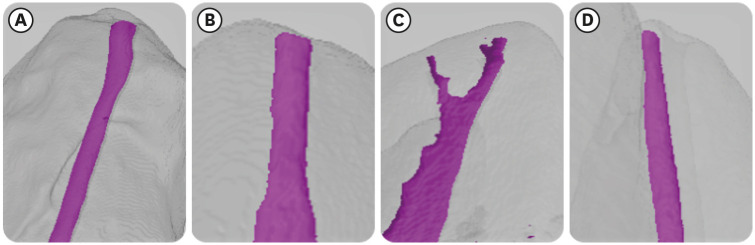
Figure 1 Apical constriction morphotypes: (A) traditional constriction, (B) parallel constriction, (C) delta constriction, and (D) tapering constriction.
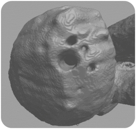
Figure 2 Complex multiple apical ramifications in a delta constriction.
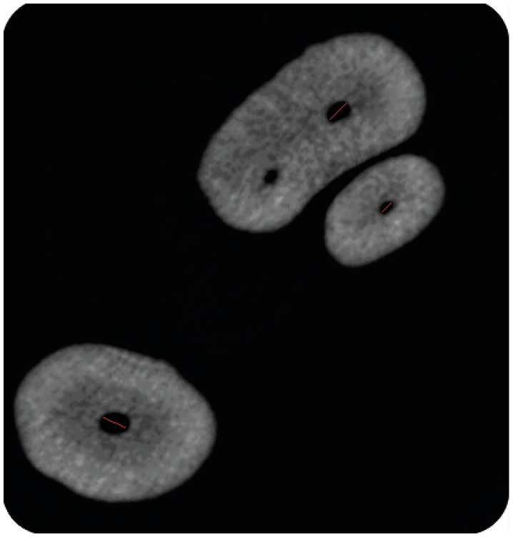
Figure 3 Apical canal dimensions: the mesio-buccal and disto-buccal canals are wider in the bucco-palatal dimension, and the palatal canal is wider in the mesio-distal dimension.
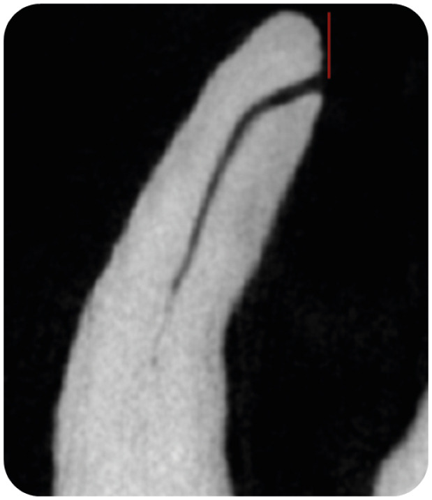
Figure 4 Lateral view of the anatomic apex showing apical foramen deviation.
Figure 5 Presence of accessory canals in a first mesio-buccal canal.
Reference
-
1. Eleazer P, Glickman G, McClanahan S. AAE glossary of endodontic terms. New York, NY: American Association of Endodontists;2020.2. Kuttler Y. Microscopic investigation of root apexes. J Am Dent Assoc. 1955; 50:544–552. PMID: 14366934.
Article3. Dummer PM, McGinn JH, Rees DG. The position and topography of the apical canal constriction and apical foramen. Int Endod J. 1984; 17:192–198. PMID: 6593303.
Article4. Gani O, Visvisian C. Apical canal diameter in the first upper molar at various ages. J Endod. 1999; 25:689–691. PMID: 10687530.
Article5. Meder-Cowherd L, Williamson AE, Johnson WT, Vasilescu D, Walton R, Qian F. Apical morphology of the palatal roots of maxillary molars by using micro-computed tomography. J Endod. 2011; 37:1162–1165. PMID: 21763914.
Article6. ElAyouti A, Hülber-J M, Judenhofer MS, Connert T, Mannheim JG, Löst C, Pichler BJ, von Ohle C. Apical constriction: location and dimensions in molars-a micro-computed tomography study. J Endod. 2014; 40:1095–1099. PMID: 25069914.
Article7. Schell S, Judenhofer MS, Mannheim JG, Hülber-J M, Löst C, Pichler BJ, ElAyouti A. Validity of longitudinal sections for determining the apical constriction. Int Endod J. 2017; 50:706–712. PMID: 27284756.
Article8. Wu MK, Wesselink PR, Walton RE. Apical terminus location of root canal treatment procedures. Oral Surg Oral Med Oral Pathol Oral Radiol Endod. 2000; 89:99–103. PMID: 10630950.9. Marceliano-Alves M, Alves FR, Mendes DM, Provenzano JC. Micro–computed tomography analysis of the root canal morphology of palatal roots of maxillary first molars. J Endod. 2016; 42:280–283. PMID: 26631299.
Article10. Wada M, Takase T, Nakanuma K, Arisue K, Nagahama F, Yamazaki M. Clinical study of refractory apical periodontitis treated by apicectomy. Part 1. Root canal morphology of resected apex. Int Endod J. 1998; 31:53–56. PMID: 9823129.
Article11. Divine KA, McClanahan SB, Fok A. Anatomic analysis of palatal roots of maxillary molars using micro–computed tomography. J Endod. 2019; 45:724–728. PMID: 31056296.
Article12. De-Deus G, Silva EJ, Marins J, Souza E, Neves AA, Gonçalves Belladonna F, Alves H, Lopes RT, Versiani MA. Lack of causal relationship between dentinal microcracks and root canal preparation with reciprocation systems. J Endod. 2014; 40:1447–1450. PMID: 25146030.
Article13. Versiani MA, Pécora JD, Sousa-Neto MD. Microcomputed tomography analysis of the root canal morphology of single-rooted mandibular canines. Int Endod J. 2013; 46:800–807. PMID: 23402296.
Article14. Verma P, Love RM. A micro CT study of the mesiobuccal root canal morphology of the maxillary first molar tooth. Int Endod J. 2011; 44:210–217. PMID: 20880136.
Article15. Wolf TG, Paqué F, Sven Patyna M, Willershausen B, Briseño-Marroquín B. Three-dimensional analysis of the physiological foramen geometry of maxillary and mandibular molars by means of micro-CT. Int J Oral Sci. 2017; 9:151–157. PMID: 28884743.
Article16. Versiani MA, Pécora JD, de Sousa-Neto MD. Root and root canal morphology of four-rooted maxillary second molars: a micro-computed tomography study. J Endod. 2012; 38:977–982. PMID: 22703664.
Article17. De-Deus G, Souza EM, Barino B, Maia J, Zamolyi RQ, Reis C, Kfir A. The self-adjusting file optimizes debridement quality in oval-shaped root canals. J Endod. 2011; 37:701–705. PMID: 21496675.
Article18. De-Deus G, Belladonna FG, de Siqueira Zuolo A, Perez R, Carvalho MS, Souza EM, Lopes RT, Silva EJ. Micro-CT comparison of XP-endo Finisher and passive ultrasonic irrigation as final irrigation protocols on the removal of accumulated hard-tissue debris from oval shaped-canals. Clin Oral Investig. 2019; 23:3087–3093.
Article19. Stein TJ, Corcoran JF. Anatomy of the root apex and its histologic changes with age. Oral Surg Oral Med Oral Pathol. 1990; 69:238–242. PMID: 2304750.
Article20. Briseño-Marroquín B, Paqué F, Maier K, Willershausen B, Wolf TG. Root canal morphology and configuration of 179 maxillary first molars by means of micro-computed tomography: an ex vivo study. J Endod. 2015; 41:2008–2013. PMID: 26518215.
Article21. Ordinola-Zapata R, Martins JN, Bramante CM, Villas-Boas MH, Duarte MH, Versiani MA. Morphological evaluation of maxillary second molars with fused roots: a micro-CT study. Int Endod J. 2017; 50:1192–1200. PMID: 28196285.
- Full Text Links
-
- Actions
-
Cited
- CITED
-
- Close
- Share
-
- Similar articles
-
- Proximity of maxillary molar apexes to the cortical bone surface and the maxillary sinus
- Generalized Pulp Stones of Primary Dentition in a Patient with Molar-Incisor Malformation : A Case Report
- Assessment of the relationship between the maxillary molars and adjacent structures using cone beam computed tomography
- Assessment of maxillary third molars with panoramic radiography and cone-beam computed tomography
- Fused roots of maxillary molars: characterization and prevalence in a Latin American sub-population: a cone beam computed tomography study
