Assessment of the relationship between the maxillary molars and adjacent structures using cone beam computed tomography
- Affiliations
-
- 1Department of Oral and Maxillofacial Radiology, College of Dentistry, Pusan National University, Yangsan, Korea. bhjo@pusan.ac.kr
- KMID: 1806787
- DOI: http://doi.org/10.5624/isd.2012.42.4.219
Abstract
- PURPOSE
This study investigated the relationship between the roots of the maxillary molars and the maxillary sinus using cone beam computed tomography (CBCT), and measured the distances between the roots of the maxillary molars and the sinus floor as well as the thickness of the bone between the root and the alveolar cortical plate.
MATERIALS AND METHODS
The study sample consisted of 83 patients with normally erupted bilateral maxillary first and second molars. A total of 332 maxillary molars were examined using CBCT images. The vertical relationship of each root with the maxillary sinus was classified into four types on CBCT cross-sectional images. The distance between the sinus floor and root and the bone thickness between the root and alveolar cortical plate were measured.
RESULTS
In the buccal roots of the maxillary molars, a root protruding into the sinus occurred most frequently. A root projecting laterally along the sinus cavity was most common in the palatal roots of the maxillary first molars. The mesiobuccal roots of the maxillary second molar were closest to the sinus. The mesiobuccal roots of the first molars were closest to the cortical plate.
CONCLUSION
The relationship between the roots of the maxillary molars and the sinus differed between the buccal and palatal roots. A root protruding into the sinus occurred more frequent in the buccal roots of the maxillary molars. The mesiobuccal root of the maxillary second molar was closest to the maxillary sinus floor and farthest from the alveolar cortical plate.
MeSH Terms
Figure
-

Fig. 1 CBCT images show 4 types of vertical relationships between the root of the maxillary molars and the sinus floor. A. Type 0, the root is separate from the sinus floor. B. Type 1, the root is in contact with the sinus floor. C. Type 2, the root is projecting laterally along the sinus cavity, but is outside the sinus borders. D. Type 3, the root is projecting into the sinus cavity.

Fig. 2 The illustrations show 3 types of the horizontal relationships between the root of the maxillary molars and the sinus floor. Type B, the lowest point of the maxillary sinus floor is located on the buccal side. Type BP, the lowest point of the sinus floor is located between the buccal and palatal roots. Type P, the lowest point of the sinus floor is located on the palatal side of the palatal root.
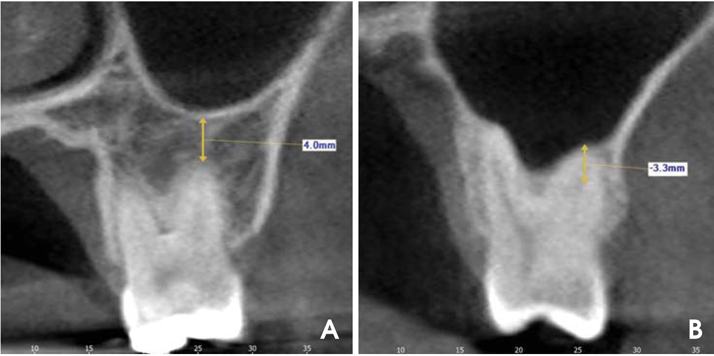
Fig. 3 The distance between the root apex and the sinus floor is measured using CBCT cross-sectional images. Apices extending below the floor of the sinus are assigned positive values (A), whereas those above the sinus floor are assigned negative values (B).
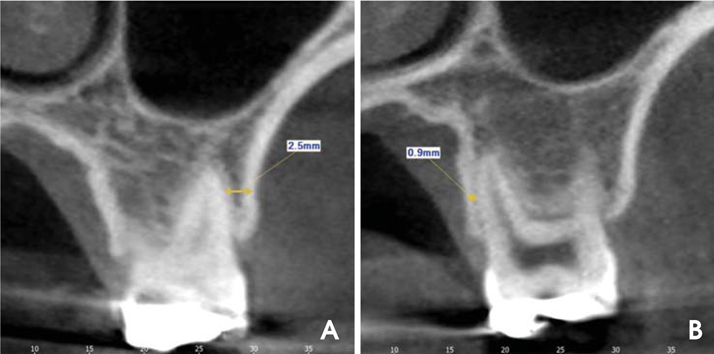
Fig. 4 The minimum bone thickness between the root and the alveolar cortical plate is measured using CBCT cross-sectional images. For the buccal roots, the distance to the buccal cortical plate is obtained (A), and for the palatal roots, the distance to the palatal cortical plate is measured (B).
Cited by 4 articles
-
Alveolar bone height according to the anatomical relationship between the maxillary molar and sinus
Yoon Joo Choi, Young Hyun Kim, Sang-Sun Han, Ui-Won Jung, Chena Lee, Ari Lee, Kug Jin Jeon
J Periodontal Implant Sci. 2020;50(1):38-47. doi: 10.5051/jpis.2020.50.1.38.Assessment of maxillary third molars with panoramic radiography and cone-beam computed tomography
Yun-Hoa Jung, Bong-Hae Cho
Imaging Sci Dent. 2015;45(4):233-240. doi: 10.5624/isd.2015.45.4.233.The use of digital periapical radiographs to study the prevalence of alveolar domes
Pedro Augusto Oliveira Santos Xambre, Claudia Scigliano Valerio, Claudia Assunção e Alves Cardoso, Antônio Luís Neto Custódio, Flávio Ricardo Manzi
Imaging Sci Dent. 2016;46(3):179-184. doi: 10.5624/isd.2016.46.3.179.Distances from the root apices of posterior teeth to the maxillary sinus and mandibular canal in patients with skeletal open bite: A cone-beam computed tomography study
Werinpimol Kosumarl, Virush Patanaporn, Dhirawat Jotikasthira, Apirum Janhom
Imaging Sci Dent. 2017;47(3):157-164. doi: 10.5624/isd.2017.47.3.157.
Reference
-
1. Kilic C, Kamburoglu K, Yuksel SP, Ozen T. An assessment of the relationship between the maxillary sinus floor and the maxillary posterior teeth root tips using dental cone-beam computerized tomography. Eur J Dent. 2010. 4:462–467.
Article2. Watzek G, Bernhart T, Ulm C. Complications of sinus perforations and their management in endodontics. Dent Clin North Am. 1997. 41:563–583.3. Hauman CH, Chandler NP, Tong DC. Endodontic implications of the maxillary sinus: a review. Int Endod J. 2002. 35:127–141.
Article4. Fuhrmann R, Bücker A, Diedrich P. Radiological assessment of artificial bone defects in the floor of the maxillary sinus. Dentomaxillofac Radiol. 1997. 26:112–116.
Article5. Engström H, Chamberlain D, Kiger R, Egelberg J. Radiographic evaluation of the effect of initial periodontal therapy on thickness of the maxillary sinus mucosa. J Periodontol. 1988. 59:604–608.
Article6. Ariji Y, Obayashi N, Goto M, Izumi M, Naitoh M, Kurita K, et al. Roots of the maxillary first and second molars in horizontal relation to alveolar cortical plates and maxillary sinus: computed tomography assessment for infection spread. Clin Oral Investig. 2006. 10:35–41.
Article7. Jung YH, Cho BH. Comparison of panoramic radiography and cone beam computed tomography for assessing the relationship between the maxillary sinus floor and maxillary molars. Korean J Oral Maxillofac Radiol. 2009. 39:69–73.8. Eberhardt JA, Torabinejad M, Christiansen EL. A computed tomographic study of the distances between the maxillary sinus floor and the apices of the maxillary posterior teeth. Oral Surg Oral Med Oral Pathol. 1992. 73:345–346.
Article9. Sharan A, Madjar D. Correlation between maxillary sinus floor topography and related root position of posterior teeth using panoramic and cross-sectional computed tomography imaging. Oral Surg Oral Med Oral Pathol Oral Radiol Endod. 2006. 102:375–381.
Article10. Yoon HR, Park CS. A radiologic study of the relationship of the maxillary sinus floor and apex of the maxillary molar. J Korean Acad Oral Maxillofac Radiol. 1998. 28:111–126.11. Kwak HH, Park HD, Yoon HR, Kang MK, Koh KS, Kim HJ. Topographic anatomy of the inferior wall of the maxillary sinus in Koreans. Int J Oral Maxillofac Surg. 2004. 33:382–388.
Article12. Georgescu CE, Rusu MC, Sandulescu M, Enache AM, Didilescu AC. Quantitative and qualitative bone analysis in the maxillary lateral region. Surg Radiol Anat. 2012. 34:551–558.
Article13. Brüllmann DD, Schmidtmann I, Hornstein S, Schulze RK. Correlation of cone beam computed tomography (CBCT) findings in the maxillary sinus with dental diagnoses: a retrospective cross-sectional study. Clin Oral Investig. 2012. 16:1023–1029.
Article14. Lu Y, Liu Z, Zhang L, Zhou X, Zheng Q, Duan X, et al. Associations between maxillary sinus mucosal thickening and apical periodontitis using cone-beam computed tomography scanning: a retrospective study. J Endod. 2012. 38:1069–1074.
Article15. Phothikhun S, Suphanantachat S, Chuenchompoonut V, Nisapakultorn K. Cone-beam computed tomographic evidence of the association between periodontal bone loss and mucosal thickening of the maxillary sinus. J Periodontol. 2012. 83:557–564.
Article
- Full Text Links
-
- Actions
-
Cited
- CITED
-
- Close
- Share
-
- Similar articles
-
- Comparison of panoramic radiography and cone beam computed tomography for assessing the relationship between the maxillary sinus floor and maxillary molars
- Assessment of maxillary third molars with panoramic radiography and cone-beam computed tomography
- Analysis of C-shaped root canal configuration in maxillary molars in a Korean population using cone-beam computed tomography
- Comparison of panoramic radiography and cone-beam computed tomography for assessing radiographic signs indicating root protrusion into the maxillary sinus
- A cone-beam computed tomographic study of C-shaped root and root canal in maxillary molars
