Restor Dent Endod.
2021 May;46(2):e16. 10.5395/rde.2021.46.e16.
Shaping ability and apical debris extrusion after root canal preparation with rotary or reciprocating instruments: a micro-CT study
- Affiliations
-
- 1Department of Endodontics, State University of Rio de Janeiro (UERJ) School of Dentistry, Rio de Janeiro, RJ, Brazil
- 2Department of Endodontics, Grande Rio University (UNIGRANRIO) School of Dentistry, Rio de Janeiro, RJ, Brazil
- 3Department of Clinical Dentistry, Federal University of Juiz de Fora (UFJF), Governador Valadares, MG, Brazil
- KMID: 2548060
- DOI: http://doi.org/10.5395/rde.2021.46.e16
Abstract
Objectives
The aim of this study was to evaluate the shaping ability of the TruShape and Reciproc Blue systems and the apical extrusion of debris after root canal instrumentation. The ProTaper Universal system was used as a reference for comparison.
Materials and Methods
Thirty-three mandibular premolars with a single canal were scanned using micro-computed tomography and were matched into 3 groups (n = 11) according to the instrumentation system: TruShape, Reciproc Blue and ProTaper Universal. The teeth were accessed and mounted in an apparatus with agarose gel, which simulated apical resistance provided by the periapical tissue and enabled the collection of apically extruded debris. During root canal preparation, 2.5% sodium hypochlorite was used as an irrigant. The samples were scanned again after instrumentation. The percentage of unprepared area, removed dentin, and volume of apically extruded debris were analyzed. The data were analyzed using 1-way analysis of variance and the Tukey test for multiple comparisons at a 5% significance level.
Results
No significant differences in the percentage of unprepared area were observed among the systems (p > 0.05). ProTaper Universal presented a higher percentage of dentin removal than the TruShape and Reciproc Blue systems (p < 0.05). The systems produced similar volumes of apically extruded debris (p > 0.05).
Conclusions
All systems caused apically extruded debris, without any significant differences among them. TruShape, Reciproc Blue, and ProTaper Universal presented similar percentages of unprepared area after root canal instrumentation; however, ProTaper Universal was associated with higher dentin removal than the other systems.
Figure
-
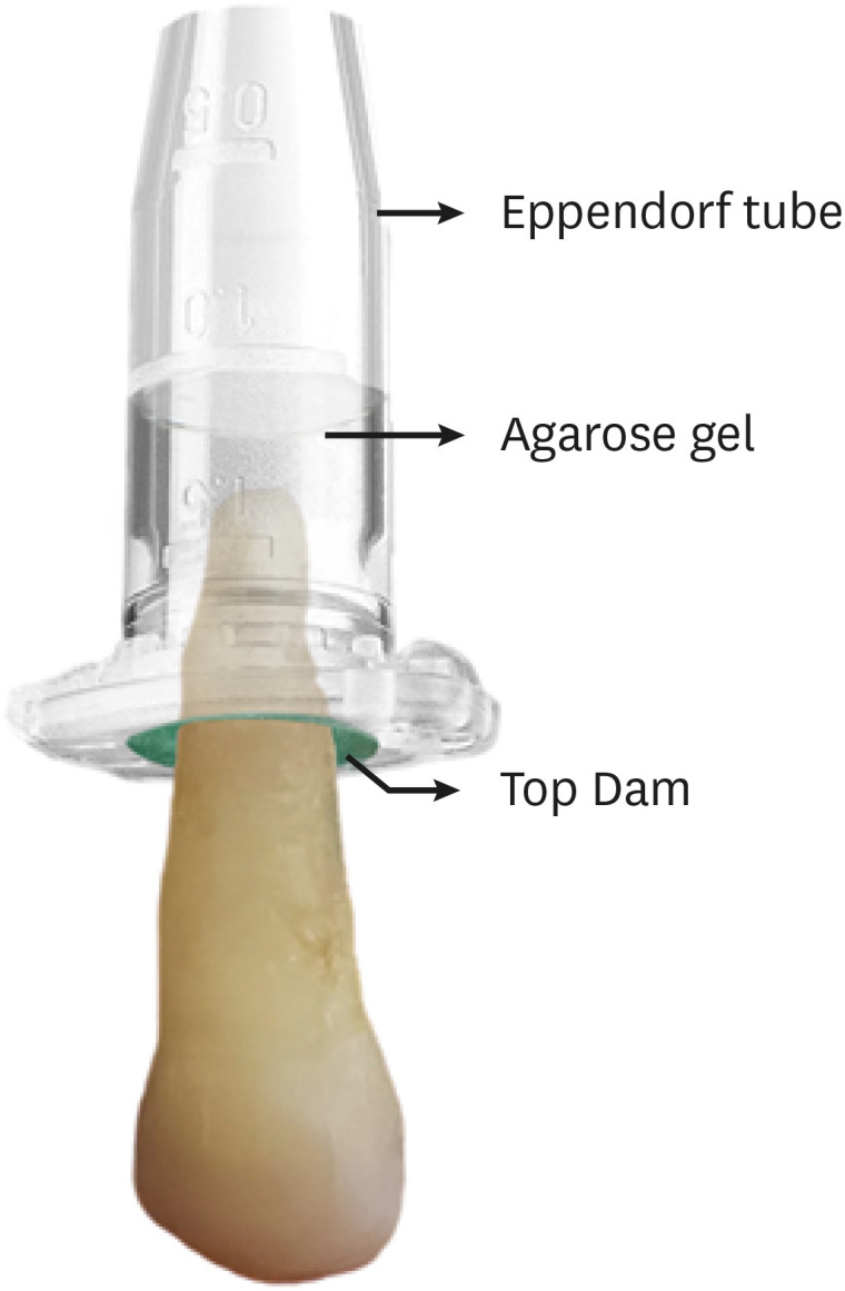
Figure 1 Eppendorf tube and agarose gel apparatus with a tooth fixed. The visible line indicates the agarose gel level inside the tube.
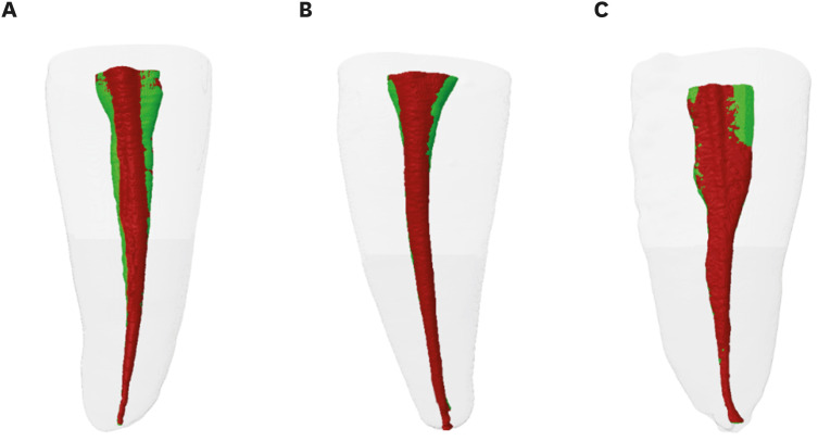
Figure 2 Representative 3-dimensional images of the root canals before (green) and after instrumentation (red) with (A) ProTaper, (B) TruShape, and (C) Reciproc Blue.

Figure 3 Micro-computed tomography 3-dimensional reconstruction of extruded debris in the (A) ProTaper, (B) TruShape, and (C) Reciproc Blue groups.
Reference
-
1. Reddy SA, Hicks ML. Apical extrusion of debris using two hand and two rotary instrumentation techniques. J Endod. 1998; 24:180–183. PMID: 9558583.
Article2. Tanalp J, Güngör T. Apical extrusion of debris: a literature review of an inherent occurrence during root canal treatment. Int Endod J. 2014; 47:211–221. PMID: 23711187.
Article3. Seltzer S, Naidorf IJ. Flare-ups in endodontics: I. Etiological factors. J Endod. 1985; 11:472–478. PMID: 3868692.
Article4. Siqueira JF Jr, Rôças IN, Favieri A, Machado AG, Gahyva SM, Oliveira JC, Abad EC. Incidence of postoperative pain after intracanal procedures based on an antimicrobial strategy. J Endod. 2002; 28:457–460. PMID: 12067129.
Article5. Torabinejad M, Walton RE. Managing endodontic emergencies. J Am Dent Assoc. 1991; 122:99–103.6. Koçak S, Koçak MM, Sağlam BC, Türker SA, Sağsen B, Er Ö. Apical extrusion of debris using self-adjusting file, reciprocating single-file, and 2 rotary instrumentation systems. J Endod. 2013; 39:1278–1280. PMID: 24041391.
Article7. Al-Omari MA, Dummer PM. Canal blockage and debris extrusion with eight preparation techniques. J Endod. 1995; 21:154–158. PMID: 7561660.
Article8. Tinoco JM, De-Deus G, Tinoco EM, Saavedra F, Fidel RA, Sassone LM. Apical extrusion of bacteria when using reciprocating single-file and rotary multifile instrumentation systems. Int Endod J. 2014; 47:560–566. PMID: 24111671.
Article9. Costa EL, Sponchiado-Júnior EC, Garcia LF, Marques AA. Effect of large instrument use on shaping ability and debris extrusion of rotary and reciprocating systems. J Investig Clin Dent. 2018; 9:1–8.
Article10. Frota MM, Bernardes RA, Vivan RR, Vivacqua-Gomes N, Duarte MA, Vasconcelos BC. Debris extrusion and foraminal deformation produced by reciprocating instruments made of thermally treated NiTi wires. J Appl Oral Sci. 2018; 26:e20170215. PMID: 29364346.
Article11. Sen OG, Bilgin B, Koçak S, Sağlam BC, Koçak MM. Evaluation of apically extruded debris using continuous rotation, reciprocation, or adaptive motion. Braz Dent J. 2018; 29:245–248. PMID: 29972449.
Article12. Silva EJ, Carapiá MF, Lopes RM, Belladonna FG, Senna PM, Souza EM, De-Deus G. Comparison of apically extruded debris after large apical preparations by full-sequence rotary and single-file reciprocating systems. Int Endod J. 2016; 49:700–705. PMID: 26174577.
Article13. Alves FR, Paiva PL, Marceliano-Alves MF, Cabreira LJ, Lima KC, Siqueira JF Jr, Rôças IN, Provenzano JC. Bacteria and hard tissue debris extrusion and intracanal bacterial reduction promoted by XP-endo Shaper and Reciproc instruments. J Endod. 2018; 44:1173–1178. PMID: 29861066.
Article14. Shen Y, Hieawy A, Huang X, Wang ZJ, Maezono H, Haapasalo M. Fatigue resistence of a 3-dimensional conforming nickel-titanium rotary instrument in double curvatures. J Endod. 2016; 42:961–964. PMID: 27106719.
Article15. Bürklein S, Hinschitza K, Dammaschke T, Schäfer E. Shaping ability and cleaning effectiveness of two single-file systems in severely curved root canals of extracted teeth: Reciproc and WaveOne versus Mtwo and ProTaper. Int Endod J. 2012; 45:449–461. PMID: 22188401.
Article16. De-Deus G, Silva EJ, Vieira VT, Belladonna FG, Elias CN, Plotino G, Grande NM. Blue thermomechanical treatment optimizes fatigue resistance and flexibility of the Reciproc files. J Endod. 2017; 43:462–466. PMID: 28131415.
Article17. Silva EJ, Hecksher F, Antunes HD, De-Deus G, Elias CN, Vieira VT. Torsional fatigue resistance of blue-treated reciprocating instruments. J Endod. 2018; 44:1038–1041. PMID: 29680726.
Article18. Prados-Privado M, Rojo R, Ivorra C, Prados-Frutos JC. Finite element analysis comparing WaveOne, WaveOne Gold, Reciproc and Reciproc Blue responses with bending and torsion tests. J Mech Behav Biomed Mater. 2019; 90:165–172. PMID: 30366307.
Article19. Uslu G, Özyürek T, Yılmaz K, Gündoğar M, Plotino G. Apically extruded debris during root canal instrumentation with Reciproc Blue, Hyflex EDM, and XP-endo shaper nickel-titanium files. J Endod. 2018; 44:856–859. PMID: 29550013.
Article20. Vertucci FJ. Root canal anatomy of the human permanent teeth. Oral Surg Oral Med Oral Pathol. 1984; 58:589–599. PMID: 6595621.
Article21. Fedorov A, Beichel R, Kalpathy-Cramer J, Finet J, Fillion-Robin JC, Pujol S, Bauer C, Jennings D, Fennessy F, Sonka M, Buatti J, Aylward S, Miller JV, Pieper S, Kikinis R. 3D Slicer as an image computing platform for the Quantitative Imaging Network. Magn Reson Imaging. 2012; 30:1323–1341. PMID: 22770690.
Article22. De-Deus G, Belladonna FG, Silva EJ, Marins JR, Souza EM, Perez R, Lopes RT, Versiani MA, Paciornik S, Neves AA. Micro-CT evaluation of non-instrumented canal areas with different enlargements performed by NiTi systems. Braz Dent J. 2015; 26:624–629. PMID: 26963207.
Article23. De-Deus G, Belladonna FG, de Siqueira Zuolo A, Perez R, Carvalho MS, Souza EM, Lopes RT, Silva EJ. Micro-CT comparison of XP-endo Finisher and passive ultrasonic irrigation as final irrigation protocols on the removal of accumulated hard-tissue debris from oval shaped-canals. Clin Oral Investig. 2019; 23:3087–3093.
Article24. Peters OA, Boessler C, Paqué F. Root canal preparation with a novel nickel-titanium instrument evaluated with micro-computed tomography: canal surface preparation over time. J Endod. 2010; 36:1068–1072. PMID: 20478468.
Article25. Lu Y, Wang R, Zhang L, Li HL, Zheng QH, Zhou XD, Huang DM. Apically extruded debris and irrigant with two Ni-Ti systems and hand files when removing root fillings: a laboratory study. Int Endod J. 2013; 46:1125–1130. PMID: 23566178.
Article26. İriboz E, Bayraktar K, Türkaydın D, Tarçın B. Comparison of apical extrusion of sodium hypochlorite using 4 different root canal irrigation techniques. J Endod. 2015; 41:380–384. PMID: 25492488.
Article27. Capar ID, Arslan H, Akcay M, Ertas H. An in vitro comparison of apically extruded debris and instrumentation times with ProTaper Universal, ProTaper Next, Twisted File Adaptive, and HyFlex instruments. J Endod. 2014; 40:1638–1641. PMID: 25260737.
Article28. Koçak MM, Çiçek E, Koçak S, Sağlam BC, Yılmaz N. Apical extrusion of debris using ProTaper Universal and ProTaper Next rotary systems. Int Endod J. 2015; 48:283–286. PMID: 24863544.
Article29. Labbaf H, Nazari Moghadam K, Shahab S, Mohammadi Bassir M, Fahimi MA. An in vitro comparison of apically extruded debris using Reciproc, ProTaper Universal, Neolix and Hyflex in curved canals. Iran Endod J. 2017; 12:307–311. PMID: 28808456.30. Zhang C, Liu J, Liu L. The influence of ProTaper and WaveOne on apically extruded debris: a systematic review and meta-analysis. J Conserv Dent. 2018; 21:474–480. PMID: 30294105.
Article31. Siqueira JF Jr, Pérez AR, Marceliano-Alves MF, Provenzano JC, Silva SG, Pires FR, Vieira GC, Rôças IN, Alves FR. What happens to unprepared root canal walls: a correlative analysis using micro-computed tomography and histology/scanning electron microscopy. Int Endod J. 2018; 51:501–508. PMID: 28196289.
Article32. Filizola de Oliveira DJ, Leoni GB, da Silva Goulart R, Sousa-Neto MD, Silva Sousa YT, Silva RG. Changes in geometry and transportation of root canals with severe curvature prepared by different heat-treated nickel-titanium instruments: a micro–computed tomographic study. J Endod. 2019; 45:768–773. PMID: 30954280.
Article33. Guimarães LS, Gomes CC, Marceliano-Alves MF, Cunha RS, Provenzano JC, Siqueira JF Jr. Preparation of oval-shaped canals with TRUShape and Reciproc Systems: a micro-computed tomography study using contralateral premolars. J Endod. 2017; 43:1018–1022. PMID: 28416315.
Article34. Serefoglu B, Piskin B. Micro computed tomography evaluation of the self-adjusting file and ProTaper Universal system on curved mandibular molars. Dent Mater J. 2017; 36:606–613. PMID: 28566670.
Article35. Zuolo ML, Zaia AA, Belladonna FG, Silva EJ, Souza EM, Versiani MA, Lopes RT, De-Deus G. Micro-CT assessment of the shaping ability of four root canal instrumentation systems in oval-shaped canals. Int Endod J. 2018; 51:564–571. PMID: 28667674.
Article36. Fayyad DM, Elhakim Elgendy AA. Cutting efficiency of twisted versus machined nickel-titanium endodontic files. J Endod. 2011; 37:1143–1146. PMID: 21763910.
Article37. Üstün Y, Çanakçi BC, Dinçer AN, Er O, Düzgün S. Evaluation of apically extruded debris associated with several Ni-Ti systems. Int Endod J. 2015; 48:701–704. PMID: 25112960.
Article38. Kirchhoff AL, Fariniuk LF, Mello I. Apical extrusion of debris in flat-oval root canals after using different instrumentation systems. J Endod. 2015; 41:237–241. PMID: 25447504.
Article39. De-Deus G, Neves A, Silva EJ, Mendonça TA, Lourenço C, Calixto C, Lima EJ. Apically extruded dentin debris by reciprocating single-file and multi-file rotary system. Clin Oral Investig. 2015; 19:357–361.
Article
- Full Text Links
-
- Actions
-
Cited
- CITED
-
- Close
- Share
-
- Similar articles
-
- Effects of the endodontic access cavity on apical debris extrusion during root canal preparation using different single-file systems
- A comparison of the shaping ability of reciprocating NiTi instruments in simulated curved canals
- Shaping ability of four rotary nickel-titanium instruments to prepare root canal at danger zone
- Micro-computed tomographic evaluation of a new system for root canal filling using calcium silicatebased root canal sealers
- A comparison of the shaping ability of four rotary nickel-titanium files in simulated root canals
