Restor Dent Endod.
2021 Feb;46(1):e5. 10.5395/rde.2021.46.e5.
Impact of root canal curvature and instrument type on the amount of extruded debris during retreatment
- Affiliations
-
- 1Department of Endodontics, Faculty of Dentistry, Ege University, Bornova, İzmir, Turkey
- 2Bornova Oral Health Center, Bornova, İzmir, Turkey
- KMID: 2548055
- DOI: http://doi.org/10.5395/rde.2021.46.e5
Abstract
Objectives
The aim of the current study was to assess whether the amount of extruded debris differs for straight and severely curved root canals during retreatment using H-files, R-Endo, Reciproc and ProTaper Universal Retreatment (PTU-R) files. Additionally, the area of residual filling material was evaluated.
Materials and Methods
Severely curved (n = 104) and straight (n = 104) root canals of maxillary molar teeth were prepared with WaveOne Primary file and obturated with guttapercha and AH Plus sealer. Root canal filling materials were removed with one of the preparation techniques: group 1: H-file; group 2: R-Endo; group 3: Reciproc; group 4: PTU-R (n = 26). The amount of extruded material and the area of the residual filling material was measured. The data were analyzed with 2-way analysis of variance (ANOVA) and 1-way ANOVA at the 0.05 significance level.
Results
Except for Reciproc group (p > 0.05), PTU-R, R-Endo, and H-file systems extruded significantly more debris in severely curved canals (p < 0.05). Each file system caused more residual filling material in severely curved canals than in straight ones (p < 0.05).
Conclusions
All instruments used in this study caused apical debris extrusion. Root canal curvature had an effect on extruded debris, except for Reciproc system. Clinicians should be aware that the difficult morphology of the severely curved root canals is a factor increasing the amount of extruded debris during the retreatment procedure.
Figure
-
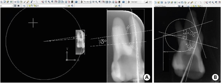
Figure 1 The root canal curvature and radius measurement according to Pruett et al. [14] using AutoCAD software. (A) Straight canal, (B) severely curved root canal.
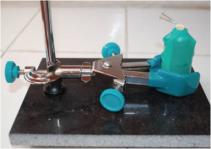
Figure 2 The experimental set-up for the collection of the extruded debris.
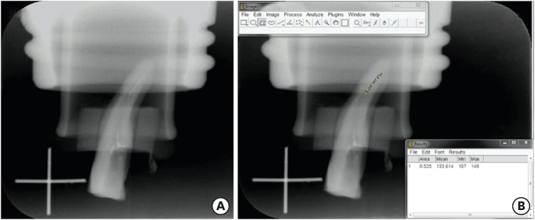
Figure 3 (A) An image showing the residual filling material on the root canal walls (B) calculation of the area of residual filling material using Image J software.
Reference
-
1. Machtou P, Reit C. Non-surgical retreatment. In : Bergenholtz G, Hørsted-Bindslev P, Reit C, editors. Textbook of endodontology. 1st ed. Oxford: Blackwell Munksgaard;2003. p. 300–310.2. Bergenholtz G, Lekholm U, Milthon R, Heden G, Odesjö B, Engström B. Retreatment of endodontic fillings. Scand J Dent Res. 1979; 87:217–224. PMID: 293884.
Article3. Çalışkan MK. Nonsurgical retreatment of teeth with periapical lesions previously managed by either endodontic or surgical intervention. Oral Surg Oral Med Oral Pathol Oral Radiol Endod. 2005; 100:242–248. PMID: 16037783.
Article4. Hülsmann M, Drebenstedt S, Holscher C. Shaping and filling root canals during root canal re-treatment. Endod Topics. 2008; 19:74–124.
Article5. Betti LV, Bramante CM. Quantec SC rotary instruments versus hand files for gutta-percha removal in root canal retreatment. Int Endod J. 2001; 34:514–519. PMID: 11601768.
Article6. Bernardes RA, Duarte MA, Vivan RR, Alcalde MP, Vasconcelos BC, Bramante CM. Comparison of three retreatment techniques with ultrasonic activation in flattened canals using micro-computed tomography and scanning electron microscopy. Int Endod J. 2016; 49:890–897. PMID: 26280904.
Article7. Hülsmann M, Bluhm V. Efficacy, cleaning ability and safety of different rotary NiTi instruments in root canal retreatment. Int Endod J. 2004; 37:468–476. PMID: 15189436.
Article8. Siqueira JF Jr, Rôças IN, Ricucci D, Hülsmann M. Causes and management of post-treatment apical periodontitis. Br Dent J. 2014; 216:305–312. PMID: 24651336.
Article9. Huang X, Ling J, Wei X, Gu L. Quantitative evaluation of debris extruded apically by using ProTaper Universal Tulsa rotary system in endodontic retreatment. J Endod. 2007; 33:1102–1105. PMID: 17931943.
Article10. Topçuoğlu HS, Aktı A, Tuncay Ö, Dinçer AN, Düzgün S, Topçuoğlu G. Evaluation of debris extruded apically during the removal of root canal filling material using ProTaper, D-RaCe, and R-Endo rotary nickel-titanium retreatment instruments and hand files. J Endod. 2014; 40:2066–2069. PMID: 25443282.
Article11. Sae-Lim V, Rajamanickam I, Lim BK, Lee HL. Effectiveness of ProFile. 04 taper rotary instruments in endodontic retreatment. J Endod. 2000; 26:100–104. PMID: 11194370.
Article12. Kaşıkçı Bilgi I, Köseler I, Güneri P, Hülsmann M, Çalışkan MK. Efficiency and apical extrusion of debris: a comparative ex vivo study of four retreatment techniques in severely curved root canals. Int Endod J. 2017; 50:910–918. PMID: 27706822.
Article13. Schirrmeister JF, Wrbas KT, Meyer KM, Altenburger MJ, Hellwig E. Efficacy of different rotary instruments for gutta-percha removal in root canal retreatment. J Endod. 2006; 32:469–472. PMID: 16631851.
Article14. Pruett JP, Clement DJ, Carnes DL Jr. Cyclic fatigue testing of nickel-titanium endodontic instruments. J Endod. 1997; 23:77–85. PMID: 9220735.
Article15. Myers GL, Montgomery S. A comparison of weights of debris extruded apically by conventional filing and Canal Master techniques. J Endod. 1991; 17:275–279. PMID: 1940753.
Article16. Ruiz-Hubard EE, Gutmann JL, Wagner MJ. A quantitative assessment of canal debris forced periapically during root canal instrumentation using two different techniques. J Endod. 1987; 13:554–558. PMID: 3482231.
Article17. Fukushima H, Yamamoto K, Hirohata K, Sagawa H, Leung KP, Walker CB. Localization and identification of root canal bacteria in clinically asymptomatic periapical pathosis. J Endod. 1990; 16:534–538. PMID: 2084210.
Article18. Bürklein S, Schäfer E. Apically extruded debris with reciprocating single-file and full-sequence rotary instrumentation systems. J Endod. 2012; 38:850–852. PMID: 22595125.
Article19. Çanakçi BC, Ustun Y, Er O, Genc Sen O. Evaluation of apically extruded debris from curved root canal filling removal using 5 nickel-titanium systems. J Endod. 2016; 42:1101–1104. PMID: 27179592.
Article20. Delai D, Boijink D, Hoppe CB, Grecca AS, Kopper PM. Apically extruded debris in filling removal of curved canals using 3 NiTi systems and hand files. Braz Dent J. 2018; 29:54–59. PMID: 29267525.
Article21. Friedman S, Stabholz A, Tamse A. Endodontic retreatment--case selection and technique. 3. Retreatment techniques. J Endod. 1990; 16:543–549. PMID: 2084213.
Article22. Hinrichs RE, Walker WA 3rd, Schindler WG. A comparison of amounts of apically extruded debris using handpiece-driven nickel-titanium instrument systems. J Endod. 1998; 24:102–106. PMID: 9641140.
Article23. Leonardi LE, Atlas DM, Raiden G. Apical extrusion of debris by manual and mechanical instrumentation. Braz Dent J. 2007; 18:16–19. PMID: 17639194.
Article24. Karataslioglu E, Arslan H, Er G, Avci E. Influence of canal curvature on the amount of apically extruded debris determined by using three-dimensional determination method. Aust Endod J. 2019; 45:216–224. PMID: 30318788.
Article25. Kirchhoff AL, Fariniuk LF, Mello I. Apical extrusion of debris in flat-oval root canals after using different instrumentation systems. J Endod. 2015; 41:237–241. PMID: 25447504.
Article26. Arslan H, Doğanay E, Alsancak M, Çapar ID, Karataş E, Gündüz HA. Comparison of apically extruded debris after root canal instrumentation using Reciproc® instruments with various kinematics. Int Endod J. 2016; 49:307–310. PMID: 25809717.
Article27. Toyoğlu M, Altunbaş D. Influence of different kinematics on apical extrusion of irrigant and debris during canal preparation using K3XF instruments. J Endod. 2017; 43:1565–1568. PMID: 28735794.
Article28. Üstün Y, Çanakçi BC, Dinçer AN, Er O, Düzgün S. Evaluation of apically extruded debris associated with several Ni-Ti systems. Int Endod J. 2015; 48:701–704. PMID: 25112960.
Article29. Silva EJ, Carapiá MF, Lopes RM, Belladonna FG, Senna PM, Souza EM, De-Deus G. Comparison of apically extruded debris after large apical preparations by full-sequence rotary and single-file reciprocating systems. Int Endod J. 2016; 49:700–705. PMID: 26174577.
Article30. Bürklein S, Benten S, Schäfer E. Quantitative evaluation of apically extruded debris with different single-file systems: Reciproc, F360 and OneShape versus Mtwo. Int Endod J. 2014; 47:405–409. PMID: 23889673.
Article31. Hassanloo A, Watson P, Finer Y, Friedman S. Retreatment efficacy of the Epiphany soft resin obturation system. Int Endod J. 2007; 40:633–643. PMID: 17627698.
Article32. Azim AA, Wang HH, Tarrosh M, Azim KA, Piasecki L. Comparison between single-file rotary systems: part 1-efficiency, effectiveness, and adverse effects in endodontic retreatment. J Endod. 2018; 44:1720–1724. PMID: 30243662.
Article33. Taşdemir T, Er K, Yildirim T, Çelik D. Efficacy of three rotary NiTi instruments in removing gutta-percha from root canals. Int Endod J. 2008; 41:191–196. PMID: 18081812.
Article34. Abramovitz I, Relles-Bonar S, Baransi B, Kfir A. The effectiveness of a self-adjusting file to remove residual gutta-percha after retreatment with rotary files. Int Endod J. 2012; 45:386–392. PMID: 22283664.
Article35. Akbulut MB, Akman M, Terlemez A, Magat G, Sener S, Shetty H. Efficacy of Twisted File Adaptive, Reciproc and ProTaper Universal Retreatment instruments for root-canal-filling removal: a cone-beam computed tomography study. Dent Mater J. 2016; 35:126–131. PMID: 26830833.
Article36. Rödig T, Reicherts P, Konietschke F, Dullin C, Hahn W, Hülsmann M. Efficacy of reciprocating and rotary NiTi instruments for retreatment of curved root canals assessed by micro-CT. Int Endod J. 2014; 47:942–948. PMID: 24386931.
Article37. Keleş A, Alcin H, Kamalak A, Versiani MA. Oval-shaped canal retreatment with self-adjusting file: a micro-computed tomography study. Clin Oral Investig. 2014; 18:1147–1153.
Article38. Monguilhott Crozeta B, Damião de Sousa-Neto M, Bianchi Leoni G, Francisco Mazzi-Chaves J, Terezinha Corrêa Silva-Sousa Y, Baratto-Filho F. A micro-computed tomography assessment of the efficacy of rotary and reciprocating techniques for filling material removal in root canal retreatment. Clin Oral Investig. 2016; 20:2235–2240.
Article39. Hülsmann M. Research that matters - canal preparation, retreatment and working length studies. Int Endod J. 2013; 46:293–295. PMID: 23464671.
Article40. Gergi R, Sabbagh C. Effectiveness of two nickel-titanium rotary instruments and a hand file for removing gutta-percha in severely curved root canals during retreatment: an ex vivo study. Int Endod J. 2007; 40:532–537. PMID: 17511787.
Article41. Takahashi CM, Cunha RS, de Martin AS, Fontana CE, Silveira CF, da Silveira Bueno CE. In vitro evaluation of the effectiveness of ProTaper universal rotary retreatment system for gutta-percha removal with or without a solvent. J Endod. 2009; 35:1580–1583. PMID: 19840652.
Article42. Gogulnath D, Rajan RM, Arathy G, Kandaswamy D. A comparative evaluation of the canal centering ability of three rotary nickel-titanium retreatment systems in the mesio-buccal canals of mandibular first molars using computed tomography. J Conserv Dent. 2015; 18:310–314. PMID: 26180417.
Article
- Full Text Links
-
- Actions
-
Cited
- CITED
-
- Close
- Share
-
- Similar articles
-
- Efficacy of retreatment NiTi files for root canals filled with calcium silicate-based sealer
- The effect of early coronal flaring about apical extrusion of debris
- Effects of the endodontic access cavity on apical debris extrusion during root canal preparation using different single-file systems
- Micro-computed tomographic evaluation of canal retreatments performed by undergraduate students using different techniques
- Shaping ability and apical debris extrusion after root canal preparation with rotary or reciprocating instruments: a micro-CT study
