J Korean Acad Conserv Dent.
2004 Mar;29(2):147-152. 10.5395/JKACD.2004.29.2.147.
The effect of early coronal flaring about apical extrusion of debris
- Affiliations
-
- 1Department of Conservative Dentistry, College of Dentistry, Chosun University, Korea. rootcanal@hanmail.net
- KMID: 1987088
- DOI: http://doi.org/10.5395/JKACD.2004.29.2.147
Abstract
- The purpose of this study was to investigate the quantity of debris which was extruded apically after canal instrumentation using different types of enlarging instrument in endodontic resin models. Five groups of 9 endodontic resin models were instrumented using each different technique : hand instrumentation without early coronal flaring, hand instrumentation after early coronal flaring, and three nickel-titanium engine-driven instrumentations (Hero 642, Protaper, K3). Debris extruded from apical foramen during instrumentation was collected on preweighed CBC bottle, desiccated and weighted using electronic balance. The results were analyzed using Kruskal-wallis test and Mann-Whitney U rank sum test at a significance level of 0.05. The results were as follows: 1. All of instrumentation techniques produced apically extruded debris. 2. Group without early coronal flaring extruded significant more debris than groups with early coronal flaring. 3. There was no significant difference among early coronal flaring groups. The early coronal flaring is very important to reduce the amount of debris extruded apically.
Keyword
MeSH Terms
Figure
-
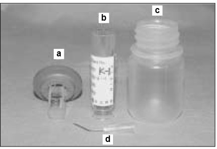
Figure 1 Resin block in stopper(a), CBC bottle(b), flask(c), and 23-gauge needle(d) before assembly
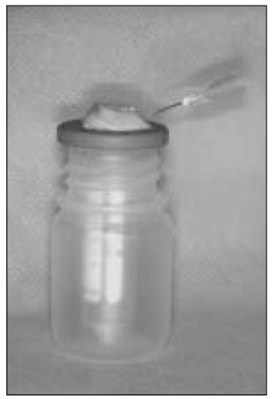
Figure 2 A device for collecting the debris extruded apically
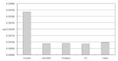
Figure 3 Average weight of apically extruded debris in each group
Reference
-
1. Becker GL, Cohen S, Borer R, Calif SF. The sequelae of accidentally injecting sodium hypochlite beyond the root apex. Oral Surg Oral Med Oral Pathol. 1974. 38:633–638.
Article2. Brown DC, Keith Moore B, Brown CE, Newton CW. An in vitro study of apical extrusion of sodium hypochlorite during endodontic canal preparation. J Endod. 1995. 21:587–591.
Article3. Gatot A, Arbelle J, Leiberman A, Yanai-Inbar I. Effects of sodium hypochlorite on soft tissues after its inadvertent injection beyond the root apex. J Endod. 1991. 17:573–574.
Article4. Marshall JG, Liesinger AW. Factors associated with endodontic posttreatment pain. J Endod. 1993. 19:573–575.
Article5. Naidorf IJ. Endodontic flare-ups. Bacteriological and immunological mechanisms. J Endod. 1985. 11:462–464.
Article6. Seltzer S, Naidorf IJ. Flare-ups in Endodontics. 1. etiological factors. J Endod. 1985. 11:472–478.7. Vande Visse JE, Brilliant JD. Effect of irrigation on the production of extruded material at the root apex during instrumentation. J Endod. 1975. 1:243–246.
Article8. Fukushima H, Yamamoto K, Hirohata K, Sagawa H, Leung K-P, Walker CB. Localization and identification of root canal bacteria in clinically asymptomatic periapical pathosis. J Endod. 1990. 16:534–538.
Article9. Abou-Rass M, Jastrab RJ. The use of rotary instruments as auxiliary aids to root canal preparation of molars. J Endod. 1982. 8:78–82.
Article10. Chapman CE, Collee JG, Beagrie GS. A preliminary report on the correlation between apical infection and instrumentation in endodontics. J Br Endod Soc. 1968. 2:7–11.
Article11. Chapman CE. The correlation between apical infection and instrumentation in endodontics. J Br Endod Soc. 1971. 5:76–80.
Article12. Martin H, Cunningham WT. The effect of endosonic and hand manipulation on the amount of root canal material extruded. Oral Surg Oral Med Oral Pathol. 1982. 53:611–613.
Article13. Seltzer S, Soltanoff W, Sinai I, Goldenberg A, Bender IB. Biologic aspects of endodontics. Part III. periapical tissue reactions to root canal instrumentation. Oral Surg Oral Med Oral Pathol. 1968. 26:534–546. 694–705.
Article14. Fairbourn DR, McWalter GM, Montgomery S. The effect of four preparation techniques on the amount of apically extruded debris. J Endod. 1987. 13:102–108.
Article15. Ruiz-Hubard EE, Gutmann JL, Wagner MJ. A quantitative assessment of canal debris forced periapically during root canal instrumentation using two different techniques. J Endod. 1987. 13:554–558.
Article16. Ferraz CC, Gomes NV, Gomes BPFA, Zaia AA, Teixeira FB, Souza-Filho FJ. Apical extrusion of debris and irrigants using two hand and three engine-driven instrumentation techniques. Int Endod J. 2001. 34:354–358.
Article17. Coffae KP, Brilliant JD. The effect of serial preparation versus nonserial preparation on tissue removal in the root canals of extracted mandibular human molars. J Endod. 1975. 1:211–214.
Article18. Goerig AC, Michelich RJ, Schultz CHH. Instrumentation of root canals in molar using the step-down technique. J Endod. 1982. 8:550–554.
Article19. Leeb J. Canal orifice enlargement as related to biomechanical preparation. J Endod. 1983. 9:463–471.
Article20. Morgan LF, Montgomery S. An evaluation of the crown-down pressureless technique. J Endod. 1984. 10:491–498.
Article21. Swindle RB, Neaverth EJ, Pantera EA, Ringle RD. Effect of coronal-radicular flaring on apical transportation. J Endod. 1991. 17:147–150.
Article22. Cohen S, Burns RC. Pathways of the Pulp. 8th ed. 246–247.23. Gambarini G. Shaping, cleaning the root canal system. A scanning electron microscopic evaluation of a new instrumentation and irrigation technique. J Endod. 1999. 25:800–803.
Article24. Myers GL, Mongomery S. A comparison of weights of debris extruded apically by conventional filling and canal master techniques. J Endod. 1991. 17:275–279.
Article25. Hinrichs RE, Walker WA III, Schindler WG. A Comparison of amounts of apically extruded debris using handpiece-driven nickel-titanium instrument systems. J Endod. 1998. 24:102–106.
Article26. Lim KC, Webber J. The validity of simulated root canals for the investigation of the prepared root canal shape. Int Endod J. 1985. 18:240–246.
Article27. Gerstein H. Techniques in clinical endodontics. 1983. Philadelphia: WB Saunders;324–331.28. Montgomery S. Root canal wall thickness of mandibular molars after biomechanical preparation. J Endod. 1985. 11:257–263.
Article29. Reddy SA, Hicks ML. Apical extrusion of debris using two hand two rotary instrumentation techniques. J Endod. 1998. 24:180–183.
Article30. Beeson TJ, Hartwell GR, Thornton JD, Gunsolley JC. Comparison of debris extruded apically in straight canals: conventional filing versus profile .04 taper series 29. J Endod. 1998. 24:18–22.
Article31. al-Omari MA, Dummer PMH. Canal blockage and debris extrusion with eight preparation techniques. J Endod. 1995. 21:154–158.
Article32. Mckendry DJ. Comparison of balanced forces, endosonic, and step-back filing instrumentation techniques: Quantification of extruded apical debris. J Endod. 1990. 16:24–27.
Article
- Full Text Links
-
- Actions
-
Cited
- CITED
-
- Close
- Share
-
- Similar articles
-
- The analysis of initial apical file size before and after coronal flaring
- Effects of the endodontic access cavity on apical debris extrusion during root canal preparation using different single-file systems
- A comparison of the length between mesio-buccal and mesio-lingual canals of the mandibular molar
- Apical prepration size in infected root canals
- An accuracy of the several electronic apex locators on the mesial root canal of the mandibular molar
