Restor Dent Endod.
2021 Feb;46(1):e2. 10.5395/rde.2021.46.e2.
A micro-computed tomographic study using a novel test model to assess the filling ability and volumetric changes of bioceramic root repair materials
- Affiliations
-
- 1Department of Restorative Dentistry, São Paulo State University (UNESP), School of Dentistry, Araraquara, SP, Brazil
- KMID: 2548052
- DOI: http://doi.org/10.5395/rde.2021.46.e2
Abstract
Objectives
New premixed bioceramic root repair materials require moisture for setting. Using micro-computed tomography (micro-CT), this study evaluated the filling ability and volumetric changes of calcium silicate-based repair materials (mineral trioxide aggregate repair high-plasticity [MTA HP] and Bio-C Repair, Angelus), in comparison with a zinc oxide and eugenol-based material (intermediate restorative material [IRM]; Dentsply DeTrey).
Materials and Methods
Gypsum models with cavities 3 mm deep and 1 mm in diameter were manufactured and scanned using micro-CT (SkyScan 1272. Bruker). The cavities were filled with the cements and scanned again to evaluate their filling capacity. Another scan was performed after immersing the samples in distilled water for 7 days to assess the volumetric changes of the cements. The statistical significance of differences in the data was evaluated using analysis of variance and the Tukey test with a 5% significance level.
Results
Bio-C Repair had a greater filling ability than MTA HP (p < 0.05). IRM was similar to Bio-C and MTA HP (p > 0.05). MTA HP presented the largest volumetric change (p < 0.05), showing more volume loss than Bio-C and IRM, which were similar (p > 0.05).
Conclusions
Bio-C Repair is a new endodontic material with excellent filling capacity and low volumetric change. The gypsum model proposed for evaluating filling ability and volumetric changes by micro-CT had appropriate and reproducible results. This model may enhance the physicochemical evaluation of premixed bioceramic materials, which need moisture for setting.
Figure
-
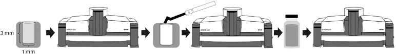
Figure 1 Schematic figure of the assessments of filling ability and volumetric changes. Gypsum–based models with cavities measuring 3 mm deep and 1 mm in diameter were manufactured and scanned using micro-computed tomography before and after filling. The samples were immersed in distilled water for 7 days, and another scan was performed.
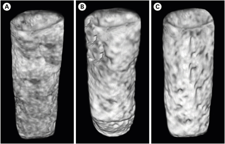
Figure 2 Three-dimensional models illustrating microtomographic images of the filling ability of Bio-C Repair (A), mineral trioxide aggregate repair high-plasticity (B), and intermediate restorative material (C) in CTVox software.
Reference
-
1. Torabinejad M, Parirokh M, Dummer PMH. Mineral trioxide aggregate and other bioactive endodontic cements: an updated overview - part II: other clinical applications and complications. Int Endod J. 2018; 51:284–317. PMID: 28846134.
Article2. Akbulut MB, Bozkurt DA, Terlemez A, Akman M. The push-out bond strength of BIOfactor mineral trioxide aggregate, a novel root repair material. Restor Dent Endod. 2019; 44:e5. PMID: 30834227.
Article3. Prati C, Gandolfi MG. Calcium silicate bioactive cements: biological perspectives and clinical applications. Dent Mater. 2015; 31:351–370. PMID: 25662204.
Article4. Ferreira CMA, Sassone LM, Gonçalves AS, de Carvalho JJ, Tomás-Catalá CJ, García-Bernal D, Oñate-Sánchez RE, Rodríguez-Lozano FJ, Silva EJ. Physicochemical, cytotoxicity and in vivo biocompatibility of a high-plasticity calcium-silicate based material. Sci Rep. 2019; 9:3933. PMID: 30850648.5. Jiménez-Sánchez MDC, Segura-Egea JJ, Díaz-Cuenca A. MTA HP Repair stimulates in vitro an homogeneous calcium phosphate phase coating deposition. J Clin Exp Dent. 2019; 11:e322–e326. PMID: 31110610.6. Cintra LTA, Benetti F, de Azevedo Queiroz IO, de Araújo Lopes JM, Penha de Oliveira SH, Sivieri Araújo G, Gomes-Filho JE. Cytotoxicity, biocompatibility, and biomineralization of the new high-plasticity MTA material. J Endod. 2017; 43:774–778. PMID: 28320539.
Article7. Tomás-Catalá CJ, Collado-González M, García-Bernal D, Oñate-Sánchez RE, Forner L, Llena C, Lozano A, Castelo-Baz P, Moraleda JM, Rodríguez-Lozano FJ. Comparative analysis of the biological effects of the endodontic bioactive cements MTA-Angelus, MTA Repair HP and NeoMTA Plus on human dental pulp stem cells. Int Endod J. 2017; 50(Supplement 2):e63–e72. PMID: 28891221.8. Benetti F, Queiroz ÍOA, Cosme-Silva L, Conti LC, Oliveira SHP, Cintra LTA. Cytotoxicity, biocompatibility and biomineralization of a new ready-for-use Bioceramic Repair material. Braz Dent J. 2019; 30:325–332. PMID: 31340221.
Article9. López-García S, Lozano A, García-Bernal D, Forner L, Llena C, Guerrero-Gironés J, Moraleda JM, Murcia L, Rodríguez-Lozano FJ. Biological effects of new hydraulic materials on human periodontal ligament stem cells. J Clin Med. 2019; 8:1216.
Article10. Biočanin V, Antonijević Đ, Poštić S, Ilić D, Vuković Z, Milić M, Fan Y, Li Z, Brković B, Đurić M. Marginal gaps between 2 calcium silicate and glass ionomer cements and apical root dentin. J Endod. 2018; 44:816–821. PMID: 29336880.
Article11. Chang SW. Chemical characteristics of mineral trioxide aggregate and its hydration reaction. Restor Dent Endod. 2012; 37:188–193. PMID: 23429542.
Article12. Torres FFE, Guerreiro-Tanomaru JM, Chavez-Andrade GM, Pinto JC, Berbert FL, Tanomaru-Filho M. Micro-computed tomographic evaluation of the flow and filling ability of endodontic materials using different test models. Restor Dent Endod. 2020; 45:e11. PMID: 32483530.
Article13. Torres FFE, Zordan-Bronzel CL, Guerreiro-Tanomaru JM, Chávez-Andrade GM, Pinto JC, Tanomaru-Filho M. Effect of immersion in distilled water or phosphate-buffered saline on the solubility, volumetric change and presence of voids within new calcium silicate-based root canal sealers. Int Endod J. 2020; 53:385–391. PMID: 31566768.
Article14. Zordan-Bronzel CL, Esteves Torres FF, Tanomaru-Filho M, Chávez-Andrade GM, Bosso-Martelo R, Guerreiro-Tanomaru JM. Evaluation of physicochemical properties of a new calcium silicate-based sealer, Bio-C sealer. J Endod. 2019; 45:1248–1252. PMID: 31447172.
Article15. Darvell BW, Wu RC. “MTA”-an hydraulic silicate cement: review update and setting reaction. Dent Mater. 2011; 27:407–422. PMID: 21353694.
Article16. Silva Almeida LH, Moraes RR, Morgental RD, Pappen FG. Are premixed calcium silicate-based endodontic sealers comparable to conventional materials? a systematic review of in vitro studies. J Endod. 2017; 43:527–535. PMID: 28216270.
Article17. International Organization for Standardization. ISO 6876: dental root canal sealing materials. Geneva: International Organization for Standardization;2012.18. Torres FFE, Bosso-Martelo R, Espir CG, Cirelli JA, Guerreiro-Tanomaru JM, Tanomaru-Filho M. Evaluation of physicochemical properties of root-end filling materials using conventional and micro-CT tests. J Appl Oral Sci. 2017; 25:374–380. PMID: 28877275.
Article19. Guo YJ, Du TF, Li HB, Shen Y, Mobuchon C, Hieawy A, Wang ZJ, Yang Y, Ma J, Haapasalo M. Physical properties and hydration behavior of a fast-setting bioceramic endodontic material. BMC Oral Health. 2016; 16:23. PMID: 26897651.
Article20. Camilleri J, Grech L, Galea K, Keir D, Fenech M, Formosa L, Damidot D, Mallia B. Porosity and root dentine to material interface assessment of calcium silicate-based root-end filling materials. Clin Oral Investig. 2014; 18:1437–1446.
Article21. Gandolfi MG, Siboni F, Botero T, Bossù M, Riccitiello F, Prati C. Calcium silicate and calcium hydroxide materials for pulp capping: biointeractivity, porosity, solubility and bioactivity of current formulations. J Appl Biomater Funct Mater. 2015; 13:43–60. PMID: 25199071.
Article22. Kim S, Kratchman S. Modern endodontic surgery concepts and practice: a review. J Endod. 2006; 32:601–623. PMID: 16793466.
Article23. Torres FF, Jacobs R, EzEldeen M, Guerreiro-Tanomaru JM, Dos Santos BC, Lucas-Oliveira É, Bonagamba TJ, Tanomaru-Filho M. Micro-computed tomography high resolution evaluation of dimensional and morphological changes of 3 root-end filling materials in simulated physiological conditions. J Mater Sci Mater Med. 2020; 31:14. PMID: 31965336.
Article24. Hashem AA, Hassanien EE. ProRoot MTA, MTA-Angelus and IRM used to repair large furcation perforations: sealability study. J Endod. 2008; 34:59–61. PMID: 18155494.
Article25. Övsay E, Kaptan RF, Şahin F. The repair of furcal perforations in different diameters with Biodentine, MTA, and IRM repair materials: a laboratory study using an E. Faecalis leakage model. BioMed Res Int. 2018; 2018:5478796. PMID: 29568756.26. Lertmalapong P, Jantarat J, Srisatjaluk RL, Komoltri C. Bacterial leakage and marginal adaptation of various bioceramics as apical plug in open apex model. J Investig Clin Dent. 2019; 10:e12371.
Article27. Tanomaru-Filho M, Torres FFE, Bosso-Martelo R, Chávez-Andrade GM, Bonetti-Filho I, Guerreiro-Tanomaru JM. A novel model for evaluating the flow of endodontic materials using micro-computed tomography. J Endod. 2017; 43:796–800. PMID: 28268019.
Article28. Dorileo MC, Pedro FL, Bandeca MC, Guedes OA, Villa RD, Borges AH. Comparative analysis of physicochemical properties of root perforation sealer materials. Restor Dent Endod. 2014; 39:201–209. PMID: 25110644.
Article29. Elyassi Y, Moinzadeh AT, Kleverlaan CJ. Characterization of leachates from 6 root canal sealers. J Endod. 2019; 45:623–627. PMID: 30905572.
Article30. Song YS, Choi Y, Lim MJ, Yu MK, Hong CU, Lee KW, Min KS. In vitro evaluation of a newly produced resin-based endodontic sealer. Restor Dent Endod. 2016; 41:189–195. PMID: 27508160.
Article
- Full Text Links
-
- Actions
-
Cited
- CITED
-
- Close
- Share
-
- Similar articles
-
- A micro-computed tomographic study of remaining filling materials of two bioceramic sealers and epoxy resin sealer after retreatment
- Micro-computed tomographic evaluation of the flow and filling ability of endodontic materials using different test models
- Micro-computed tomographic evaluation of a new system for root canal filling using calcium silicatebased root canal sealers
- Bacterial leakage and micro-computed tomography evaluation in round-shaped canals obturated with bioceramic cone and sealer using matched single cone technique
- Comparison of the sealing ability of various bioceramic materials for endodontic surgery
