Nasojejunal tube-related duodenal perforations: a multicenter experience
- Affiliations
-
- 1Department of Gastroenterology and Advanced Endoscopy, Ansh Clinic, Ahmedabad, Gujarat, India
- 2Department of Gastroenterology and Advanced Endoscopy, Prime Hospital, Rajkot, Gujarat, India
- 3Department of Gastroenterology and Advanced Endoscopy, Mission Gastro Hospital, Ahmedabad, Gujarat, India
- 4Department of Gastroenterology and Endoscopy, Gujarat Gastro Hub, Mehsana, India
- KMID: 2547905
- DOI: http://doi.org/10.5946/ce.2023.071
Figure
-
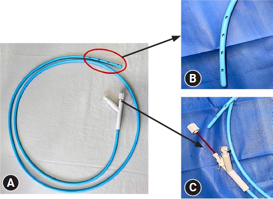
Fig. 1. Nasojejunal feeding tube (16 Fr) from Shaili endoscopy. (A) Whole tube with removal guidewire. (B) Tip of nasojejunal tube. (C) Removable guidewire.
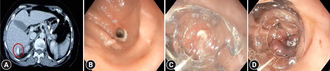
Fig. 2. Detection of perforation on computed tomography scan and closure with over-the-scope clip (OTSC) in case 1. (A) Contrast-enhanced computed tomography of the abdomen showed the tube had perforated the duodenum and lying in the subhepatic area (red circle). (B) Full-thickness defect in the duodenum in D2. (C) Mild suction was applied, and the defect was pulled in cap of the OTSC device. (D) OTSC was applied, and complete closure of the defect was achieved.
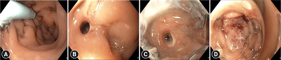
Fig. 3. Nasojejunal tube-related duodenal perforation closure was performed with over-the-scope clip (OTSC) in case 2. (A) Tip of the tube was seen penetrating the duodenal wall. (B) After withdrawal of the tube, full-thickness defect was noticed. (C) OTSC device was taken, and the defect was sucked into the cap with mild suction. (D) Complete closure was achieved with the OTSC clip.
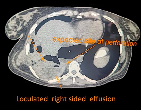
Fig. 4. Chest computed tomography showing dilated esophagus with possible site of perforation and right-sided loculated pleural effusion.
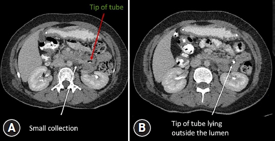
Fig. 5. Detection of perforation on contrast-enhanced computed tomography (CT) abdomen in case 4. (A) CT abdomen axial image showed small loculated collection around the tip at the third to fourth part of the duodenum. (B) CT abdomen axial image showed the tip of the tube penetrating the wall and lying just outside the lumen.
Reference
-
1. Pearce CB, Duncan HD. Enteral feeding. Nasogastric, nasojejunal, percutaneous endoscopic gastrostomy, or jejunostomy: its indications and limitations. Postgrad Med J. 2002; 78:198–204.
Article2. Sajid MS, Harper A, Hussain Q, et al. An integrated systematic review and meta-analysis of published randomized controlled trials evaluating nasogastric against postpyloris (nasoduodenal and nasojejunal) feeding in critically ill patients admitted in intensive care unit. Eur J Clin Nutr. 2014; 68:424–432.
Article3. Blumenstein I, Shastri YM, Stein J. Gastroenteric tube feeding: techniques, problems and solutions. World J Gastroenterol. 2014; 20:8505–8524.
Article4. Halkic N, Guerid S, Blanchard A, et al. Small-bowel perforation: a consequence of feeding jejunostomy. Can J Surg. 2005; 48:161–162.5. Fang JC, Hilden K, Holubkov R, et al. Transnasal endoscopy vs. fluoroscopy for the placement of nasoenteric feeding tubes in critically ill patients. Gastrointest Endosc. 2005; 62:661–666.
Article6. ASGE Technology Committee, Kwon RS, Banerjee S, et al. Enteral nutrition access devices. Gastrointest Endosc. 2010; 72:236–248.
Article7. Schwab D, Mühldorfer S, Nusko G, et al. Endoscopic placement of nasojejunal tubes: a randomized, controlled, prospective trial comparing suitability and technical success for two different tubes. Gastrointest Endosc. 2002; 56:858–863.
Article8. Prabhakaran S, Doraiswamy VA, Nagaraja V, et al. Nasoenteric tube complications. Scand J Surg. 2012; 101:147–155.
Article9. Siegle RL, Rabinowitz JG, Sarasohn C. Intestinal perforations secondary to nasojejunal feeding tubes. AJR Am J Roentgenol. 1976; 126:1229–1232.
Article10. Merten DF, Mumford L, Filston HC, et al. Radiological observations during transpyloric tube feeding in infants of low birth weight. Perforation of the duodenum and variations in normal duodenal anatomy. Radiology. 1980; 136:67–75.
Article
- Full Text Links
-
- Actions
-
Cited
- CITED
-
- Close
- Share
-
- Similar articles
-
- The Management of Endoscopic Retrograde Cholangiopancreatography-Related Duodenal Perforation
- Two Cases of Successful ERCP during ERCP-Related Iatrogenic Duodenal Perforation
- Endoscopic Treatments of Endoscopic Retrograde Cholangiopancreatography-Related Duodenal Perforations
- Perforation on the superior side of duodenum is a risk factor of laparoscopic primary repair for duodenal ulcer perforation
- A Case of Type I Duodenal Perforation Treated with Covered Metal Stent
