Cell clusters in intervertebral disc degeneration: an attempted repair mechanism aborted via apoptosis
- Affiliations
-
- 1Department of Anatomy, Sikkim Manipal Institute of Medical Sciences, Sikkim Manipal University, Sikkim, India
- 2Centre for Clinical Anatomy, University of Bristol, Bristol, UK
- 3Biomolecular Research Centre, Sheffield Hallam University, Sheffield, UK
- KMID: 2546467
- DOI: http://doi.org/10.5115/acb.23.067
Abstract
- Cell clusters are a histological hallmark feature of intervertebral disc degeneration. Clusters arise from cell proliferation, are associated with replicative senescence, and remain metabolically, but their precise role in various stages of disc degeneration remain obscure. The aim of this study was therefore to investigate small, medium, and large size cell-clusters. For this purpose, human disc samples were collected from 55 subjects, aged 37–72 years, 21 patients had disc herniation, 10 had degenerated non-herniated discs, and 9 had degenerative scoliosis with spinal curvature <45°. 15 non-degenerated control discs were from cadavers. Clusters and matrix changes were investigated with histology, immunohistochemistry, and Sodium dodecyl sulphate polyacrylamide gel electrophoresis (SDS-PAGE). Data obtained were analyzed with spearman rank correlation and ANOVA. Results revealed, small and medium-sized clusters were positive for cell proliferation markers Ki-67 and proliferating cell nuclear antigen (PCNA) in control and slightly degenerated human discs, while large cell clusters were typically more abundant in severely degenerated and herniated discs. Large clusters associated with matrix fissures, proteoglycan loss, matrix metalloproteinase-1 (MMP-1), and Caspase-3. Spatial association findings were reconfirmed with SDS-PAGE that showed presence to these target markers based on its molecular weight. Controls, slightly degenerated discs showed smaller clusters, less proteoglycan loss, MMP-1, and Caspase-3. In conclusion, cell clusters in the early stages of degeneration could be indicative of repair, however sustained loading increases large cell clusters especially around microscopic fissures that accelerates inflammatory catabolism and alters cellular metabolism, thus attempted repair process initiated by cell clusters fails and is aborted at least in part via apoptosis.
Keyword
Figure
-
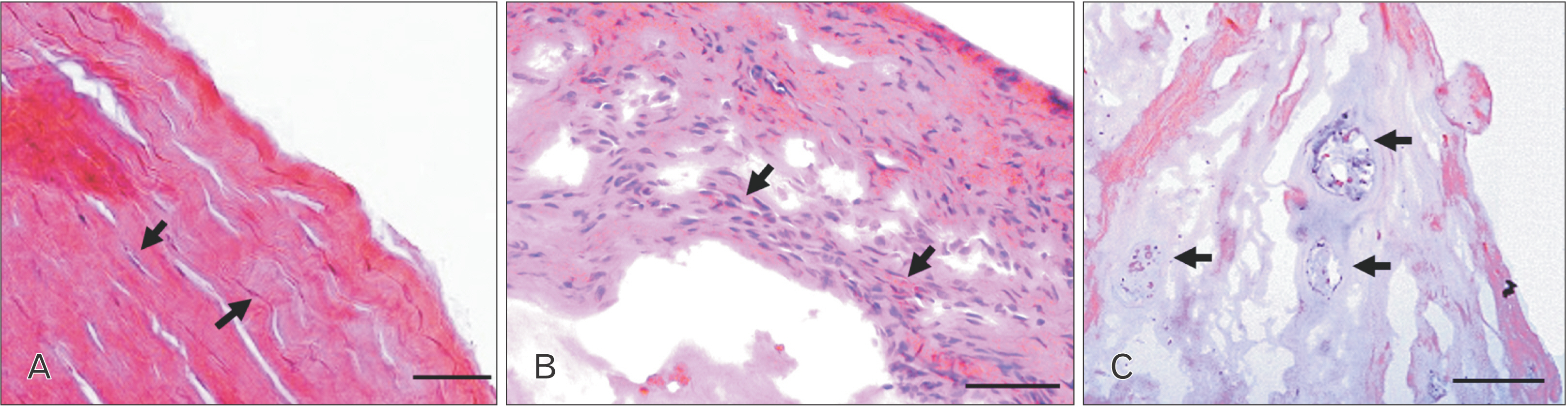
Fig. 1 Comparisons between (A) control non-degenerated cadaveric human disc with extremely flattened annulus fibrosus (AF) cells (arrows) and (B) herniated disc, arrows shows inflammatory cells, and (C) degenerated disc with displaced lamellae and large cell clusters (arrows). Scale bar: 100 μm.

Fig. 2 Cell clusters comparisons between (A) control non-degenerated human cadaveric disc with small size cell clusters (2, 3, 6 cells) and (B) herniated disc with large sized cell clusters (50–60 cells, arrow). Scale bar: 100 μm (A) and 50 μm (B). (C) Toluidine blue stained control NP/IAF region without clusters and (D) herniated disc tissue with large cluster (arrow) stained with toluidine blue. Scale bar: 50 μm. (E) Cell cluster scores (0–5) and increasing pf grade of disc degeneration. (F) Proteoglycan staining score (0–3) and increasing pf grade of disc degeneration. NP, nucleus pulposus; IAF, inner annulus fibrosus; pf, Pfirrmann grade of degeneration; PG, proteoglycans.
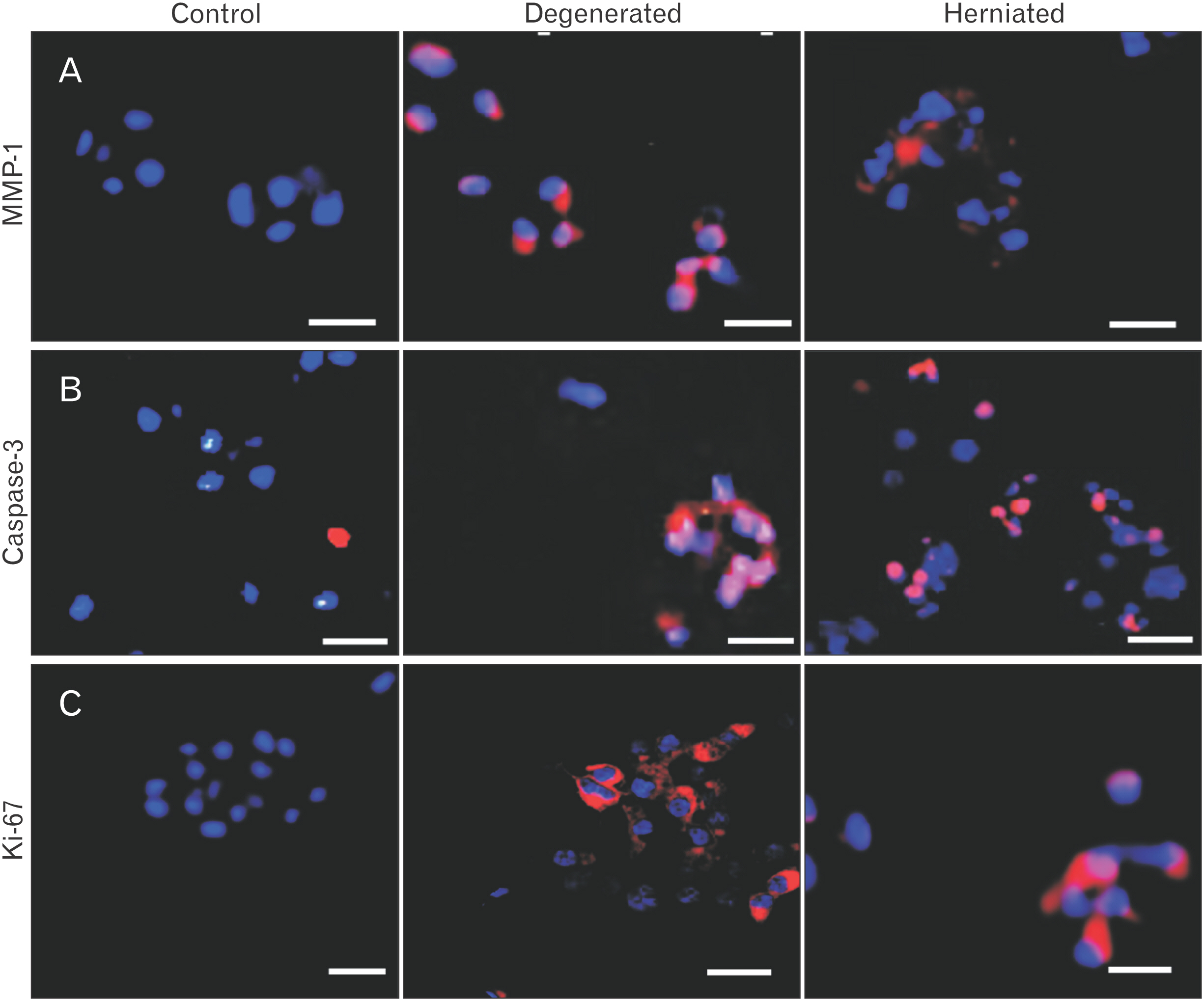
Fig. 3 Small, medium, and large sized cell clusters of the ‘control’ and its comparisons to ‘degenerated’ and ‘herniated’ discs for (A) MMP-1, (B) Caspase-3, and (C) Ki-67. Scale bar: 50 μm. MMP-1, matrix metalloproteinase-1.
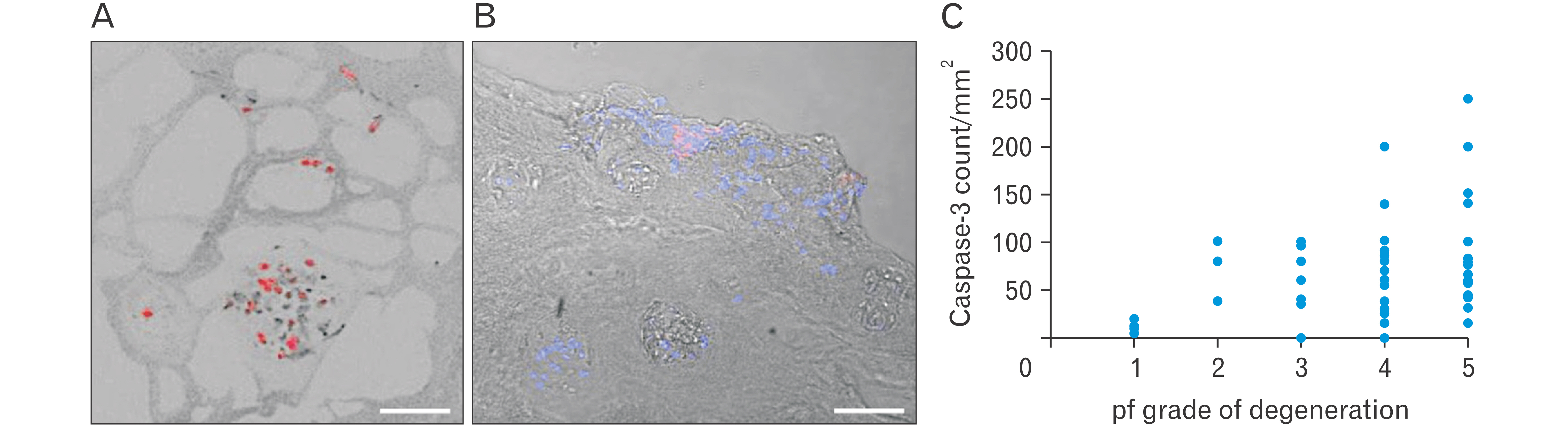
Fig. 4 Medium and large sized cell clusters stained with (A) Caspase-3 (red stain, blue indicates DAPI stained cell nuclei), (B) MMP-1 (red stain, blue indicates DAPI stain cell nuclei) with phase contrast background. Scale bar: 100 μm. (C) Caspase-3 positive cell clusters count and increasing pf grade of disc degeneration. MMP-1, matrix metalloproteinase-1; DAPI, 4’,6-diamidino-2-phenylindole; pf, Pfirrmann.
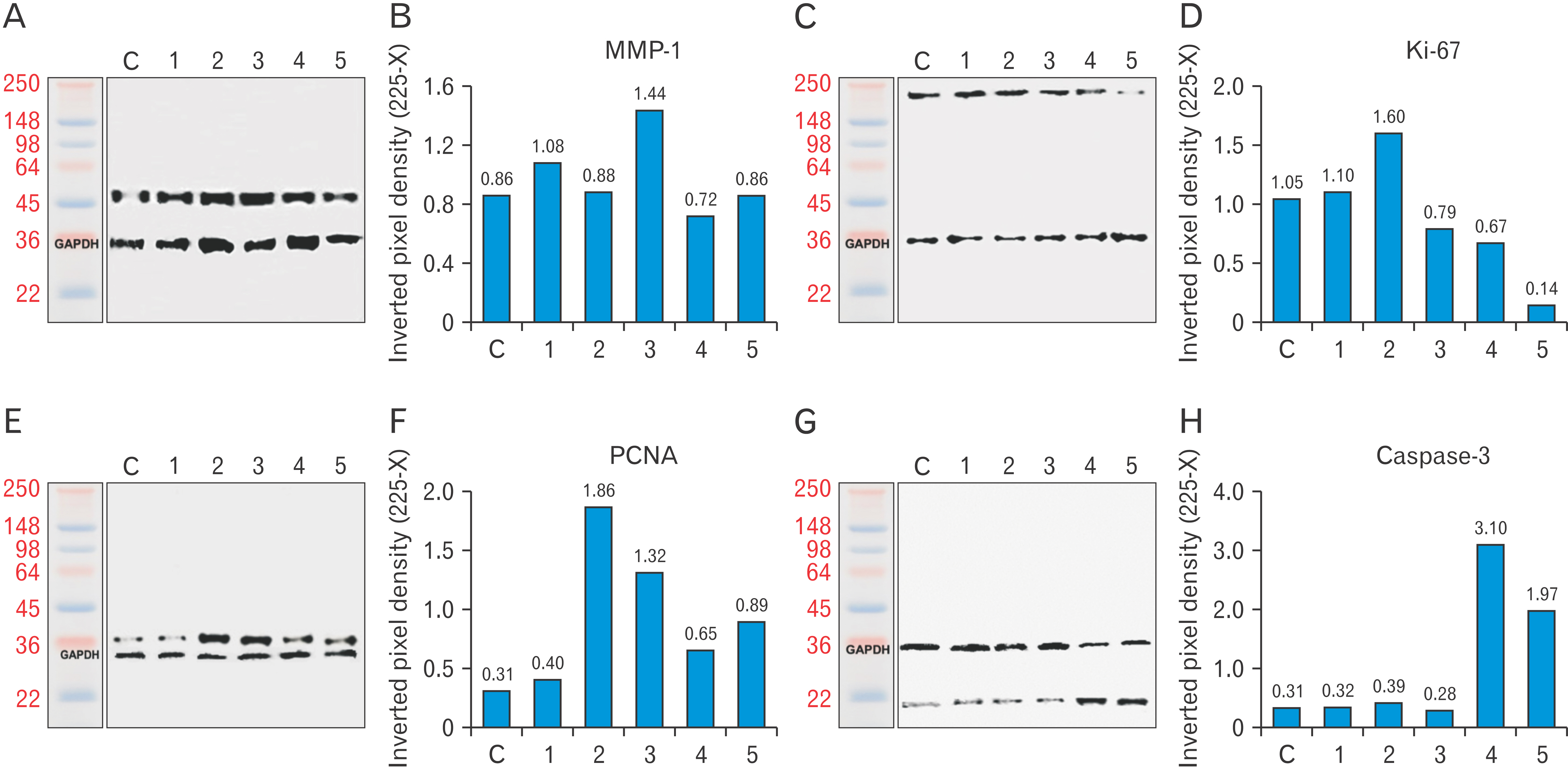
Fig. 5 Western blot analysis on a 10% gel. Lane were loaded as C, ‘non-degenerated cadaveric disc’ followed by disc specimens categorised according to pf grades degeneration in the subsequent lanes as pf grade 1, 2, 3, 4, and 5 discs. (A) MMP-1 core protein at 54 kDa. (B) Expression level of control ‘c’, and grade ‘1–5 discs’ shown with grade 3 discs at the highest expression level. (C) Ki-67 core protein with a molecular weight of 250 kDa. (D) Weak expression level for degenerated grade 3–5 discs. (E) PCNA core protein with a molecular weight of 36 kDa. (F) Bands expressed higher values for grade 2 and 3 discs in comparisons to other grades. (G) Caspase-3 core protein at 22 kDa. (H) Strongly expressed for the severely degenerated grade 4 and 5 discs. GAPDH as loading control is at 36 kDa. C, control; pf, Pfirrmann; MMP-1, matrix metalloproteinase-1; PCNA, proliferating cell nuclear antigen; GAPDH, glyceraldehyde 3-phosphate dehydrogenase.
Reference
-
References
1. Karppinen J, Shen FH, Luk KD, Andersson GB, Cheung KM, Samartzis D. 2011; Management of degenerative disk disease and chronic low back pain. Orthop Clin North Am. 42:513–28. DOI: 10.1016/j.ocl.2011.07.009. PMID: 21944588.
Article2. Newell N, Little JP, Christou A, Adams MA, Adam CJ, Masouros SD. 2017; Biomechanics of the human intervertebral disc: a review of testing techniques and results. J Mech Behav Biomed Mater. 69:420–34. DOI: 10.1016/j.jmbbm.2017.01.037. PMID: 28262607.
Article3. Adams MA, Freeman BJ, Morrison HP, Nelson IW, Dolan P. 2000; Mechanical initiation of intervertebral disc degeneration. Spine (Phila Pa 1976). 25:1625–36. DOI: 10.1097/00007632-200007010-00005. PMID: 10870137.
Article4. Adams MA, Roughley PJ. 2006; What is intervertebral disc degeneration, and what causes it? Spine (Phila Pa 1976). 31:2151–61. DOI: 10.1097/01.brs.0000231761.73859.2c. PMID: 16915105.
Article5. Adams A, Roche O, Mazumder A, Davagnanam I, Mankad K. 2014; Imaging of degenerative lumbar intervertebral discs; linking anatomy, pathology and imaging. Postgrad Med J. 90:511–9. DOI: 10.1136/postgradmedj-2013-132193. PMID: 24965489.
Article6. Adams MA, Dolan P. 2012; Intervertebral disc degeneration: evidence for two distinct phenotypes. J Anat. 221:497–506. DOI: 10.1111/j.1469-7580.2012.01551.x. PMID: 22881295. PMCID: PMC3512277.
Article7. Roberts S, Menage J, Urban JP. 1989; Biochemical and structural properties of the cartilage end-plate and its relation to the intervertebral disc. Spine (Phila Pa 1976). 14:166–74. DOI: 10.1097/00007632-198902000-00005. PMID: 2922637.
Article8. Lama P, Le Maitre CL, Dolan P, Tarlton JF, Harding IJ, Adams MA. 2013; Do intervertebral discs degenerate before they herniate, or after? Bone Joint J. 95-B:1127–33. DOI: 10.1302/0301-620X.95B8.31660. PMID: 23908431.
Article9. Johnson WE, Eisenstein SM, Roberts S. 2001; Cell cluster formation in degenerate lumbar intervertebral discs is associated with increased disc cell proliferation. Connect Tissue Res. 42:197–207. DOI: 10.3109/03008200109005650. PMID: 11913491.
Article10. Lyu FJ, Cheung KM, Zheng Z, Wang H, Sakai D, Leung VY. 2019; IVD progenitor cells: a new horizon for understanding disc homeostasis and repair. Nat Rev Rheumatol. 15:102–12. DOI: 10.1038/s41584-018-0154-x. PMID: 30643232.
Article11. Lama P, Claireaux H, Flower L, Harding IJ, Dolan T, Le Maitre CL, Adams MA. 2019; Physical disruption of intervertebral disc promotes cell clustering and a degenerative phenotype. Cell Death Discov. 5:154. DOI: 10.1038/s41420-019-0233-z. PMID: 31871771. PMCID: PMC6917743.
Article12. Le Maitre CL, Freemont AJ, Hoyland JA. 2005; The role of interleukin-1 in the pathogenesis of human intervertebral disc degeneration. Arthritis Res Ther. 7:R732–45. DOI: 10.1186/ar1732. PMID: 15987475. PMCID: PMC1175026.13. Le Maitre CL, Freemont AJ, Hoyland JA. 2007; Accelerated cellular senescence in degenerate intervertebral discs: a possible role in the pathogenesis of intervertebral disc degeneration. Arthritis Res Ther. 9:R45. DOI: 10.1186/ar2198. PMID: 17498290. PMCID: PMC2206356.
Article14. Horner HA, Roberts S, Bielby RC, Menage J, Evans H, Urban JP. 2002; Cells from different regions of the intervertebral disc: effect of culture system on matrix expression and cell phenotype. Spine (Phila Pa 1976). 27:1018–28. DOI: 10.1097/00007632-200205150-00004. PMID: 12004167.15. Trout JJ, Buckwalter JA, Moore KC. 1982; Ultrastructure of the human intervertebral disc: II. Cells of the nucleus pulposus. Anat Rec. 204:307–14. DOI: 10.1002/ar.1092040403. PMID: 7181135.
Article16. Sharp CA, Roberts S, Evans H, Brown SJ. 2009; Disc cell clusters in pathological human intervertebral discs are associated with increased stress protein immunostaining. Eur Spine J. 18:1587–94. DOI: 10.1007/s00586-009-1053-2. PMID: 19517141. PMCID: PMC2899395.
Article17. Kim KW, Lim TH, Kim JG, Jeong ST, Masuda K, An HS. 2003; The origin of chondrocytes in the nucleus pulposus and histologic findings associated with the transition of a notochordal nucleus pulposus to a fibrocartilaginous nucleus pulposus in intact rabbit intervertebral discs. Spine (Phila Pa 1976). 28:982–90. DOI: 10.1097/01.BRS.0000061986.03886.4F. PMID: 12768135.
Article18. Kim JH, Deasy BM, Seo HY, Studer RK, Vo NV, Georgescu HI, Sowa GA, Kang JD. 2009; Differentiation of intervertebral notochordal cells through live automated cell imaging system in vitro. Spine (Phila Pa 1976). 34:2486–93. DOI: 10.1097/BRS.0b013e3181b26ed1. PMID: 19841610.19. Phillips KL, Chiverton N, Michael AL, Cole AA, Breakwell LM, Haddock G, Bunning RA, Cross AK, Le Maitre CL. 2013; The cytokine and chemokine expression profile of nucleus pulposus cells: implications for degeneration and regeneration of the intervertebral disc. Arthritis Res Ther. 15:R213. DOI: 10.1186/ar4408. PMID: 24325988. PMCID: PMC3979161.
Article20. Ashton IK, Eisenstein SM. 1996; The effect of substance P on proliferation and proteoglycan deposition of cells derived from rabbit intervertebral disc. Spine (Phila Pa 1976). 21:421–6. DOI: 10.1097/00007632-199602150-00004. PMID: 8658244.
Article21. Henriksson HB, Svala E, Skioldebrand E, Lindahl A, Brisby H. 2012; Support of concept that migrating progenitor cells from stem cell niches contribute to normal regeneration of the adult mammal intervertebral disc: a descriptive study in the New Zealand white rabbit. Spine (Phila Pa 1976). 37:722–32. DOI: 10.1097/BRS.0b013e318231c2f7. PMID: 21897341.22. Gardner DL. 1994; Problems and paradigms in joint pathology. J Anat. 184(Pt 3):465–76. PMID: 7928636. PMCID: PMC1259955.23. Lotz MK, Otsuki S, Grogan SP, Sah R, Terkeltaub R, D'Lima D. 2010; Cartilage cell clusters. Arthritis Rheum. 62:2206–18. DOI: 10.1002/art.27528. PMID: 20506158. PMCID: PMC2921934.
Article24. Bryant PJ. 2001; Growth factors controlling imaginal disc growth in Drosophila. Novartis Found Symp. 237:182–94. discussion 194–202. DOI: 10.1002/0470846666.ch14. PMID: 11444043.25. Li FX, Xu F, Lin X, Wu F, Zhong JY, Wang Y, Guo B, Zheng MH, Shan SK, Yuan LQ. 2020; The role of substance P in the regulation of bone and cartilage metabolic activity. Front Endocrinol (Lausanne). 11:77. DOI: 10.3389/fendo.2020.00077. PMID: 32180759. PMCID: PMC7059306. PMID: 487345bbe61c448dbdff7a6b8b563d3e.
Article26. Opolka A, Straub RH, Pasoldt A, Grifka J, Grässel S. 2012; Substance P and norepinephrine modulate murine chondrocyte proliferation and apoptosis. Arthritis Rheum. 64:729–39. DOI: 10.1002/art.33449. PMID: 22042746.
Article27. Ogunlade B, Fidelis OP, Adelakun SA, Adedotun OA. 2020; Grape seed extract inhibits nucleus pulposus cell apoptosis and attenuates annular puncture induced intervertebral disc degeneration in rabbit model. Anat Cell Biol. 53:313–24. DOI: 10.5115/acb.20.047. PMID: 32782235. PMCID: PMC7527127.
Article28. Horner HA, Urban JP. 2001; 2001 Volvo Award Winner in Basic Science Studies: effect of nutrient supply on the viability of cells from the nucleus pulposus of the intervertebral disc. Spine (Phila Pa 1976). 26:2543–9. DOI: 10.1097/00007632-200112010-00006. PMID: 11725234.
Article29. Mavrogonatou E, Papadimitriou K, Urban JP, Papadopoulos V, Kletsas D. 2015; Deficiency in the α1 subunit of Na+/K+-ATPase enhances the anti-proliferative effect of high osmolality in nucleus pulposus intervertebral disc cells. J Cell Physiol. 230:3037–48. DOI: 10.1002/jcp.25040. PMID: 25967398.30. Sakai D, Nakamura Y, Nakai T, Mishima T, Kato S, Grad S, Alini M, Risbud MV, Chan D, Cheah KS, Yamamura K, Masuda K, Okano H, Ando K, Mochida J. 2012; Exhaustion of nucleus pulposus progenitor cells with ageing and degeneration of the intervertebral disc. Nat Commun. 3:1264. DOI: 10.1038/ncomms2226. PMID: 23232394. PMCID: PMC3535337.
Article31. Tessier S, Risbud MV. 2021; Understanding embryonic development for cell-based therapies of intervertebral disc degeneration: toward an effort to treat disc degeneration subphenotypes. Dev Dyn. 250:302–17. DOI: 10.1002/dvdy.217. PMID: 32564440.
Article32. Mizrahi O, Sheyn D, Tawackoli W, Ben-David S, Su S, Li N, Oh A, Bae H, Gazit D, Gazit Z. 2013; Nucleus pulposus degeneration alters properties of resident progenitor cells. Spine J. 13:803–14. DOI: 10.1016/j.spinee.2013.02.065. PMID: 23578990. PMCID: PMC3759825.
Article33. Sakai D, Schol J, Bach FC, Tekari A, Sagawa N, Nakamura Y, Chan SCW, Nakai T, Creemers LB, Frauchiger DA, May RD, Grad S, Watanabe M, Tryfonidou MA, Gantenbein B. 2018; Successful fishing for nucleus pulposus progenitor cells of the intervertebral disc across species. JOR Spine. 1:e1018. DOI: 10.1002/jsp2.1018. PMID: 31463445. PMCID: PMC6686801. PMID: f4cae7a3c9014c9f88a26f0e692748ea.
Article34. Pfirrmann CW, Metzdorf A, Zanetti M, Hodler J, Boos N. 2001; Magnetic resonance classification of lumbar intervertebral disc degeneration. Spine (Phila Pa 1976). 26:1873–8. DOI: 10.1097/00007632-200109010-00011. PMID: 11568697.
Article35. Schmitz N, Laverty S, Kraus VB, Aigner T. 2010; Basic methods in histopathology of joint tissues. Osteoarthritis Cartilage. 18 Suppl 3:S113–6. DOI: 10.1016/j.joca.2010.05.026. PMID: 20864017.
Article36. Boos N, Weissbach S, Rohrbach H, Weiler C, Spratt KF, Nerlich AG. 2002; Classification of age-related changes in lumbar intervertebral discs: 2002 Volvo Award in basic science. Spine (Phila Pa 1976). 27:2631–44. DOI: 10.1097/00007632-200212010-00002. PMID: 12461389.37. Baschong W, Suetterlin R, Laeng RH. 2001; Control of autofluorescence of archival formaldehyde-fixed, paraffin-embedded tissue in confocal laser scanning microscopy (CLSM). J Histochem Cytochem. 49:1565–72. DOI: 10.1177/002215540104901210. PMID: 11724904.
Article38. Bouayad D, Pederzoli-Ribeil M, Mocek J, Candalh C, Arlet JB, Hermine O, Reuter N, Davezac N, Witko-Sarsat V. 2012; Nuclear-to-cytoplasmic relocalization of the proliferating cell nuclear antigen (PCNA) during differentiation involves a chromosome region maintenance 1 (CRM1)-dependent export and is a prerequisite for PCNA antiapoptotic activity in mature neutrophils. J Biol Chem. 287:33812–25. DOI: 10.1074/jbc.M112.367839. PMID: 22846997. PMCID: PMC3460476.
Article39. Faratian D, Munro A, Twelves C, Bartlett JM. 2009; Membranous and cytoplasmic staining of Ki67 is associated with HER2 and ER status in invasive breast carcinoma. Histopathology. 54:254–7. DOI: 10.1111/j.1365-2559.2008.03191.x. PMID: 19207951.
Article40. Cserni G. 2018; Analysis of membranous Ki-67 staining in breast cancer and surrounding breast epithelium. Virchows Arch. 473:145–53. DOI: 10.1007/s00428-018-2343-z. PMID: 29594352.
Article41. Remnant L, Kochanova NY, Reid C, Cisneros-Soberanis F, Earnshaw WC. 2021; The intrinsically disorderly story of Ki-67. Open Biol. 11:210120. DOI: 10.1098/rsob.210120. PMID: 34375547. PMCID: PMC8354752. PMID: 35ca6220cccf483eae229c9aafbdb243.
Article42. Le Maitre CL, Freemont AJ, Hoyland JA. 2004; Localization of degradative enzymes and their inhibitors in the degenerate human intervertebral disc. J Pathol. 204:47–54. DOI: 10.1002/path.1608. PMID: 15307137.
Article43. Miller I, Min M, Yang C, Tian C, Gookin S, Carter D, Spencer SL. 2018; Ki67 is a graded rather than a binary marker of proliferation versus quiescence. Cell Rep. 24:1105–12.e5. DOI: 10.1016/j.celrep.2018.06.110. PMID: 30067968. PMCID: PMC6108547.
Article44. Sasaki K, Kurose A, Ishida Y. 1993; Flow cytometric analysis of the expression of PCNA during the cell cycle in HeLa cells and effects of the inhibition of DNA synthesis on it. Cytometry. 14:876–82. DOI: 10.1002/cyto.990140805. PMID: 7904555.
Article45. Van Doren SR. 2015; Matrix metalloproteinase interactions with collagen and elastin. Matrix Biol. 44-46:224–31. DOI: 10.1016/j.matbio.2015.01.005. PMID: 25599938. PMCID: PMC4466143.
Article46. Vo NV, Hartman RA, Yurube T, Jacobs LJ, Sowa GA, Kang JD. 2013; Expression and regulation of metalloproteinases and their inhibitors in intervertebral disc aging and degeneration. Spine J. 13:331–41. DOI: 10.1016/j.spinee.2012.02.027. PMID: 23369495. PMCID: PMC3637842.47. Tschoeke SK, Hellmuth M, Hostmann A, Robinson Y, Ertel W, Oberholzer A, Heyde CE. 2008; Apoptosis of human intervertebral discs after trauma compares to degenerated discs involving both receptor-mediated and mitochondrial-dependent pathways. J Orthop Res. 26:999–1006. DOI: 10.1002/jor.20601. PMID: 18302283.
Article48. Jiang LB, Liu HX, Zhou YL, Sheng SR, Xu HZ, Xue EX. 2017; An ultrastructural study of chondroptosis: programmed cell death in degenerative intervertebral discs in vivo. J Anat. 231:129–39. DOI: 10.1111/joa.12618. PMID: 28436567. PMCID: PMC5472521.49. Lama P, Kulkarni JP, Tamang BK. 2017; The role of cell clusters in intervertebral disc degeneration and its relevance behind repair. Spine Res. 3:15. DOI: 10.21767/2471-8173.100035.
Article50. Sandell LJ, Aigner T. 2001; Articular cartilage and changes in arthritis. An introduction: cell biology of osteoarthritis. Arthritis Res. 3:107–13. DOI: 10.1186/ar148. PMID: 11178118. PMCID: PMC128887.51. Pearle AD, Warren RF, Rodeo SA. 2005; Basic science of articular cartilage and osteoarthritis. Clin Sports Med. 24:1–12. DOI: 10.1016/j.csm.2004.08.007. PMID: 15636773.
Article52. Melrose J, Roberts S, Smith S, Menage J, Ghosh P. 2002; Increased nerve and blood vessel ingrowth associated with proteoglycan depletion in an ovine anular lesion model of experimental disc degeneration. Spine (Phila Pa 1976). 27:1278–85. DOI: 10.1097/00007632-200206150-00007. PMID: 12065974.
Article53. Buckwalter JA. 1995; Aging and degeneration of the human intervertebral disc. Spine (Phila Pa 1976). 20:1307–14. DOI: 10.1097/00007632-199506000-00022. PMID: 7660243.
Article54. Urban JP, Maroudas A. 1981; Swelling of the intervertebral disc in vitro. Connect Tissue Res. 9:1–10. DOI: 10.3109/03008208109160234. PMID: 6456121.55. Urban JP, Smith S, Fairbank JC. 2004; Nutrition of the intervertebral disc. Spine (Phila Pa 1976). 29:2700–9. DOI: 10.1097/01.brs.0000146499.97948.52. PMID: 15564919.
Article56. Zhao CQ, Wang LM, Jiang LS, Dai LY. 2007; The cell biology of intervertebral disc aging and degeneration. Ageing Res Rev. 6:247–61. DOI: 10.1016/j.arr.2007.08.001. PMID: 17870673.
Article57. Lotz JC, Chin JR. 2000; Intervertebral disc cell death is dependent on the magnitude and duration of spinal loading. Spine (Phila Pa 1976). 25:1477–83. DOI: 10.1097/00007632-200006150-00005. PMID: 10851095.
Article58. Le Maitre CL, Frain J, Millward-Sadler J, Fotheringham AP, Freemont AJ, Hoyland JA. 2009; Altered integrin mechanotransduction in human nucleus pulposus cells derived from degenerated discs. Arthritis Rheum. 60:460–9. DOI: 10.1002/art.24248. PMID: 19180480.
Article59. Wuertz K, Vo N, Kletsas D, Boos N. 2012; Inflammatory and catabolic signalling in intervertebral discs: the roles of NF-κB and MAP kinases. Eur Cell Mater. 23:103–19. discussion 119–20. DOI: 10.22203/eCM.v023a08. PMID: 22354461.
Article60. Vogl T, Eisenblätter M, Völler T, Zenker S, Hermann S, van Lent P, Faust A, Geyer C, Petersen B, Roebrock K, Schäfers M, Bremer C, Roth J. 2014; Alarmin S100A8/S100A9 as a biomarker for molecular imaging of local inflammatory activity. Nat Commun. 5:4593. DOI: 10.1038/ncomms5593. PMID: 25098555. PMCID: PMC4143994.
Article61. Haglund L, Bernier SM, Onnerfjord P, Recklies AD. 2008; Proteomic analysis of the LPS-induced stress response in rat chondrocytes reveals induction of innate immune response components in articular cartilage. Matrix Biol. 27:107–18. DOI: 10.1016/j.matbio.2007.09.009. PMID: 18023983.
Article
- Full Text Links
-
- Actions
-
Cited
- CITED
-
- Close
- Share
-
- Similar articles
-
- Biochemical Factors of Intervertebral Disc Degeneration: Implications for Disc Regeneration
- Spontaneous Total Resolution of Severe Lumbar Disc Herniation
- Detection of O-Linked-N-Acetylglucosamine Modification and Its Associated Enzymes in Human Degenerated Intervertebral Discs
- Correlation between Bone Mineral Density and Interverterbral Disc Degeneration
- Disparity between MR Imaging and Histochemical Grading in Human Intervertebral Disc Degeneration
