Detecting colorectal lesions with image-enhanced endoscopy: an updated review from clinical trials
- Affiliations
-
- 1Department of Gastroenterology, International University of Health and Welfare Ichikawa Hospital, Chiba, Japan
- 2Department of Gastrointestinal Endoscopy, NTT Medical Center Tokyo, Tokyo, Japan
- KMID: 2546133
- DOI: http://doi.org/10.5946/ce.2023.055
Abstract
- Colonoscopy plays an important role in reducing the incidence and mortality of colorectal cancer by detecting adenomas and other precancerous lesions. Image-enhanced endoscopy (IEE) increases lesion visibility by enhancing the microstructure, blood vessels, and mucosal surface color, resulting in the detection of colorectal lesions. In recent years, various IEE techniques have been used in clinical practice, each with its unique characteristics. Numerous studies have reported the effectiveness of IEE in the detection of colorectal lesions. IEEs can be divided into two broad categories according to the nature of the image: images constructed using narrowband wavelength light, such as narrowband imaging and blue laser imaging/blue light imaging, or color images based on white light, such as linked color imaging, texture and color enhancement imaging, and i-scan. Conversely, artificial intelligence (AI) systems, such as computer-aided diagnosis systems, have recently been developed to assist endoscopists in detecting colorectal lesions during colonoscopy. To better understand the features of each IEE, this review presents the effectiveness of each type of IEE and their combination with AI for colorectal lesion detection by referencing the latest research data.
Figure
-
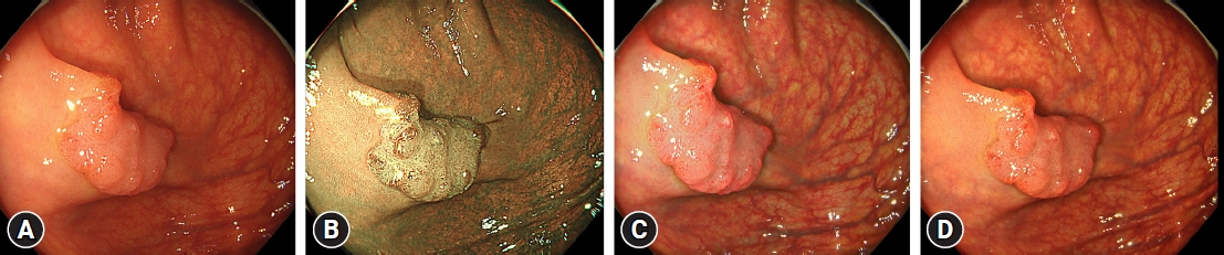
Fig. 1. Imaging of colon cancer under white light endoscopy (WLE), narrow-band imaging (NBI), texture and color enhancement imaging (TXI) mode 1, and TXI mode 2. A type IIa+IIc early cancer is identified using (A) WLE, (B) NBI, (C) TXI mode 1, and (D) TXI mode 2.

Fig. 2. Imaging of a sessile serrated lesion under white light endoscopy (WLE), blue laser imaging/blue light imaging (BLI), and linked color imaging (LCI). Flat, elevated sessile serrated lesions are shown on (A) WLE, (B) BLI, and (C) LCI.

Fig. 3. Imaging of an adenoma under i-scan. A protruding adenoma lesion is shown with i-scan: (A) white-light endoscopy, (B) surface enhancement, (C) contrast enhancement, (D) tone enhancement, (E) optical enhancement mode 1, and (F) optical enhancement mode 2.
Reference
-
1. Sung H, Ferlay J, Siegel RL, et al. Global cancer statistics 2020: GLOBOCAN estimates of incidence and mortality worldwide for 36 cancers in 185 countries. CA Cancer J Clin. 2021; 71:209–249.2. Zauber AG, Winawer SJ, O'Brien MJ, et al. Colonoscopic polypectomy and long-term prevention of colorectal-cancer deaths. N Engl J Med. 2012; 366:687–696.3. Erichsen R, Baron JA, Hamilton-Dutoit SJ, et al. Increased risk of colorectal cancer development among patients with serrated polyps. Gastroenterology. 2016; 150:895–902.4. Li D, Doherty AR, Raju M, et al. Risk stratification for colorectal cancer in individuals with subtypes of serrated polyps. Gut. 2022; 71:2022–2029.5. Sekiguchi M, Matsuda T, Hotta K, et al. Post-polypectomy surveillance: the present and the future. Clin Endosc. 2022; 55:489–495.6. Kim SY, Kwak MS, Yoon SM, et al. Korean guidelines for postpolypectomy colonoscopic surveillance: 2022 revised edition. Clin Endosc. 2022; 55:703–725.7. Saito Y, Oka S, Kawamura T, et al. Colonoscopy screening and surveillance guidelines. Dig Endosc. 2021; 33:486–519.8. Gupta S, Lieberman D, Anderson JC, et al. Recommendations for follow-up after colonoscopy and polypectomy: a consensus update by the US Multi-Society Task Force on Colorectal Cancer. Am J Gastroenterol. 2020; 115:415–434.9. Patel SG, May FP, Anderson JC, et al. Updates on age to start and stop colorectal cancer screening: recommendations from the U.S. Multi-Society Task Force on Colorectal Cancer. Am J Gastroenterol. 2022; 117:57–69.10. Chang WY, Chiu HM. Can image-enhanced endoscopy improve adenoma detection rate? Dig Endosc. 2022; 34:284–296.11. Aminalai A, Rösch T, Aschenbeck J, et al. Live image processing does not increase adenoma detection rate during colonoscopy: a randomized comparison between FICE and conventional imaging (Berlin Colonoscopy Project 5, BECOP-5). Am J Gastroenterol. 2010; 105:2383–2388.12. Omata F, Ohde S, Deshpande GA, et al. Image-enhanced, chromo, and cap-assisted colonoscopy for improving adenoma/neoplasia detection rate: a systematic review and meta-analysis. Scand J Gastroenterol. 2014; 49:222–237.13. Murakami T, Kurosawa T, Fukushima H, et al. Sessile serrated lesions: clinicopathological characteristics, endoscopic diagnosis, and management. Dig Endosc. 2022; 34:1096–1109.14. Maeda Y, Kudo SE, Ogata N, et al. Use of advanced endoscopic technology for optical characterization of neoplasia in patients with ulcerative colitis: systematic review. Dig Endosc. 2022; 34:1297–1310.15. Lee W. Application of current image-enhanced endoscopy in gastric diseases. Clin Endosc. 2021; 54:477–487.16. Lee YN, Moon JH, Choi HJ. Role of image-enhanced endoscopy in pancreatobiliary diseases. Clin Endosc. 2018; 51:541–546.17. Park SB, Cha JM. Quality indicators in colonoscopy: the chasm between ideal and reality. Clin Endosc. 2022; 55:332–338.18. Kaminski MF, Regula J, Kraszewska E, et al. Quality indicators for colonoscopy and the risk of interval cancer. N Engl J Med. 2010; 362:1795–1803.19. Waldmann E, Kammerlander AA, Gessl I, et al. Association of adenoma detection rate and adenoma characteristics with colorectal cancer mortality after screening colonoscopy. Clin Gastroenterol Hepatol. 2021; 19:1890–1898.20. Atkinson NS, Ket S, Bassett P, et al. Narrow-band imaging for detection of neoplasia at colonoscopy: a meta-analysis of data from individual patients in randomized controlled trials. Gastroenterology. 2019; 157:462–471.21. Sakamoto T, Ikematsu H, Tamai N, et al. Detection of colorectal adenomasa with texture and color enhancement imaging: multicenter observational study. Dig Endosc. 2023; 35:529–537.22. Ikematsu H, Sakamoto T, Togashi K, et al. Detectability of colorectal neoplastic lesions using a novel endoscopic system with blue laser imaging: a multicenter randomized controlled trial. Gastrointest Endosc. 2017; 86:386–394.23. Suzuki S, Aniwan S, Chiu HM, et al. Linked-color imaging detects more colorectal adenoma and serrated lesions: an international randomized controlled trial. Clin Gastroenterol Hepatol. 2023; 21:1439–1502.24. Aziz M, Ahmed Z, Haghbin H, et al. Does i-scan improve adenoma detection rate compared to high-definition colonoscopy? A systematic review and meta-analysis. Endosc Int Open. 2022; 10:E824–E831.25. Emura F, Saito Y, Ikematsu H. Narrow-band imaging optical chromocolonoscopy: advantages and limitations. World J Gastroenterol. 2008; 14:4867–4872.26. Machida H, Sano Y, Hamamoto Y, et al. Narrow-band imaging in the diagnosis of colorectal mucosal lesions: a pilot study. Endoscopy. 2004; 36:1094–1098.27. Bürger M, Weber M, Petersen I, et al. Adenoma detection rate using narrow-band imaging is inferior to high-definition white light colonoscopy in screening and surveillance colonoscopies in daily clinical care: a randomized controlled trial. Medicine (Baltimore). 2022; 101:e29858.28. Kim H, Goong HJ, Ko BM, et al. Randomized, back-to-back trial of a new generation NBI with a high-definition white light (HQ290) for detecting colorectal polyps. Scand J Gastroenterol. 2019; 54:1058–1063.29. Staudenmann D, Liu K, Varma P, et al. Narrow band imaging versus white light for detecting sessile serrated lesion: a prospective randomized multicenter study. DEN Open. 2021; 2:e44.30. Jung Y, Moon JR, Jeon SR, et al. Usefulness of narrow-band imaging for the detection of remnant sessile-serrated adenoma (SSA) tissue after endoscopic resection: the KASID multicenter study. Surg Endosc. 2021; 35:5217–5224.31. Netinatsunton N, Cheewasereechon N, Pattarapuntakul T, et al. Optical diagnosis by near-focus versus normal-focus narrow band imaging colonoscopy in colorectal polyps based on combined NICE and WASP classification: a randomized controlled trial. Clin Endosc. 2022; 55:645–654.32. Yamashina T, Setoyama T, Sakamoto A, et al. Prospective comparison of diagnostic performance of magnifying endoscopy and biopsy for sessile serrated adenoma/polyp. Ann Gastroenterol. 2022; 35:414–419.33. Lee BI, Matsuda T. Estimation of invasion depth: the first key to successful colorectal ESD. Clin Endosc. 2019; 52:100–106.34. Yoshida N, Inoue K, Yasuda R, et al. An additional 30-s observation of the right-sided colon with narrow band imaging decreases missed polyps: a pilot study. Dig Dis Sci. 2018; 63:3457–3464.35. Yoshida N, Inoue K, Dohi O, et al. Analysis of texture and color enhancement imaging for improving the visibility of non-polypoid colorectal lesions. Dig Dis Sci. 2022; 67:5657–5665.36. Abe S, Makiguchi ME, Nonaka S, et al. Emerging texture and color enhancement imaging in early gastric cancer. Dig Endosc. 2022; 34:714–720.37. Dobashi A, Ono S, Furuhashi H, et al. Texture and color enhancement imaging increases color changes and improves visibility for squamous cell carcinoma suspicious lesions in the pharynx and esophagus. Diagnostics (Basel). 2021; 11:1971.38. Tamai N, Horiuchi H, Matsui H, et al. Visibility evaluation of colorectal lesion using texture and color enhancement imaging with video. DEN Open. 2022; 2:e90.39. Nishizawa T, Toyoshima O, Yoshida S, et al. TXI (Texture and Color Enhancement Imaging) for serrated colorectal lesions. J Clin Med. 2021; 11:119.40. Sakamoto T, Cho H, Saito Y. Clinical applications of linked color imaging and blue laser/light imaging in the screening, diagnosis, and treatment of superficial colorectal tumors. Clin Endosc. 2021; 54:488–493.41. Higurashi T, Ashikari K, Tamura S, et al. Comparison of the diagnostic performance of NBI, Laser-BLI and LED-BLI: a randomized controlled noninferiority trial. Surg Endosc. 2022; 36:7577–7587.42. Yoshida N, Dohi O, Inoue K, et al. Blue laser imaging, blue light imaging, and linked color imaging for the detection and characterization of colorectal tumors. Gut Liver. 2019; 13:140–148.43. Subramaniam S, Hayee B, Aepli P, et al. Optical diagnosis of colorectal polyps with blue light imaging using a new international classification. United European Gastroenterol J. 2019; 7:316–325.44. Bisschops R, Hassan C, Bhandari P, et al. BASIC (BLI Adenoma Serrated International Classification) classification for colorectal polyp characterization with blue light imaging. Endoscopy. 2018; 50:211–220.45. Chang A, Munjit P, Sriprayoon T, et al. Comparison of blue laser imaging and narrow band imaging for the differentiation of diminutive colorectal polyps: a randomized controlled trial. Surg Endosc. 2022; 36:5743–5752.46. Shimoda R, Sakata Y, Fujise T, et al. The adenoma miss rate of blue-laser imaging vs. white-light imaging during colonoscopy: a randomized tandem trial. Endoscopy. 2017; 49:186–190.47. Oliveira Dos Santos CE, Malaman D, Pereira-Lima JC, et al. Impact of linked-color imaging on colorectal adenoma detection. Gastrointest Endosc. 2019; 90:826–834.48. Dos Santos CE, Malaman D, Arciniegas Sanmartin ID, et al. Effect of linked-color imaging on the detection of adenomas in screening colonoscopies. J Clin Gastroenterol. 2022; 56:e268–e272.49. Ang TL, Li JW, Wong YJ, et al. A prospective randomized study of colonoscopy using blue laser imaging and white light imaging in detection and differentiation of colonic polyps. Endosc Int Open. 2019; 7:E1207–E1213.50. Yamasaki Y, Harada K, Yamamoto S, et al. Blue laser imaging and linked color imaging improve the color difference value and visibility of colorectal polyps in underwater conditions. Dig Endosc. 2020; 32:791–800.51. Yoshida N, Hisabe T, Ikematsu H, et al. Comparison between linked color imaging and blue laser imaging for improving the visibility of flat colorectal polyps: a multicenter pilot study. Dig Dis Sci. 2020; 65:2054–2062.52. Yoshida N, Naito Y, Yasuda R, et al. Linked color imaging improves the visibility of various featured colorectal polyps in an endoscopist's visibility and color difference value. Int J Colorectal Dis. 2017; 32:1253–1260.53. Yoshida N, Hayashi Y, Kashida H, et al. Images of laser and light-emitting diode colonoscopy for comparing large colorectal lesion visibility with linked color imaging and white-light imaging. Dig Endosc. 2022; 34:1413–1421.54. Wang J, Ye C, Wu K, et al. The effect of linked color imaging for adenoma detection. A meta-analysis of randomized controlled studies. J Gastrointestin Liver Dis. 2022; 31:67–73.55. Shinozaki S, Kobayashi Y, Hayashi Y, et al. Colon polyp detection using linked color imaging compared to white light imaging: systematic review and meta-analysis. Dig Endosc. 2020; 32:874–881.56. Murakami T, Kamba E, Nomura K, et al. Linked color imaging improves visibility of colorectal serrated lesion by high color contrast to surrounding mucosa. Dig Endosc. 2022; 34:1422–1432.57. Li J, Zhang D, Wei Y, et al. Colorectal sessile serrated lesion detection using linked color imaging: a multicenter, parallel randomized controlled trial. Clin Gastroenterol Hepatol. 2023; 21:328–336.58. Kodashima S, Fujishiro M. Novel image-enhanced endoscopy with i-scan technology. World J Gastroenterol. 2010; 16:1043–1049.59. Lee JS, Jeon SW, Kwon YH. Comparative study of narrow-band imaging and i-scan for predicting the histology of intermediate-to-large colorectal polyps: a prospective, randomized pilot study. Clin Endosc. 2021; 54:881–887.60. Neumann H, Fujishiro M, Wilcox CM, et al. Present and future perspectives of virtual chromoendoscopy with i-scan and optical enhancement technology. Dig Endosc. 2014; 26 Suppl 1:43–51.61. Kandiah K, Subramaniam S, Thayalasekaran S, et al. Multicentre randomized controlled trial on virtual chromoendoscopy in the detection of neoplasia during colitis surveillance high-definition colonoscopy (the VIRTUOSO trial). Gut. 2021; 70:1684–1690.62. Milluzzo SM, Cesaro P, Grazioli LM, et al. Artificial intelligence in lower gastrointestinal endoscopy: the current status and future perspective. Clin Endosc. 2021; 54:329–339.63. Racz I, Horvath A, Kranitz N, et al. Artificial intelligence-based colorectal polyp histology prediction by using narrow-band image-magnifying colonoscopy. Clin Endosc. 2022; 55:113–121.64. Sivananthan A, Nazarian S, Ayaru L, et al. Does computer-aided diagnostic endoscopy improve the detection of commonly missed polyps? A meta-analysis. Clin Endosc. 2022; 55:355–364.65. Hassan C, Spadaccini M, Iannone A, et al. Performance of artificial intelligence in colonoscopy for adenoma and polyp detection: a systematic review and meta-analysis. Gastrointest Endosc. 2021; 93:77–85.66. Barua I, Vinsard DG, Jodal HC, et al. Artificial intelligence for polyp detection during colonoscopy: a systematic review and meta-analysis. Endoscopy. 2021; 53:277–284.67. Spadaccini M, Iannone A, Maselli R, et al. Computer-aided detection versus advanced imaging for detection of colorectal neoplasia: a systematic review and network meta-analysis. Lancet Gastroenterol Hepatol. 2021; 6:793–802.68. Mori Y, Wang P, Løberg M, et al. Impact of artificial intelligence on colonoscopy surveillance after polyp removal: a pooled analysis of randomized trials. Clin Gastroenterol Hepatol. 2023; 21:949–959.69. Neumann H, Kreft A, Sivanathan V, et al. Evaluation of novel LCI CAD EYE system for real time detection of colon polyps. PLoS One. 2021; 16:e0255955.70. Yoshida N, Inoue K, Tomita Y, et al. An analysis about the function of a new artificial intelligence, CAD EYE with the lesion recognition and diagnosis for colorectal polyps in clinical practice. Int J Colorectal Dis. 2021; 36:2237–2245.
- Full Text Links
-
- Actions
-
Cited
- CITED
-
- Close
- Share
-
- Similar articles
-
- Equipment-Based Image-Enhanced Endoscopy for Differentiating Colorectal Polyps
- Current status of image-enhanced endoscopy in inflammatory bowel disease
- Colon Cancer Screening with Image-Enhanced Endoscopy
- Image-Enhanced Endoscopy in Lower Gastrointestinal Diseases: Present and Future
- Can Computed Tomography Colonography Replace Optical Colonoscopy in Detecting Colorectal Lesions?: State of the Art
