Equipment-Based Image-Enhanced Endoscopy for Differentiating Colorectal Polyps
- Affiliations
-
- 1Division of Gastroenterology and Hepatology, Department of Internal Medicine, Korea University College of Medicine, Seoul, Korea. jskoo@korea.ac.kr
- KMID: 1907933
- DOI: http://doi.org/10.5946/ce.2014.47.4.330
Abstract
- The use of colonoscopy for the screening and surveillance of colorectal cancer has increased. However, the miss rate of advanced colorectal neoplasm is known to be 2% to 6%, which could be affected by the image intensity of colorectal lesions. Image-enhanced endoscopy (IEE) is capable of highlighting lesions, which can improve the colorectal adenoma detection rate and diagnostic accuracy. Equipment-based IEE methods, such as narrow band imaging (NBI), Fujinon intelligent color enhancement (FICE), and i-Scan, are used to observe the mucosal epithelium of the microstructure and capillaries of the lesion, and are helpful in the detection and differential diagnosis of colorectal tumors. Although NBI is similar to chromoendoscopy in terms of adenoma detection rates, NBI can be used to differentiate colorectal polyps and to predict the submucosal invasion of malignant tumors. It is also known that FICE and i-Scan are similar to NBI in their detection rates of colorectal lesions. Through more effective and advanced endoscopic equipment, diagnostic accuracy could be improved and new treatment paradigms developed.
MeSH Terms
Figure
-
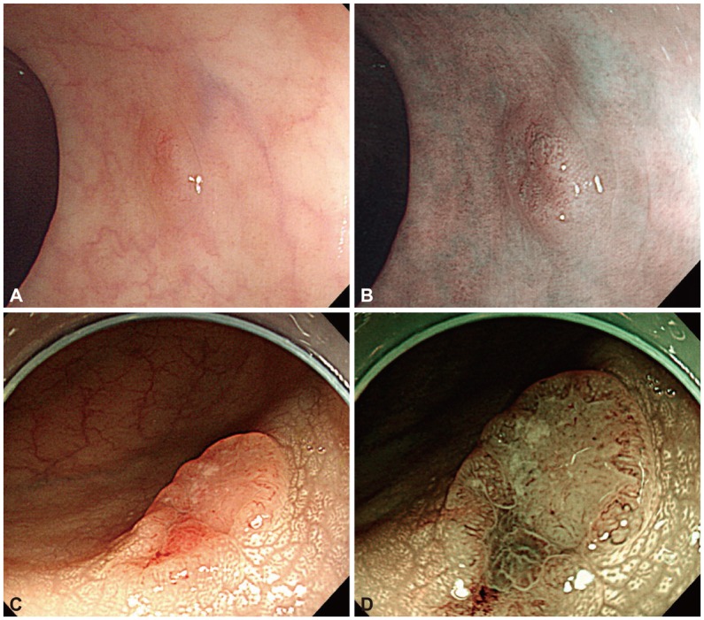
Fig. 1 (A, C) White light (WLE) and (B, D) narrow-band imaging (NBI) of two colonic neoplastic lesions. (A) WLE showing a 6-mm lesion in the sigmoid colon. (B) NBI demonstrating thick brown vessels and a branched surface, compatible with NBI international colorectal endoscopic (NICE) type 2. After snare polypectomy of the lesion, microscopic evaluation showed tubular adenoma with low-grade dysplasia. (C) WLE showing a 1.5-cm slightly elevated lesion with a central depression on the mid ascending colon. (D) NBI showing a distorted surface and missing vessels, compatible with NICE type 3. On histologic evaluation of the resected lesion, an adenocarcinoma that invaded into the submucosal layer was found.
Cited by 2 articles
-
Increased Detection of Colorectal Polyps in Screening Colonoscopy Using High Definition i-SCAN Compared with Standard White Light
Woo Jung Kim, Sang Young Park, Iksoo Park, Wook Jin Lee, Jaechan Park, Nuri Chon, Tak Geun Oh, Kwang Hyun Kim
Clin Endosc. 2016;49(1):69-75. doi: 10.5946/ce.2016.49.1.69.Highlights from the 50th Seminar of the Korean Society of Gastrointestinal Endoscopy
Eun Young Kim, Il Ju Choi, Kwang An Kwon, Ji Kon Ryu, Seok Ho Dong, Ki Baik Hahm
Clin Endosc. 2014;47(4):285-294. doi: 10.5946/ce.2014.47.4.285.
Reference
-
1. Bressler B, Paszat LF, Chen Z, Rothwell DM, Vinden C, Rabeneck L. Rates of new or missed colorectal cancers after colonoscopy and their risk factors: a population-based analysis. Gastroenterology. 2007; 132:96–102. PMID: 17241863.
Article2. Kaltenbach T, Soetikno R. Image-enhanced endoscopy is critical in the detection, diagnosis, and treatment of non-polypoid colorectal neoplasms. Gastrointest Endosc Clin N Am. 2010; 20:471–485. PMID: 20656245.
Article3. Gono K, Igarashi M, Obi T, Yamaguchi M, Ohyama N. Multiple-discriminant analysis for light-scattering spectroscopy and imaging of two-layered tissue phantoms. Opt Lett. 2004; 29:971–973. PMID: 15143644.
Article4. Kuznetsov K, Lambert R, Rey JF. Narrow-band imaging: potential and limitations. Endoscopy. 2006; 38:76–81. PMID: 16429359.
Article5. Emura F, Saito Y, Ikematsu H. Narrow-band imaging optical chromocolonoscopy: advantages and limitations. World J Gastroenterol. 2008; 14:4867–4872. PMID: 18756593.
Article6. Hirata M, Tanaka S, Oka S, et al. Magnifying endoscopy with narrow band imaging for diagnosis of colorectal tumors. Gastrointest Endosc. 2007; 65:988–995. PMID: 17324407.
Article7. East JE, Suzuki N, Stavrinidis M, Guenther T, Thomas HJ, Saunders BP. Narrow band imaging for colonoscopic surveillance in hereditary non-polyposis colorectal cancer. Gut. 2008; 57:65–70. PMID: 17682000.
Article8. Rastogi A, Bansal A, Wani S, et al. Narrow-band imaging colonoscopy: a pilot feasibility study for the detection of polyps and correlation of surface patterns with polyp histologic diagnosis. Gastrointest Endosc. 2008; 67:280–286. PMID: 18155210.9. Inoue T, Murano M, Murano N, et al. Comparative study of conventional colonoscopy and pan-colonic narrow-band imaging system in the detection of neoplastic colonic polyps: a randomized, controlled trial. J Gastroenterol. 2008; 43:45–50. PMID: 18297435.
Article10. van den Broek FJ, Reitsma JB, Curvers WL, Fockens P, Dekker E. Systematic review of narrow-band imaging for the detection and differentiation of neoplastic and nonneoplastic lesions in the colon (with videos). Gastrointest Endosc. 2009; 69:124–135. PMID: 19111693.
Article11. Aihara H, Saito S, Tajiri H. Rationale for and clinical benefits of colonoscopy with narrow band imaging: pathological prediction and colorectal screening. Int J Colorectal Dis. 2013; 28:1–7. PMID: 23053681.
Article12. Ng SC, Lau JY. Narrow-band imaging in the colon: limitations and potentials. J Gastroenterol Hepatol. 2011; 26:1589–1596. PMID: 21793916.
Article13. Hewett DG, Kaltenbach T, Sano Y, et al. Validation of a simple classification system for endoscopic diagnosis of small colorectal polyps using narrow-band imaging. Gastroenterology. 2012; 143:599–607.e1. PMID: 22609383.
Article14. Hayashi N, Tanaka S, Hewett DG, et al. Endoscopic prediction of deep submucosal invasive carcinoma: validation of the narrow-band imaging international colorectal endoscopic (NICE) classification. Gastrointest Endosc. 2013; 78:625–632. PMID: 23910062.
Article15. Fujiya M, Kohgo Y. Image-enhanced endoscopy for the diagnosis of colon neoplasms. Gastrointest Endosc. 2013; 77:111–118.e5. PMID: 23148965.
Article16. Sano Y, Ikematsu H, Fu KI, et al. Meshed capillary vessels by use of narrow-band imaging for differential diagnosis of small colorectal polyps. Gastrointest Endosc. 2009; 69:278–283. PMID: 18951131.
Article17. Kanao H, Tanaka S, Oka S, Hirata M, Yoshida S, Chayama K. Narrow-band imaging magnification predicts the histology and invasion depth of colorectal tumors. Gastrointest Endosc. 2009; 69(3 Pt 2):631–636. PMID: 19251003.
Article18. Wada Y, Kudo SE, Kashida H, et al. Diagnosis of colorectal lesions with the magnifying narrow-band imaging system. Gastrointest Endosc. 2009; 70:522–531. PMID: 19576581.
Article19. ASGE Technology Committee. Song LM, Adler DG, et al. Narrow band imaging and multiband imaging. Gastrointest Endosc. 2008; 67:581–589. PMID: 18374021.20. Pohl J, Nguyen-Tat M, Pech O, May A, Rabenstein T, Ell C. Computed virtual chromoendoscopy for classification of small colorectal lesions: a prospective comparative study. Am J Gastroenterol. 2008; 103:562–569. PMID: 18070234.
Article21. Pohl J, Lotterer E, Balzer C, et al. Computed virtual chromoendoscopy versus standard colonoscopy with targeted indigocarmine chromoscopy: a randomised multicentre trial. Gut. 2009; 58:73–78. PMID: 18838485.
Article
- Full Text Links
-
- Actions
-
Cited
- CITED
-
- Close
- Share
-
- Similar articles
-
- Image-Enhanced Endoscopy in Lower Gastrointestinal Diseases: Present and Future
- Classification and endoscopic diagnosis of colorectal polyps
- Resection of Diminutive and Small Colorectal Polyps: What Is the Optimal Technique?
- Detecting colorectal lesions with image-enhanced endoscopy: an updated review from clinical trials
- Clinical Applications of Linked Color Imaging and Blue Laser/Light Imaging in the Screening, Diagnosis, and Treatment of Superficial Colorectal Tumors
