Anat Cell Biol.
2023 Jun;56(2):185-190. 10.5115/acb.22.247.
Accuracy verification of dental cone-beam computed tomography of mandibular incisor root canals and assessment of its morphology and aging-related changes
- Affiliations
-
- 1Department of Morphological Biology, Ohu University School of Dentistry, Fukushima, Japan
- 2Department of Oral Radiology and Diagnosis, Ohu University School of Dentistry, Fukushima, Japan
- KMID: 2544079
- DOI: http://doi.org/10.5115/acb.22.247
Abstract
- The root canal morphology undergoes aging-related changes, and relevant quantitative analyses have not yet been reported. We compared the cone beam computed tomography (CBCT) and micro-computed tomography (microCT) scans of extracted mandibular incisors to check the accuracy of morphological measurements. Thereafter, the root canal morphology and aging-related changes in the mandibular incisors of Japanese individuals were assessed using CBCT. Six extracted teeth were fixed in a phantom head and imaged using CBCT and micro-CT. The correlation between the findings of the two imaging modalities was examined. Further, CBCT reconstructed images of the mandibular incisors of 81 individuals were observed. Age-related changes of the root canals were compared between participants aged <30 years and those aged ≥30 years. The CBCT and micro-CT findings regarding the root canals of the extracted teeth coincided in 94.4% of the cases. Mandibular incisors exhibiting two root canals in either cross-section accounted for 9.9% of central incisors and 12.4% of lateral incisors. Mandibular central incisors with two root canals were observed in two (6.3%) individuals aged <30 years and six (12.2%) aged ≥30 years. Mandibular lateral incisors with two root canals were observed in one (3.1%) individual aged <30 years and nine (18.4%) aged ≥30 years. CBCT allows accurate evaluation of complex root canal morphologies and is useful for endodontic preoperative assessment. Mandibular incisors have more frequent occurrence of two root canals with aging.
Keyword
Figure
-
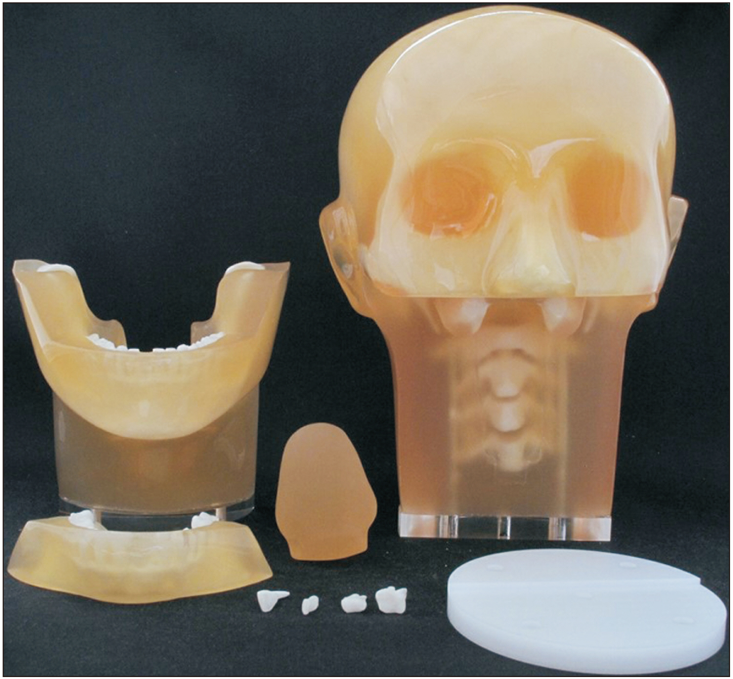
Fig. 1 Tooth extraction specifications for the dental head phantom. The maxillary/mandibular incisor, maxillary premolar, and maxillary molar models are removable, allowing fixation of the extracted teeth.
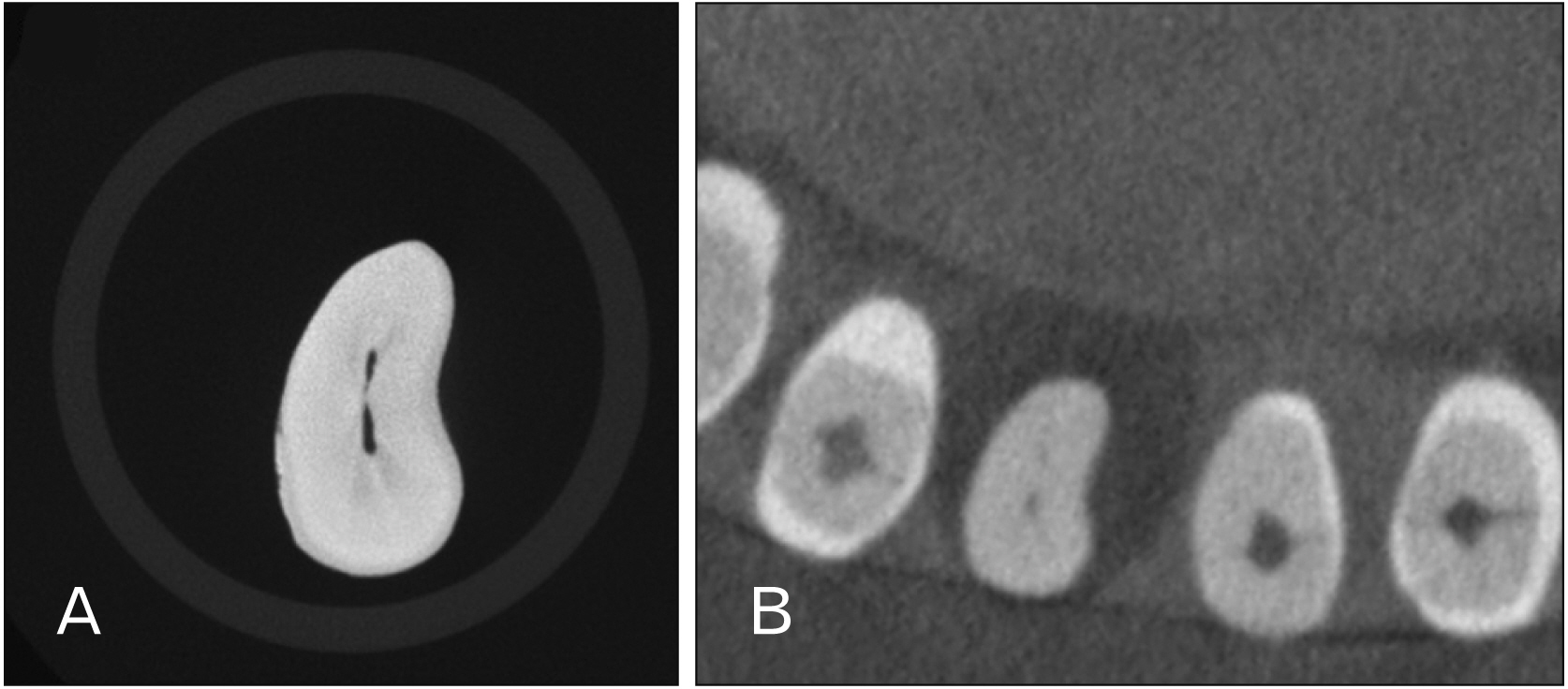
Fig. 2 The observed slice images of the discordant evaluation. (A) Micro-CT images, (B) CBCT images. CT, computed tomography; CBCT, cone beam computed tomography.

Fig. 3 Incidence of root canal morphology by age (<30 and ≥30 years old) mandibular central incisors (A) and mandibular lateral incisors (B).
Reference
-
References
1. Vertucci FJ. 1984; Root canal anatomy of the human permanent teeth. Oral Surg Oral Med Oral Pathol. 58:589–99. DOI: 10.1016/0030-4220(84)90085-9. PMID: 6595621.
Article2. Martins JNR, Gu Y, Marques D, Francisco H, Caramês J. 2018; Differences on the root and root canal morphologies between Asian and White Ethnic groups analyzed by cone-beam computed tomography. J Endod. 44:1096–104. DOI: 10.1016/j.joen.2018.04.001. PMID: 29861062.
Article3. Baxter S, Jablonski M, Hülsmann M. 2020; Cone-beam-computed-tomography of the symmetry of root canal anatomy in mandibular incisors. J Oral Sci. 62:180–3. DOI: 10.2334/josnusd.19-0113. PMID: 32224571.
Article4. Aung NM, Myint KK. 2020; Evidence of second canal between permanent mandibular central and lateral incisors in China; a systematic review on CBCT studies. Int J Dent. 2020:8849609. DOI: 10.1155/2020/8849609. PMID: 33343667. PMCID: PMC7728484. PMID: ddc8e9de561c4353801707952472d383.
Article5. Mahmood Talabani R. 2021; Assessment of root canal morphology of mandibular permanent anterior teeth in an Iraqi subpopulation by cone-beam computed tomography. J Dent Sci. 16:1182–90. DOI: 10.1016/j.jds.2021.02.010. PMID: 34484586. PMCID: PMC8403811.
Article6. Verma GR, Bhadage C, Bhoosreddy AR, Vedpathak PR, Mehrotra GP, Nerkar AC, Bhandari A, Chaubey S. 2017; Cone beam computed tomography study of root canal morphology of permanent mandibular incisors in Indian subpopulation. Pol J Radiol. 82:371–5. DOI: 10.12659/PJR.901840. PMID: 28794810. PMCID: PMC5513681.
Article7. Tolentino ES, Amoroso-Silva PA, Alcalde MP, Yamashita FC, Iwaki LCV, Rubira-Bullen IRF, Duarte MAH. 2021; Comparison of limited- and large-volume cone-beam computed tomography using a small voxel size for detecting isthmuses in mandibular molars. Imaging Sci Dent. 51:27–34. DOI: 10.5624/isd.20200144. PMID: 33828958. PMCID: PMC8007390.
Article8. Kim SY, Yang SE. 2012; Cone-beam computed tomography study of incidence of distolingual root and distance from distolingual canal to buccal cortical bone of mandibular first molars in a Korean population. J Endod. 38:301–4. DOI: 10.1016/j.joen.2011.10.023. PMID: 22341064.
Article9. Aytugar E, Özeren C, Lacin N, Veli I, Çene E. 2019; Cone-beam computed tomographic evaluation of accessory mental foramen in a Turkish population. Anat Sci Int. 94:257–65. DOI: 10.1007/s12565-019-00481-7. PMID: 30790181.
Article10. Vizzotto MB, Silveira PF, Arús NA, Montagner F, Gomes BP, da Silveira HE. 2013; CBCT for the assessment of second mesiobuccal (MB2) canals in maxillary molar teeth: effect of voxel size and presence of root filling. Int Endod J. 46:870–6. DOI: 10.1111/iej.12075. PMID: 23442087.
Article11. Blattner TC, George N, Lee CC, Kumar V, Yelton CD. 2010; Efficacy of cone-beam computed tomography as a modality to accurately identify the presence of second mesiobuccal canals in maxillary first and second molars: a pilot study. J Endod. 36:867–70. DOI: 10.1016/j.joen.2009.12.023. PMID: 20416435.
Article12. Morikage N, Hamada T, Usami A, Takada S. 2017; Topographical relationship between positions of lingual foramina and attachment of mylohyoid muscle in mental region. Surg Radiol Anat. 39:735–9. DOI: 10.1007/s00276-016-1804-9. PMID: 28078367.
Article13. Yoza T, Serikawa M, Sugita T, Harada T, Usami A. 2021; Cone-beam computed tomography observation of maxillary first premolar canal shapes. Anat Cell Biol. 54:424–30. DOI: 10.5115/acb.21.110. PMID: 34465669. PMCID: PMC8693140.
Article14. Shigefuji R, Serikawa M, Usami A. 2022; Observation of mandibular second molar roots and root canal morphology using dental cone-beam computed tomography. Anat Cell Biol. 55:155–60. DOI: 10.5115/acb.22.050. PMID: 35773218. PMCID: PMC9256481.
Article15. Suomalainen A, Pakbaznejad Esmaeili E, Robinson S. 2015; Dentomaxillofacial imaging with panoramic views and cone beam CT. Insights Imaging. 6:1–16. DOI: 10.1007/s13244-014-0379-4. PMID: 25575868. PMCID: PMC4330237.
Article16. Katsumata A, Hirukawa A, Okumura S, Naitoh M, Fujishita M, Ariji E, Langlais RP. 2007; Effects of image artifacts on gray-value density in limited-volume cone-beam computerized tomography. Oral Surg Oral Med Oral Pathol Oral Radiol Endod. 104:829–36. DOI: 10.1016/j.tripleo.2006.12.005. PMID: 17448704.
Article17. Bootsma GJ, Verhaegen F, Jaffray DA. 2011; The effects of compensator and imaging geometry on the distribution of x-ray scatter in CBCT. Med Phys. 38:897–914. DOI: 10.1118/1.3539575. PMID: 21452727.
Article18. Arai Y. 2021; Local cone beam CT: how did it all start? Dentomaxillofac Radiol. 50:20210276. DOI: 10.1259/dmfr.20210276. PMID: 34739304. PMCID: PMC8611279.
Article19. Scarfe WC, Farman AG. 2008; What is cone-beam CT and how does it work? Dent Clin North Am. 52:707–30. vDOI: 10.1016/j.cden.2008.05.005. PMID: 18805225.
Article20. Asami R, Aboshi H, Iwawaki A, Ohtaka Y, Odaka K, Abe S, Saka H. 2019; Age estimation based on the volume change in the maxillary premolar crown using micro CT. Leg Med (Tokyo). 37:18–24. DOI: 10.1016/j.legalmed.2018.12.001. PMID: 30597413.
Article21. Pineda F, Kuttler Y. 1972; Mesiodistal and buccolingual roentgenographic investigation of 7,275 root canals. Oral Surg Oral Med Oral Pathol. 33:101–10. DOI: 10.1016/0030-4220(72)90214-9. PMID: 4500261.
Article22. Nelson S. Nelson S, editor. 2015. Pulp chambers and canals. Wheeler's Dental Anatomy, Physiology, and Occlusion. 10th ed. Elsevier;p. 203–30.23. Berkovitz BKB. Standring S, editor. 2016. Oral cavity. Gray's Anatomy. 41st ed. Elsevier;p. 507–33. DOI: 10.37019/e-anatomy/826010.24. Special Committee to Revise the Joint AAE/AAOMR Position Statement on use of CBCT in Endodontics. 2015; AAE and AAOMR joint position statement: use of cone beam computed tomography in endodontics 2015 update. Oral Surg Oral Med Oral Pathol Oral Radiol. 120:508–12. DOI: 10.1016/j.oooo.2015.07.033. PMID: 26346911.25. Rabiee H, McDonald NJ, Jacobs R, Aminlari A, Inglehart MR. 2018; Endodontics program directors', residents', and endodontists' considerations about CBCT-related graduate education. J Dent Educ. 82:989–99. DOI: 10.21815/JDE.018.098. PMID: 30173196.
Article26. Miyashita M, Kasahara E, Yasuda E, Yamamoto A, Sekizawa T. 1997; Root canal system of the mandibular incisor. J Endod. 23:479–84. DOI: 10.1016/S0099-2399(97)80305-6. PMID: 9587315.
Article
- Full Text Links
-
- Actions
-
Cited
- CITED
-
- Close
- Share
-
- Similar articles
-
- Observation of mandibular second molar roots and root canal morphology using dental cone-beam computed tomography
- Nutrient canals on mandibular anterior region in cone beam computed tomography
- Endodontic treatment of maxillary lateral incisors with anatomical variations
- Endodontic management of a maxillary first molar with three roots and seven root canals with the aid of cone-beam computed tomography
- Asymmetry in mesial root number and morphology in mandibular second molars: a case report
