Change of Femoral Anteversion Angle in Children With Intoeing Gait Measured by Three-Dimensional Computed Tomography Reconstruction: 3-Year Follow-Up Study
- Affiliations
-
- 1Department of Rehabilitation Medicine, Gyeongsang National University Hospital, Gyeongsang National University School of Medicine, Jinju, Korea
- 2Department of Preventive Medicine, Institute of Health Sciences, Gyeongsang National University College of Medicine, Jinju, Korea
- KMID: 2543415
- DOI: http://doi.org/10.5535/arm.23043
Abstract
Objective
To investigate long-term changes in femoral anteversion angle (FAA) in children with intoeing gait and to identify factors that affect FAA changes.
Methods
We retrospectively analyzed three-dimensional computed tomography data from 2006 to 2022 of children with intoeing gait with ≥3 years of follow-up without active treatment. The study examined the mean changes in FAA, the effects of sex, age, and initial FAA on FAA change, and mean FAAs by age. Changes in FAA severity up to eight years of age were also observed and analyzed by sex.
Results
A total of 126 lower limbs of 63 children (30 males, 33 females) with intoeing gait were included, with a mean age of 5.11±1.05 years and a mean follow-up period of 43.59±7.74 months. The initial FAA was 41.42°±8.29° and the follow-up FAA was 33.25°±9.19°, indicating a significant decrease (p<0.001). Significant correlations were observed between age and changes in FAA, as well as between initial FAA and changes in FAA (r=0.248, p=0.005; r=-0.333, p<0.001). At age 8 years, only 22 limbs were classified as having mild FAA severity.
Conclusion
During the follow-up period, children with intoeing gait had a significant decreased in FAA. No significant difference in FAA change was found between sex, but younger children and those with greater initial FAA were more likely to have decreased FAA. However, most children retained moderate to severe severity of increased FAA. Further studies are required to validate these findings.
Figure
-
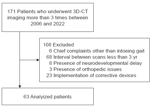
Fig. 1. Flowchart of patient inclusion. 3D-CT, three-dimensional computed tomography.
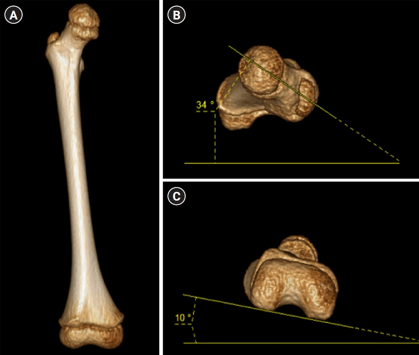
Fig. 2. (A) Anteroposterior view of right femur obtained from three-dimensional computed tomography. (B) Looking down view of the femur and the measurement of femoral neck angle. (C) Upside-down view of the femur neck and the measurement of trans-condylar axis.
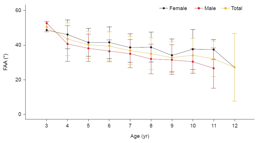
Fig. 3. Changes in femoral anteversion angle (FAA) measured from total three-dimensional computed tomography images according to age.

Fig. 4. Femoral anteversion angle (FAA) change was greater in younger children (r=0.248, p=0.005) (A) and in those with higher initial FAA (r=-0.333, p<0.001) (B), according to correlations with age and initial FAA, respectively.

Fig. 5. The change in femoral anteversion angle severity between the initial measurement and at age 8.

Reference
-
1. Staheli LT. Rotational problems in children. Instr Course Lect. 1994; 43:199–209.
Article2. Staheli LT. Torsion--treatment indications. Clin Orthop Relat Res. 1989; (247):61–6.
Article3. Fabry G, Cheng LX, Molenaers G. Normal and abnormal torsional development in children. Clin Orthop Relat Res. 1994; (302):22–6.
Article4. Li YH, Leong JC. Intoeing gait in children. Hong Kong Med J. 1999; 5:360–6.5. Naqvi G, Stohr K, Rehm A. Proximal femoral derotation osteotomy for idiopathic excessive femoral anteversion and intoeing gait. SICOT J. 2017; 3:49.
Article6. Ito K, Minka MA 2nd, Leunig M, Werlen S, Ganz R. Femoroacetabular impingement and the cam-effect. A MRI-based quantitative anatomical study of the femoral head-neck offset. J Bone Joint Surg Br. 2001; 83:171–6.7. Ejnisman L, Philippon MJ, Lertwanich P, Pennock AT, Herzog MM, Briggs KK, et al. Relationship between femoral anteversion and findings in hips with femoroacetabular impingement. Orthopedics. 2013; 36:e293–300.
Article8. Stevens PM, Gililland JM, Anderson LA, Mickelson JB, Nielson J, Klatt JW. Success of torsional correction surgery after failed surgeries for patellofemoral pain and instability. Strategies Trauma Limb Reconstr. 2014; 9:5–12.
Article9. Uden H, Kumar S. Non-surgical management of a pediatric "intoed" gait pattern- a systematic review of the current best evidence. J Multidiscip Healthc. 2012; 5:27–35.10. Nelitz M. Femoral derotational osteotomies. Curr Rev Musculoskelet Med. 2018; 11:272–9.
Article11. Hartigan DE, Perets I, Walsh JP, Domb BG. Femoral derotation osteotomy technique for excessive femoral anteversion. Arthrosc Tech. 2017; 6:e1405–10.
Article12. Kim JS, Park TS, Park SB, Kim JS, Kim IY, Kim SI. Measurement of femoral neck anteversion in 3D. Part 1: 3D imaging method. Med Biol Eng Comput. 2000; 38:603–9.
Article13. Scorcelletti M, Reeves ND, Rittweger J, Ireland A. Femoral anteversion: significance and measurement. J Anat. 2020; 237:811–26.
Article14. Byun HY, Shin H, Lee ES, Kong MS, Lee SH, Lee CH. The availability of radiological measurement of femoral anteversion angle: three-dimensional computed tomography reconstruction. Ann Rehabil Med. 2016; 40:237–43.
Article15. Riccio AI, Carney CD, Hammel LC, Stanley M, Cassidy J, Davids JR. Three-dimensional computed tomography for determination of femoral anteversion in a cerebral palsy model. J Pediatr Orthop. 2015; 35:167–71.
Article16. Shalaby MH, Samir S, Deif A. CT measurement of femoral anteversion angle in patients with unilateral developmental hip dysplasia: a comparative study between 2D and 3D techniques. Egypt J Radiol Nucl Med. 2017; 48:639–43.
Article17. Fabry G, MacEwen GD, Shands AR Jr. Torsion of the femur. A follow-up study in normal and abnormal conditions. J Bone Joint Surg Am. 1973; 55:1726–38.18. Svenningsen S, Apalset K, Terjesen T, Anda S. Regression of femoral anteversion. A prospective study of intoeing children. Acta Orthop Scand. 1989; 60:170–3.
Article19. Kong M, Jo H, Lee CH, Chun SW, Yoon C, Shin H. Change of femoral anteversion angle in children with intoeing gait measured by three-dimensional computed tomography reconstruction: one-year follow-up study. Ann Rehabil Med. 2018; 42:137–44.
Article20. Gose S, Sakai T, Shibata T, Murase T, Yoshikawa H, Sugamoto K. Morphometric analysis of the femur in cerebral palsy: 3-dimensional CT study. J Pediatr Orthop. 2010; 30:568–74.21. Jia J, Li L, Zhang L, Zhao Q, Liu X. Three dimensional-CT evaluation of femoral neck anteversion, acetabular anteversion and combined anteversion in unilateral DDH in an early walking age group. Int Orthop. 2012; 36:119–24.
Article22. Wissing H, Spira G. [Determination of rotational defects of the femur by computer tomographic determination of the antetorsion angle of the femoral neck]. Unfallchirurgie. 1986; 12:1–11. German.
Article23. Yan W, Xu X, Xu Q, Yan W, Sun Z, Jiang Q, et al. Femoral and tibial torsion measurements based on EOS imaging compared to 3D CT reconstruction measurements. Ann Transl Med. 2019; 7:460.
Article24. Decker S, Suero EM, Hawi N, Müller CW, Krettek C, Citak M. The physiological range of femoral antetorsion. Skeletal Radiol. 2013; 42:1501–5.
Article25. Nakahara I, Takao M, Sakai T, Nishii T, Yoshikawa H, Sugano N. Gender differences in 3D morphology and bony impingement of human hips. J Orthop Res. 2011; 29:333–9.
Article26. Jani L. Idiopathic anteversion of the femoral neck follow-up study of 51 patients on completion of growth. Int Orthop. 1978; 2:283–92.
Article27. Chodick G, Kim KP, Shwarz M, Horev G, Shalev V, Ron E. Radiation risks from pediatric computed tomography scanning. Pediatr Endocrinol Rev. 2009; 7:29–36.28. Brody AS, Frush DP, Huda W, Brent RL; American Academy of Pediatrics Section on Radiology. Radiation risk to children from computed tomography. Pediatrics. 2007; 120:677–82.
Article29. Muhamad AR, Freitas JM, Bomar JD, Dwek J, Hosalkar HS. CT and MRI lower extremity torsional profile studies: measurement reproducibility. J Child Orthop. 2012; 6:391–6.
Article30. Obara H, Takahashi M, Kudou K, Mariya Y, Takai Y, Kashiwakura I. Estimation of effective doses in pediatric X-ray computed tomography examination. Exp Ther Med. 2017; 14:4515–20.
Article31. Pearce MS, Salotti JA, Little MP, McHugh K, Lee C, Kim KP, et al. Radiation exposure from CT scans in childhood and subsequent risk of leukaemia and brain tumours: a retrospective cohort study. Lancet. 2012; 380:499–505.
Article
- Full Text Links
-
- Actions
-
Cited
- CITED
-
- Close
- Share
-
- Similar articles
-
- Relationship between Femoral Anteversion and Tibial Torsion in Intoeing Gait
- Change of Femoral Anteversion Angle in Children With Intoeing Gait Measured by Three-Dimensional Computed Tomography Reconstruction: One-Year Follow-Up Study
- Relationship between Physical Examinations and Two-Dimensional Computed Tomographic Findings in Children with Intoeing Gait
- The Availability of Radiological Measurement of Femoral Anteversion Angle: Three-Dimensional Computed Tomography Reconstruction
- Physical Examination and Computed Tomography in Children with Toe in Gait
