Ann Rehabil Med.
2023 Apr;47(2):98-107. 10.5535/arm.23005.
Trunk Impairment Scale for Predicting Lumbar Spine Bone Mineral Density in Young Male Patients With Subacute Stroke
- Affiliations
-
- 1Department of Physical Medicine and Rehabilitation, Soonchunhyang University Bucheon Hospital, Soonchunhyang University College of Medicine, Bucheon, Korea
- KMID: 2541843
- DOI: http://doi.org/10.5535/arm.23005
Abstract
Objective
To investigate the relationship between Trunk Impairment Scale (TIS) and lumbar spine bone mineral density (BMD) in subacute stroke patients.
Methods
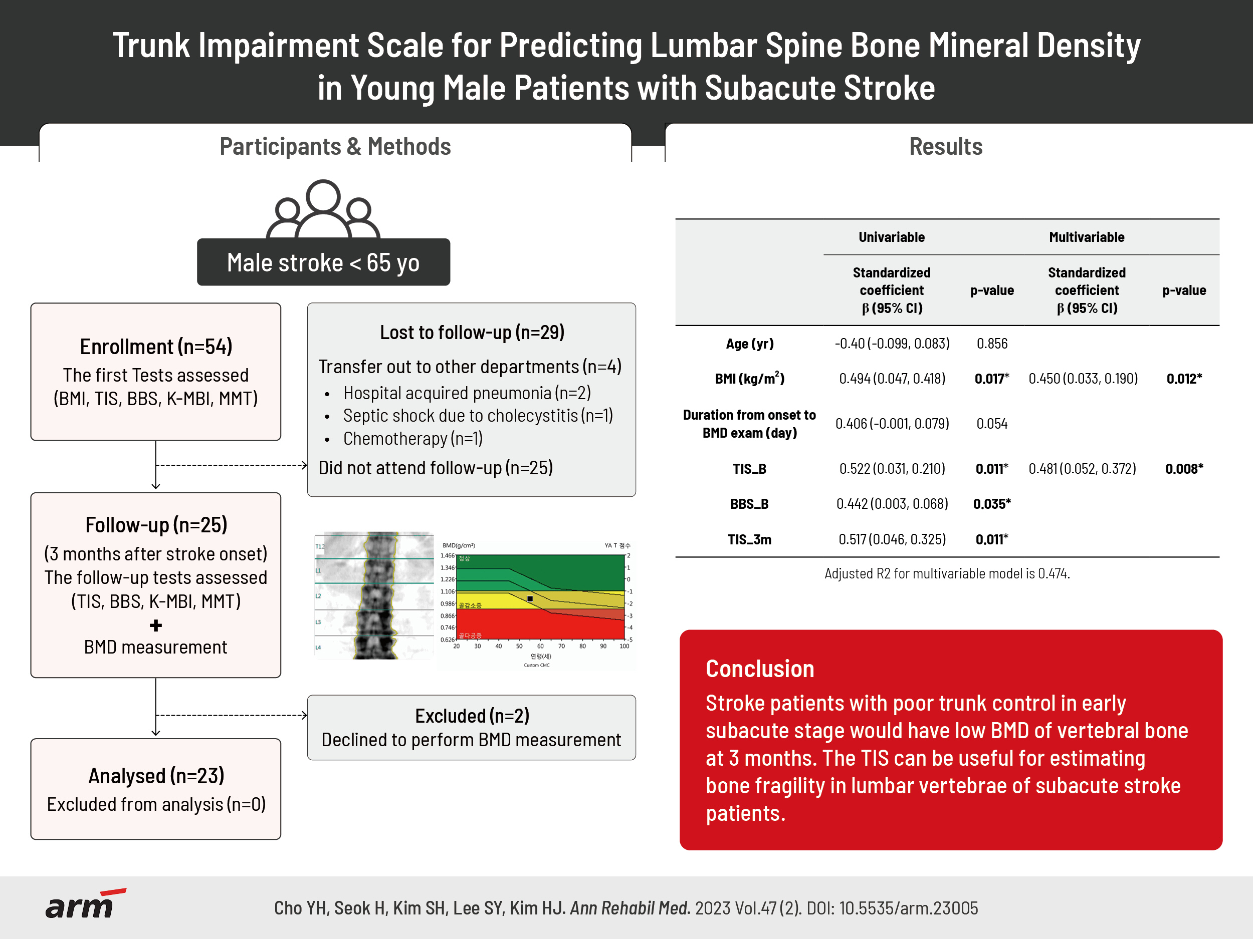
Twenty-three subacute male stroke patients under the age of 65 were prospectively enrolled to exclude both postmenopausal and senile effects on BMD. The TIS, Berg Balance Scale, the Korean version of the Modified Barthel Index, and manual muscle test were measured at admission and 3 months after stroke onset. BMD of the bilateral lower extremities and lumbar vertebrae was measured by dual-energy X-ray absorptiometry 3 months after stroke onset.
Results
TIS at baseline (TIS_B) and TIS at 3 months after stroke (TIS_3m) showed significant correlations with lumbar BMD (TIS_B, r=0.522; TIS_3m, r=0.517). Through multiple regression analysis, the TIS_B was associated with lumbar BMD (adjusted R2=0.474). However, BMD of the bilateral lower extremities was not correlated with any clinical measurements except body mass index.
Conclusion
We found a relationship between TIS_B and lumbar BMD in subacute young male stroke patients. Stroke patients with poor trunk control in the early subacute stage would have low BMD of vertebral bones at 3 months. The TIS can be useful for estimating bone fragility in the lumbar vertebrae of subacute stroke patients.
Keyword
Figure
-

Fig. 1. Flowchart for this study. BMI, body mass index; TIS, Trunk Impairment Scale; BBS, Berg Balance Scale; K-MBI, Korean version of Modified Barthel Index; MMT, manual muscle test; BMD, bone mineral density.

Fig. 2. Scatter plot showing the correlation of the bone mineral density (BMD) of lumbar spine and Trunk Impairment Scale (TIS) scores (n=23). TIS scores showed a strong positive correlation with BMD of lumbar spine at baseline (TIS_base) and 3 months after stroke (TIS_3m) (r=0.522, p<0.05; r=0.517, p<0.05, respectively).
Reference
-
1. Beaupre GS, Lew HL. Bone-density changes after stroke. Am J Phys Med Rehabil. 2006; 85:464–72.
Article2. Tinetti ME, Liu WL, Claus EB. Predictors and prognosis of inability to get up after falls among elderly persons. JAMA. 1993; 269:65–70.
Article3. Kanis J, Oden A, Johnell O. Acute and long-term increase in fracture risk after hospitalization for stroke. Stroke. 2001; 32:702–6.
Article4. Pasco JA, Henry MJ, Korn S, Nicholson GC, Kotowicz MA. Morphometric vertebral fractures of the lower thoracic and lumbar spine, physical function and quality of life in men. Osteoporos Int. 2009; 20:787–92.
Article5. Carda S, Cisari C, Invernizzi M, Bevilacqua M. Osteoporosis after stroke: a review of the causes and potential treatments. Cerebrovasc Dis. 2009; 28:191–200.
Article6. del Puente A, Pappone N, Mandes MG, Mantova D, Scarpa R, Oriente P. Determinants of bone mineral density in immobilization: a study on hemiplegic patients. Osteoporos Int. 1996; 6:50–4.
Article7. Hamdy RC, Moore SW, Cancellaro VA, Harvill LM. Long-term effects of strokes on bone mass. Am J Phys Med Rehabil. 1995; 74:351–6.
Article8. Ramnemark A, Nyberg L, Lorentzon R, Englund U, Gustafson Y. Progressive hemiosteoporosis on the paretic side and increased bone mineral density in the nonparetic arm the first year after severe stroke. Osteoporos Int. 1999; 9:269–75.
Article9. Kim HD, Kim SH, Kim DK, Jeong HJ, Sim YJ, Kim GC. Change of bone mineral density and relationship to clinical parameters in male stroke patients. Ann Rehabil Med. 2016; 40:981–8.
Article10. Lee HY, Park JH, Lee H, Kim TW, Yoo SD. Does hip bone density differ between paretic and non-paretic sides in hemiplegic stroke patients? and its relationship with physical impairment. J Bone Metab. 2020; 27:237–46.
Article11. Haruyama K, Kawakami M, Otsuka T. Effect of core stability training on trunk function, standing balance, and mobility in stroke patients. Neurorehabil Neural Repair. 2017; 31:240–9.
Article12. McDonnell P, McHugh PE, O'Mahoney D. Vertebral osteoporosis and trabecular bone quality. Ann Biomed Eng. 2007; 35:170–89.
Article13. Chen H, Zhou X, Fujita H, Onozuka M, Kubo KY. Age-related changes in trabecular and cortical bone microstructure. Int J Endocrinol. 2013; 2013:213234.
Article14. Legrand E, Chappard D, Pascaretti C, Duquenne M, Krebs S, Rohmer V, et al. Trabecular bone microarchitecture, bone mineral density, and vertebral fractures in male osteoporosis. J Bone Miner Res. 2000; 15:13–9.
Article15. Dalle Carbonare L, Giannini S. Bone microarchitecture as an important determinant of bone strength. J Endocrinol Invest. 2004; 27:99–105.
Article16. Jørgensen L, Jacobsen BK, Wilsgaard T, Magnus JH. Walking after stroke: does it matter? Changes in bone mineral density within the first 12 months after stroke. A longitudinal study. Osteoporos Int. 2000; 11:381–7.
Article17. Demirbag D, Ozdemir F, Kokino S, Berkarda S. The relationship between bone mineral density and immobilization duration in hemiplegic limbs. Ann Nucl Med. 2005; 19:695–700.
Article18. Arceo-Mendoza RM, Camacho PM. Postmenopausal osteoporosis: latest guidelines. Endocrinol Metab Clin North Am. 2021; 50:167–78.19. Vilaca T, Eastell R, Schini M. Osteoporosis in men. Lancet Diabetes Endocrinol. 2022; 10:273–83.
Article20. Rinonapoli G, Ruggiero C, Meccariello L, Bisaccia M, Ceccarini P, Caraffa A. Osteoporosis in men: a review of an underestimated bone condition. Int J Mol Sci. 2021; 22:2105.
Article21. Verheyden G, Nieuwboer A, Mertin J, Preger R, Kiekens C, De Weerdt W. The Trunk Impairment Scale: a new tool to measure motor impairment of the trunk after stroke. Clin Rehabil. 2004; 18:326–34.
Article22. Kleerekoper M, Nelson DA. Which bone density measurement? J Bone Miner Res. 1997; 12:712–4.
Article23. Shuhart CR, Yeap SS, Anderson PA, Jankowski LG, Lewiecki EM, Morse LR, et al. Executive summary of the 2019 ISCD position development conference on monitoring treatment, DXA cross-calibration and least significant change, spinal cord injury, peri-prosthetic and orthopedic bone health, transgender medicine, and pediatrics. J Clin Densitom. 2019; 22:453–71.
Article24. Akoglu H. User’s guide to correlation coefficients. Turk J Emerg Med. 2018; 18:91–3.
Article25. Jang A, Bae CH, Han SJ, Bae H. Association between length of stay in the intensive care unit and sarcopenia among hemiplegic stroke patients. Ann Rehabil Med. 2021; 45:49–56.
Article26. Wall BT, Dirks ML, van Loon LJ. Skeletal muscle atrophy during short-term disuse: implications for agerelated sarcopenia. Ageing Res Rev. 2013; 12:898–906.
Article27. Krabak B, Kennedy DJ. Functional rehabilitation of lumbar spine injuries in the athlete. Sports Med Arthrosc Rev. 2008; 16:47–54.
Article28. Oftadeh R, Perez-Viloria M, Villa-Camacho JC, Vaziri A, Nazarian A. Biomechanics and mechanobiology of trabecular bone: a review. J Biomech Eng. 2015; 137:0108021–01080215.
Article29. Banijamali SM, Oftadeh R, Nazarian A, Goebel R, Vaziri A, Nayeb-Hashemi H. Effects of different loading patterns on the trabecular bone morphology of the proximal femur using adaptive bone remodeling. J Biomech Eng. 2015; 137:011011.30. Osterhoff G, Morgan EF, Shefelbine SJ, Karim L, McNamara LM, Augat P. Bone mechanical properties and changes with osteoporosis. Injury. 2016; 47(Suppl 2):S11–20.
Article31. Fukuoka H, Nishimura Y, Haruna M, Suzuki Y, Oyama K, Igawa S, et al. Effect of bed rest immobilization on metabolic turnover of bone and bone mineral density. J Gravit Physiol. 1997; 4:S75–81.32. Burr DB, Robling AG, Turner CH. Effects of biomechanical stress on bones in animals. Bone. 2002; 30:781–6.
Article33. Ehrlich PJ, Noble BS, Jessop HL, Stevens HY, Mosley JR, Lanyon LE. The effect of in vivo mechanical loading on estrogen receptor alpha expression in rat ulnar osteocytes. J Bone Miner Res. 2002; 17:1646–55.
Article34. Tan SD, Bakker AD, Semeins CM, Kuijpers-Jagtman AM, Klein-Nulend J. Inhibition of osteocyte apoptosis by fluid flow is mediated by nitric oxide. Biochem Biophys Res Commun. 2008; 369:1150–4.
Article35. Bansod YD, Kebbach M, Kluess D, Bader R, van Rienen U. Computational analysis of bone remodeling in the proximal tibia under electrical stimulation considering the piezoelectric properties. Front Bioeng Biotechnol. 2021; 9:705199.
Article36. Lazoura O, Groumas N, Antoniadou E, Papadaki PJ, Papadimitriou A, Thriskos P, et al. Bone mineral density alterations in upper and lower extremities 12 months after stroke measured by peripheral quantitative computed tomography and DXA. J Clin Densitom. 2008; 11:511–7.37. Ramnemark A, Nyberg L, Lorentzon R, Olsson T, Gustafson Y. Hemiosteoporosis after severe stroke, independent of changes in body composition and weight. Stroke. 1999; 30:755–60.
Article38. Palle S, Vico L, Bourrin S, Alexandre C. Bone tissue response to four-month antiorthostatic bedrest: a bone histomorphometric study. Calcif Tissue Int. 1992; 51:189–94.
Article39. Ishiwatari M, Tani M, Isayama R, Honaga K, Hayakawa M, Takakura T, et al. Prediction of gait independence using the Trunk Impairment Scale in patients with acute stroke. Ther Adv Neurol Disord. 2022; 15:17562864221140180.
Article
- Full Text Links
-
- Actions
-
Cited
- CITED
-
- Close
- Share
-
- Similar articles
-
- Trunk Muscle Strength and Bone Mineral Density in Women
- The Effect of Chronic Low Back Pain on Bone Mineral Density and Trunk Muscle Strength in Women
- Lumbar Spine Degenerative Disease: Effect on Bone Mineral Density Measurements in the Lumbar Spine and Femoral Neck
- The relationship of maturation value of vaginal epithelium and bone mineral density in postmenopausal women
- Correlation between Bone Mineral Density and Interverterbral Disc Degeneration
