Anat Cell Biol.
2023 Mar;56(1):137-140. 10.5115/acb.22.009.
Concomitant variations of the tibialis anterior, and extensor hallucis longus, and extensor hallucis brevis muscles
- Affiliations
-
- 1Tulane University School of Medicine, New Orleans, LA, USA
- 2Katsuta Osteopathic Clinic, Itoshima, Fukuoka, Japan
- 3Kyushu Medical Sports Vocational School, Kitakyushu, Fukuoka, Japan
- 4Department of Neurosurgery, Tulane Center for Clinical Neurosciences, Tulane University School of Medicine, New Orleans, LA, USA
- 5Department of Neurology, Tulane Center for Clinical Neurosciences, Tulane University School of Medicine, New Orleans, LA, USA
- 6Division of Gross and Clinical Anatomy, Department of Anatomy, Kurume University School of Medicine, Kurume, Fukuoka, Japan
- 7Department of Anatomical Dissection and Donation, Medical University of Łódź, Poland
- 8Department of Anatomical Sciences, St. George’s University, St. George’s, Grenada, West Indies
- 9Department of Structural & Cellular Biology, Tulane University School of Medicine, New Orleans, LA, USA
- 10Department of Surgery, Tulane University School of Medicine, New Orleans, LA, USA
- 11Department of Neurosurgery and Ochsner Neuroscience Institute, Ochsner Health System, New Orleans, LA, USA
- 12University of Queensland, Brisbane, Australia
- KMID: 2540992
- DOI: http://doi.org/10.5115/acb.22.009
Abstract
- Tibialis anterior (TA) muscle originates from the lateral surface of tibia and its tendon attaches to the medial cuneiform and base of the first metatarsal. The TA muscle is responsible for both dorsiflexion and inversion of the foot. We present a case of bilateral TA muscle variations that diverge slightly from the current classification systems of this muscle. Recognizing variations such as these may be important for anatomists, surgeons, podiatrists, and physicians. Following routine dissection, an accessory tendon of the TA muscle was found on both sides. Accessory tendons of the extensor hallucis longus and extensor hallucis brevis joined to form a common tendon on both sides. We believe that this unique case will help further the classification systems for the tendons of the TA and also be informative for clinical anatomists as well as physicians treating patients with pathology in this region.
Figure
-
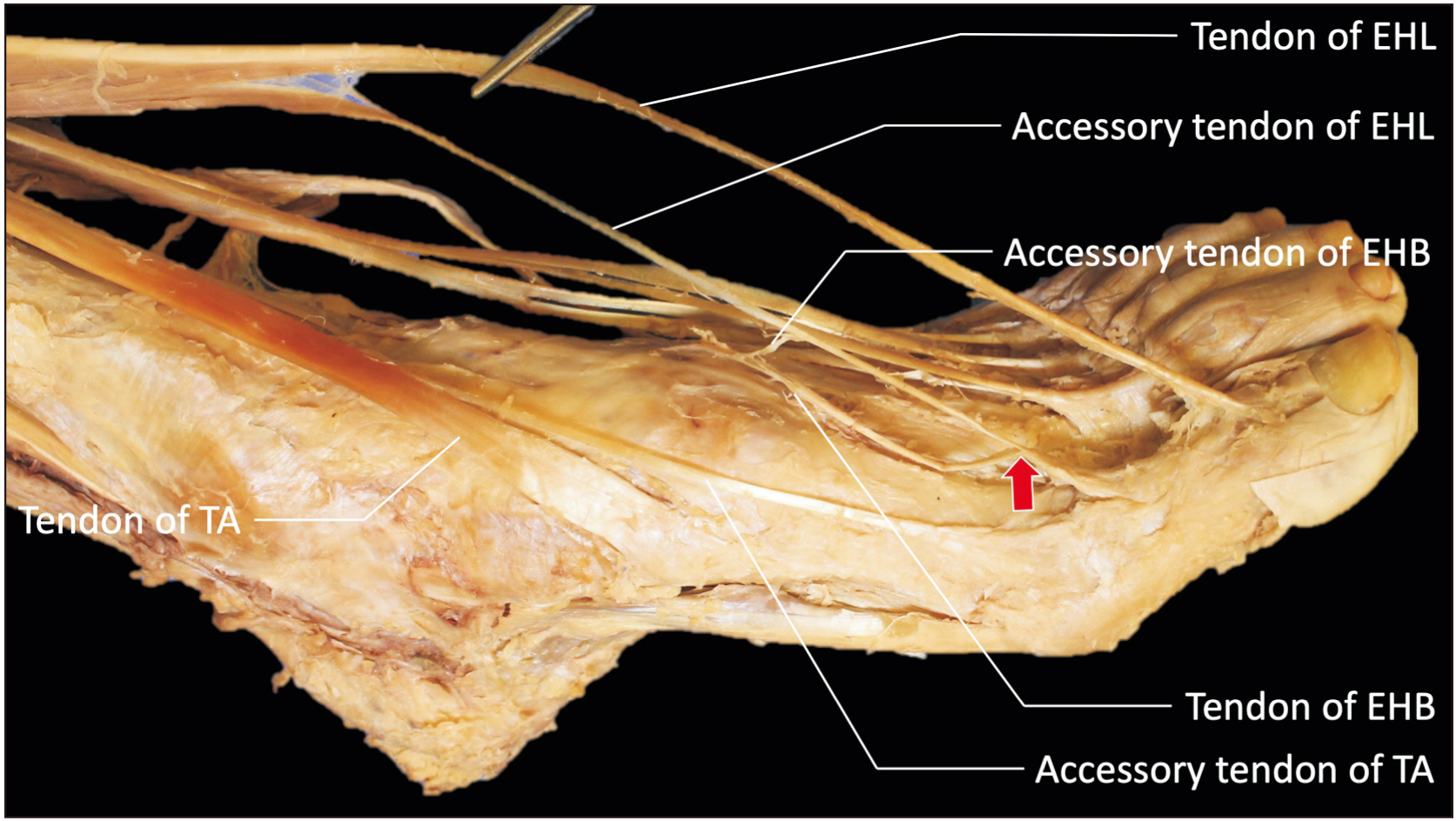
Fig. 1 Variant anatomy on the left foot. EHB, extensor hallucis brevis; EHL, extensor hallucis longus; TA, tibialis anterior.
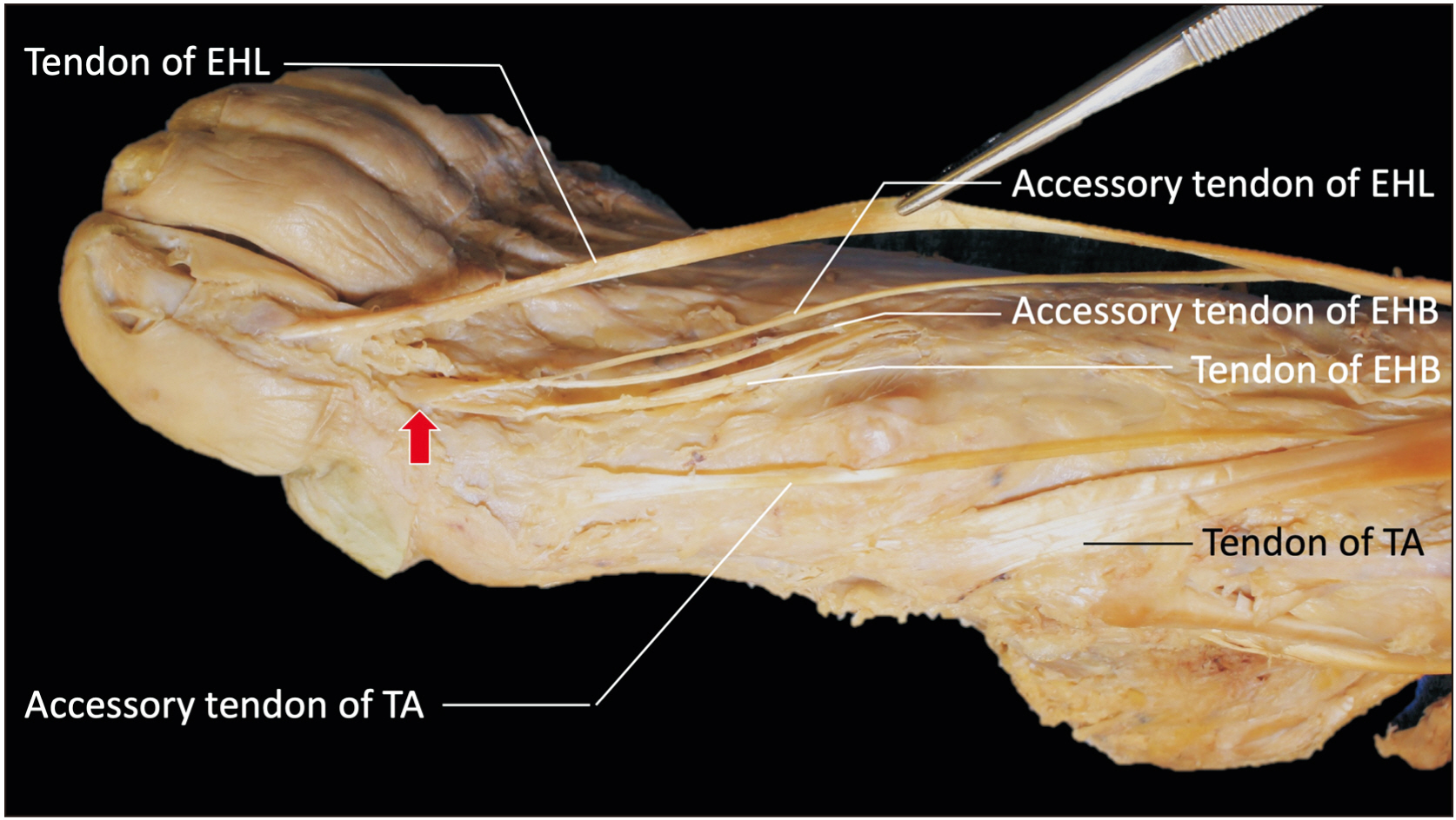
Fig. 2 Variant anatomy on the right foot. EHB, extensor hallucis brevis; EHL, extensor hallucis longus; TA, tibialis anterior.
Reference
-
References
1. Karauda P, Podgórski M, Paulsen F, Polguj M, Olewnik Ł. 2021; Anatomical variations of the tibialis anterior tendon. Clin Anat. 34:397–404. DOI: 10.1002/ca.23663. PMID: 32713016.
Article2. Drake RL, Vogl AW, Mitchell AWM. Drake RL, Vogl AW, Mitchell AWM, editors. 2020. Lower limb/leg. Gray's Anatomy for Students. 4th ed. Elsevier;Philadelphia: p. 612–27.3. Htwe O, Swarhib M, Pei TS, Naicker AS, Das S. 2012; Congenital bilateral agenesis of the tibialis anterior muscles: a rare case report. Rom J Morphol Embryol. 53:657–9. PMID: 22990563.4. Drake RL, Vogl AW, Mitchell AWM. Drake RL, Vogl AW, Mitchell AWM, editors. 2020. Lower limb/foot. Gray's Anatomy for Students. 4th ed. Elsevier;Philadelphia: p. 627–59.5. DeLuca M, Boucher L. 2019; Morphological variations and accessory ossicles in the peroneal and tibialis muscles. Anat Cell Biol. 52:344–8. DOI: 10.5115/acb.19.030. PMID: 31598366. PMCID: PMC6773904.
Article6. Olewnik Ł, Podgórski M, Polguj M, Topol M. 2019; A cadaveric and sonographic study of the morphology of the tibialis anterior tendon - a proposal for a new classification. J Foot Ankle Res. 12:9. DOI: 10.1186/s13047-019-0319-0. PMID: 30733832. PMCID: PMC6359855. PMID: b635b5eda22148dc836d413a2ea51a35.
Article7. Olewnik Ł, Podgórski M, Polguj M, Ruzik K, Topol M. 2019; A cadaveric study of the morphology of the extensor hallucis longus - a proposal for a new classification. BMC Musculoskelet Disord. 20:310. DOI: 10.1186/s12891-019-2688-8. PMID: 31266496. PMCID: PMC6607556. PMID: 303dc2ed5e6c4623b63768e75a71ace8.
Article8. Musiał W. 1963; Variations of the terminal insertions of the anterior and posterior tibial muscles in man. Folia Morphol. 26:237–47.9. Arthornthurasook A, Gaew Im K. 1990; Anterior tibial tendon insertion: an anatomical study. J Med Assoc Thai. 73:692–6. PMID: 2086718.10. Brenner E. 2002; Insertion of the tendon of the tibialis anterior muscle in feet with and without hallux valgus. Clin Anat. 15:217–23. DOI: 10.1002/ca.10021. PMID: 11948958.
Article11. DiDomenico LA, Williams K, Petrolla AF. 2008; Spontaneous rupture of the anterior tibial tendon in a diabetic patient: results of operative treatment. J Foot Ankle Surg. 47:463–7. DOI: 10.1053/j.jfas.2008.05.007. PMID: 18725129.
Article12. Constantinou M, Wilson A. 2004; Traumatic tear of tibialis anterior during a Gaelic football game: a case report. Br J Sports Med. 38:e30. DOI: 10.1136/bjsm.2003.007625. PMID: 15562145. PMCID: PMC1724965.
Article13. Jerome JT, Varghese M, Sankaran B, Thomas S, Thirumagal SK. 2008; Tibialis anterior tendon rupture in gout--case report and literature review. Foot Ankle Surg. 14:166–9. DOI: 10.1016/j.fas.2007.12.001. PMID: 19083637.
Article14. Iwanaga J, Singh V, Ohtsuka A, Hwang Y, Kim HJ, Moryś J, Ravi KS, Ribatti D, Trainor PA, Sañudo JR, Apaydin N, Şengül G, Albertine KH, Walocha JA, Loukas M, Duparc F, Paulsen F, Del Sol M, Adds P, Hegazy A, Tubbs RS. 2021; Acknowledging the use of human cadaveric tissues in research papers: recommendations from anatomical journal editors. Clin Anat. 34:2–4. DOI: 10.1002/ca.23671. PMID: 32808702.
Article
- Full Text Links
-
- Actions
-
Cited
- CITED
-
- Close
- Share
-
- Similar articles
-
- The Checkrein Deformity of Extensor Hallucis Longus Tendon and Extensor Retinaculum Syndrome with Deep Peroneal Nerve Entrapment after Triplane Fracture: A Case Report
- Reconstruction of Chronic Extensor Hallucis Longus Tendon Rupture Using Interposed Scar Tissue: A Case Report
- Muscular Variations of Extensor Digitorum Brevis Muscle Related with Anterior Tarsal Tunnel Syndrome
- Spontaneous Degenerative Rupture of Extensor Hallucis Longus Treated with a Split Tibialis Anterior Tendon Autograft: A Case Report
- Split Transfer of Extensor Digitorum Longus of the Second Toe for Elongation of EHB after Modified Jones' Procedure
