The Checkrein Deformity of Extensor Hallucis Longus Tendon and Extensor Retinaculum Syndrome with Deep Peroneal Nerve Entrapment after Triplane Fracture: A Case Report
- Affiliations
-
- 1Department of Orthopedic Surgery, Kyung Hee University Hospital at Gangdong, Seoul, Korea
- KMID: 2520106
- DOI: http://doi.org/10.14193/jkfas.2021.25.3.145
Abstract
- A checkrein deformity can occur after a distal tibiofibular fracture. Usually, a checkrein deformity due to a dysfunction of the extensor hallucis longus muscle is rarer than that of the flexor hallucis longus. Only a few related studies have been reported. The authors encountered an extensor hallucis longus checkrein deformity due to extensor retinaculum syndrome while managing a triplane fracture. In magnetic resonance imaging, an increase in the heterogeneous signal was observed on the T2-weighted images suggesting muscle necrosis or ischemic changes in a part of the extensor hallucis muscle. Postoperative great toe motor weakness, unintentional movement, sensory changes, and weakness improved spontaneously during the follow-up.
Keyword
Figure
-
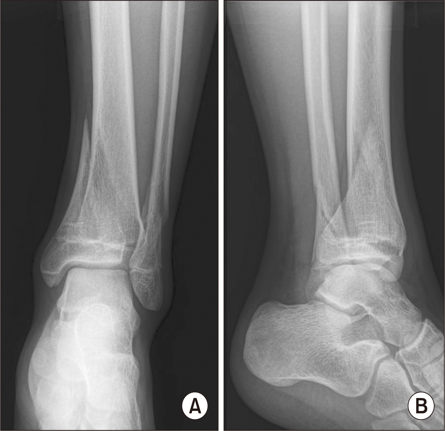
Figure. 1 Preoperative anteroposterior (A) and lateral (B) view of plain radiographs. A 14-year-old male with triplane fracture, which showed Salter–Harris type IV (epiphysis, minimally involved) fracture configuration.
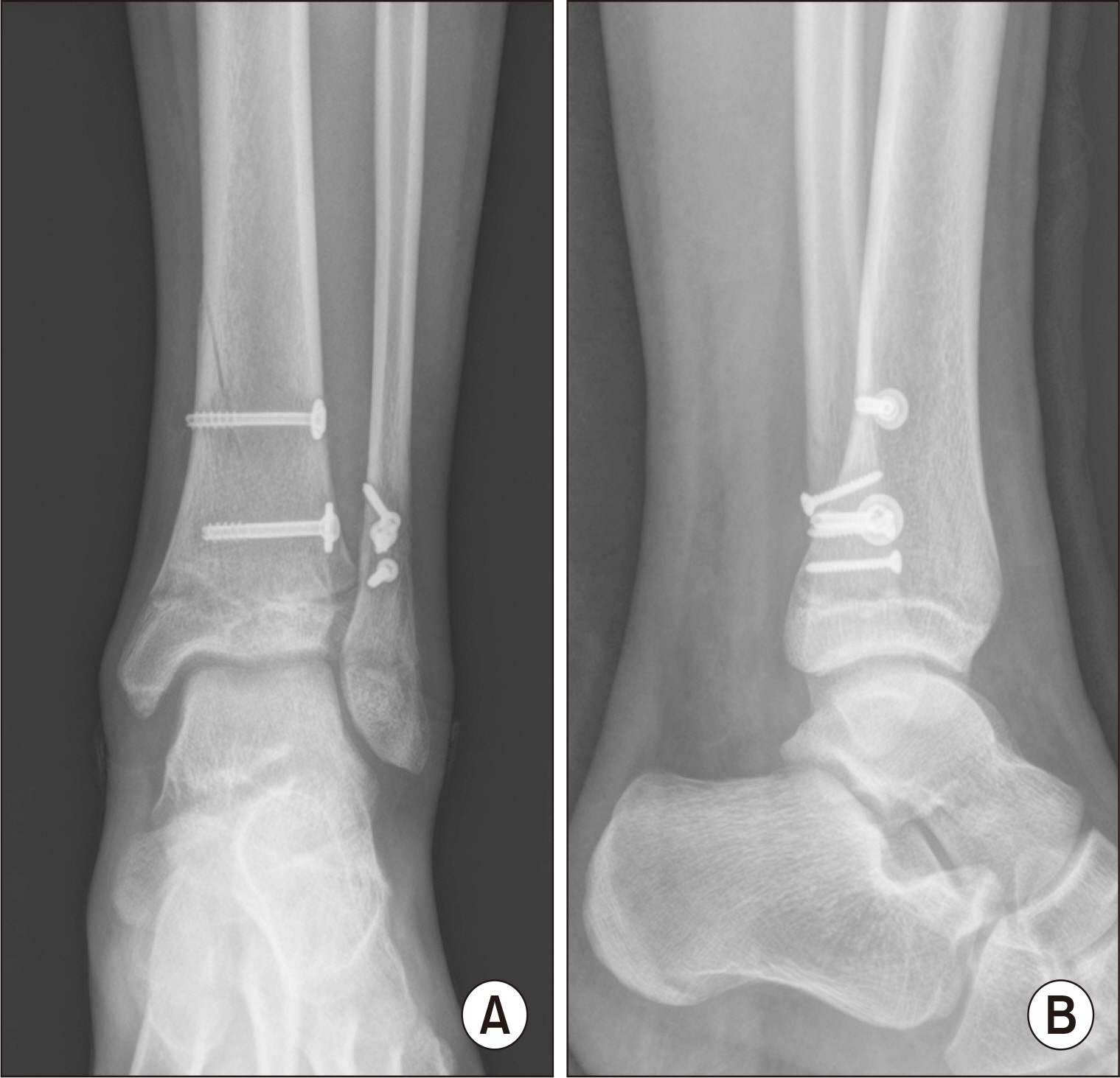
Figure. 2 Postoperative state; anteroposterior (A) and lateral (B) view. Anatomical reduction of fracture was obtained.

Figure. 3 Involuntary extension of great toe during ankle plantar flexion was observed.
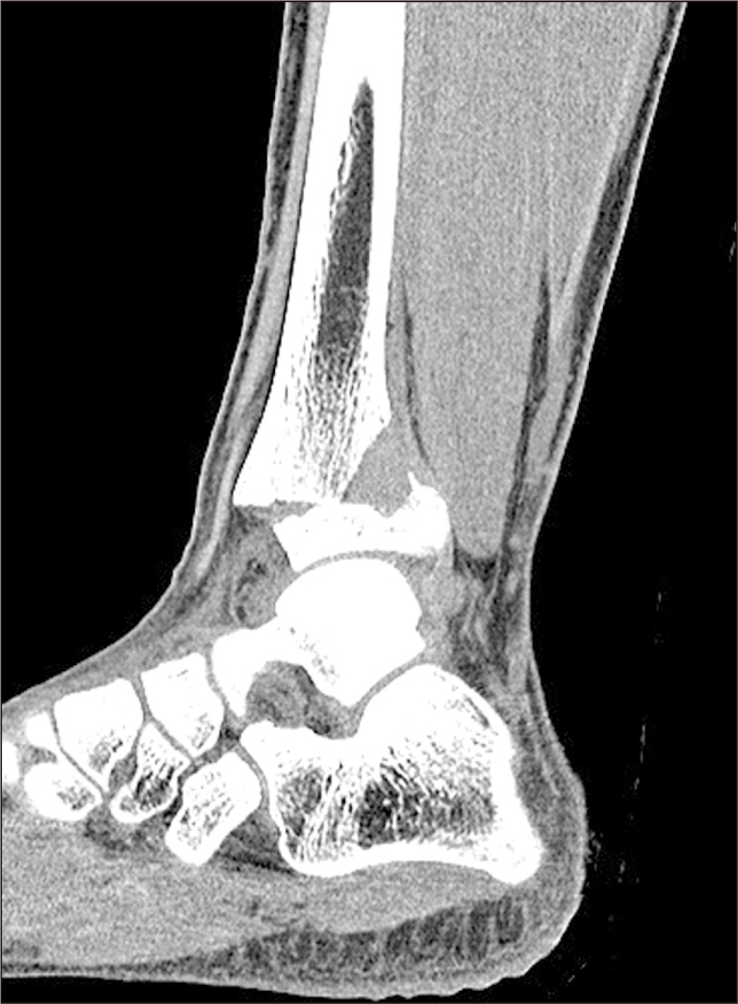
Figure. 4 Preoperative computed tomography (CT) sagittal image shows slight tenting of extensor hallucis longus tendon, but there is no incarcerations on CT image.
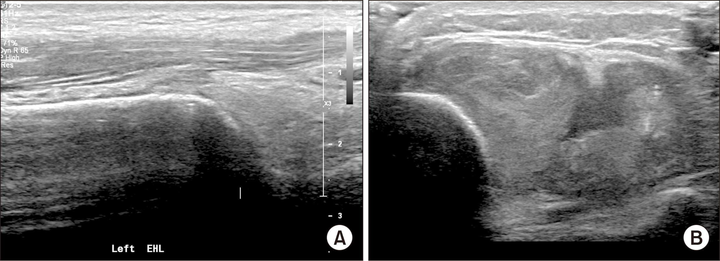
Figure. 5 The sonography of ankle and lower leg was evaluated. (A) The abnormality of integrity and continuity of extensor hallucis longus (EHL) were not identified. (B) Abnormal findings of a 5~6 cm long hypoechoic edema pattern were observed in the anterior muscle compartment.

Figure. 6 Postoperative magnetic resonance imaging of anterior compartment muscles. (A) Diffuse and heterogeneously increased signal intensity at anterior compartment muscles on T2-weighted image involving extensor digitorum longus, extensor hallucis longus, and tibialis anterior. (B) Geographic and thick, peripheral enhancements, and internal non-enhancing or decreased enhancing portions in the muscles on contrast enhanced T1-weighted image.
Reference
-
1. Wuerz TH, Gurd DP. 2013; Pediatric physeal ankle fracture. J Am Acad Orthop Surg. 21:234–44. doi: 10.5435/JAAOS-21-04-234. DOI: 10.5435/JAAOS-21-04-234. PMID: 23545729.
Article2. D¡¯Angelo F, Solarino G, Tanas D, Zani A, Cherubino P, Moretti B. 2017; Outcome of distal tibia physeal fractures: a review of cases as related to risk factors. Injury. 48(Suppl 3):S7–11. doi: 10.1016/S0020-1383(17)30650-2. DOI: 10.1016/S0020-1383(17)30650-2.
Article3. Holcomb TM, Temple EW, Barp EA, Smith HL. 2014; Surgical correction of checkrein deformity after malunited distal tibia fracture: a case report. J Foot Ankle Surg. 53:631–4. doi: 10.1053/j.jfas.2014.04.028. DOI: 10.1053/j.jfas.2014.04.028. PMID: 24942372.
Article4. Abolfotouh SM, Mahmoud KM, Mekhaimar MM, Said N, Al Dosari MA. 2015; Checkrein deformity associated with intra-articular talar fracture: a report of three cases. JBJS Case Connect. 5:e21–5. doi: 10.2106/JBJS.CC.N.00024. DOI: 10.2106/JBJS.CC.N.00024. PMID: 29252575.5. Bae SY, Chung HJ, Kim MY. 2011; Checkrein deformity by incarcerated posterior tibial tendon and displaced flexor hallucis longus tendon following ankle dislocation: a case report. J Korean Fract Soc. 24:271–6. doi: 10.12671/jkfs.2011.24.3.271. DOI: 10.12671/jkfs.2011.24.3.271.6. Kurashige T, Kawabata K, Suzuki S. 2014; Checkrein deformity due to extensor hallucis longus hypotrophy treated with extensor digitorum longus tendon transfer. Foot Ankle Surg. 20:e30–4. doi: 10.1016/j.fas.2014.02.008. DOI: 10.1016/j.fas.2014.02.008. PMID: 24796843.
Article7. Heo YM, Roh JY, Kim SB, Kim JY, Lee JB, Kim KK. 2010; Contracture of extensor hallucis longus tendon occurring after intramedullary nailing for a tibial fracture. J Korean Orthop Assoc. 45:399–403. doi: 10.4055/jkoa.2010.45.5.399. DOI: 10.4055/jkoa.2010.45.5.399.
Article8. Mubarak SJ. 2002; Extensor retinaculum syndrome of the ankle after injury to the distal tibial physis. J Bone Joint Surg Br. 84:11–4. doi: 10.1302/0301-620x.84b1.11800. DOI: 10.1302/0301-620X.84B1.11800. PMID: 11837815.
Article9. Rosenberg GA, Sferra JJ. 1999; Checkrein deformity-an unusual complication associated with a closed Salter-Harris type II ankle fracture: a case report. Foot Ankle Int. 20:591–4. doi: 10.1177/107110079902000910. DOI: 10.1177/107110079902000910. PMID: 10509688.
Article10. Haumont T, Gauchard GC, Zabee L, Arnoux JM, Journeau P, Lascombes P. 2007; Extensor retinaculum syndrome after distal tibial fractures: anatomical basis. Surg Radiol Anat. 29:303–11. doi: 10.1007/s00276-007-0215-3. DOI: 10.1007/s00276-007-0215-3. PMID: 17502984.
Article
- Full Text Links
-
- Actions
-
Cited
- CITED
-
- Close
- Share
-
- Similar articles
-
- Muscular Variations of Extensor Digitorum Brevis Muscle Related with Anterior Tarsal Tunnel Syndrome
- Contracture of Extensor Hallucis Longus Tendon Occurring after Intramedullary Nailing for a Tibial Fracture
- Traumatic Rupture of the Extensor Pollicis Longus Tendon Associated with Smiths Fracture: A case report
- Reconstruction of Chronic Extensor Hallucis Longus Tendon Rupture Using Interposed Scar Tissue: A Case Report
- Management of Checkrein Deformity
