Spontaneous Degenerative Rupture of Extensor Hallucis Longus Treated with a Split Tibialis Anterior Tendon Autograft: A Case Report
- Affiliations
-
- 1Department of Orthopedic Surgery, Kosin University Gospel Hospital, Kosin University School of Medicine, Busan, Kore
- 2Department of Orthopedic Surgery, Kangdong Sacred Heart Hospital, Hallym University School of Medicine, Seoul, Kore
- KMID: 2536716
- DOI: http://doi.org/10.14193/jkfas.2022.26.4.192
Abstract
- Chronic extensor hallucis longus (EHL) tendon rupture is relatively rare, but in such cases, surgical repair is necessary to prevent hallux dysfunction. To the best of our knowledge, reconstruction of chronic EHL rupture using a split tibialis anterior tendon autograft has not been previously reported. Here we present a case of spontaneous EHL tendon rupture with a 5 cm gap in a healthy 57-year-old woman. At the 1-year follow-up evaluation, hallux function was restored, and the patient was well satisfied with results.
Figure
-

Figure 1 Preoperative macroscopic findings: (A) distal and proximal extensor hallucis longus stumps and (B) insufficient extension of metatarsophalangeal and interphalangeal joints of the hallux.

Figure 2 Preoperative magnetic resonance imaging (MRI) findings. Sagittal T2-weighted MRI revealed the rupture of the extensor hallucis longus tendon (arrow).

Figure 3 Intraoperative findings: (A) the proximal and distal extensor hallucis longus (EHL) stumps (arrows) following the incision of the tendon sheath and the distal part of the inferior extensor retinaculum. (B) Tibialis anterior (TA) tendon (dotted line) was found medial to the EHL (arrows). (C) TA autograft was sutured to the proximal EHL stump in a Fish-mouth technique. (D) TA autograft was sutured to the distal EHL stump in a Fish-mouth technique. Ankle joint remained at 10 degrees of dorsiflexion, the first metatarsophalangeal joint remained at 10 degrees of extension.
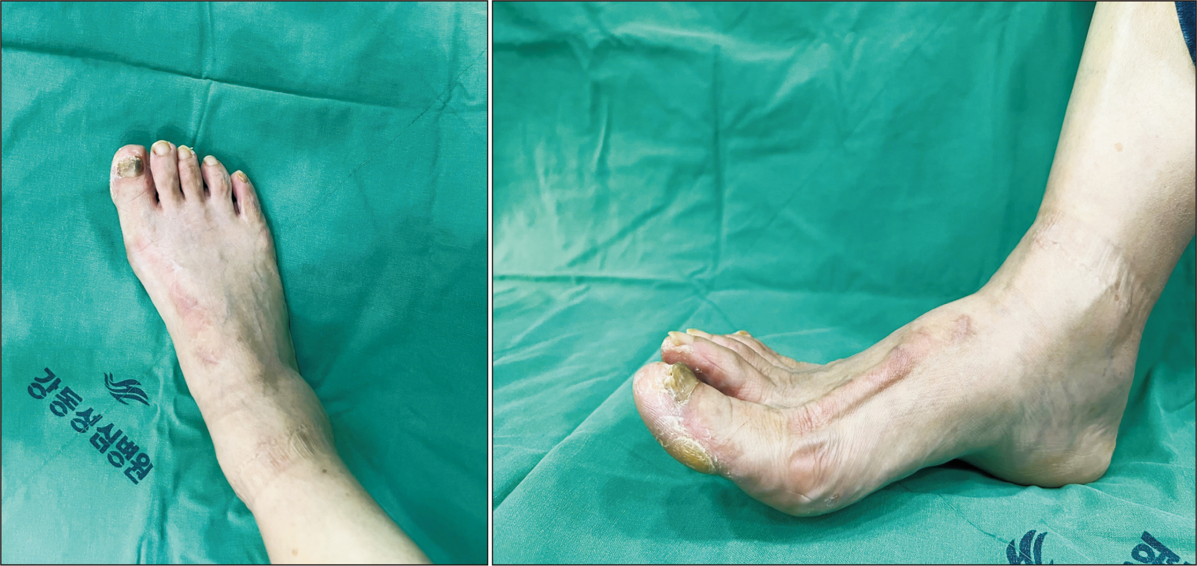
Figure 4 Clinical photograph shows extension of the active range of dorsiflexion of the hallux 1 year after reconstruction.
Reference
-
1. Kim WJ, Jung KJ, Ahn H, Yeo ED, Lee HS, Won SH, et al. 2021; Reconstruction of a neglected, extensor hallucis longus tendon rupture using interposed scar tissue: a case report and literature review. Int J Environ Res Public Health. 18:12157. doi: 10.3390/ijerph182212157. DOI: 10.3390/ijerph182212157. PMID: 34831920. PMCID: PMC8619473.2. Al-Qattan MM. 2007; Surgical treatment and results in 17 cases of open lacerations of the extensor hallucis longus tendon. J Plast Reconstr Aesthet Surg. 60:360–7. doi: 10.1016/j.bjps.2006.05.003. DOI: 10.1016/j.bjps.2006.05.003. PMID: 17349589.3. Mulcahy DM, Dolan AM, Stephens MM. 1996; Spontaneous rupture of extensor hallucis longus tendon. Foot Ankle Int. 17:162–3. doi: 10.1177/107110079601700308. DOI: 10.1177/107110079601700308. PMID: 8919621.4. Park HG, Lee BK, Sim JA. 2003; Autogenous graft repair using semitendinous tendon for a chronic multifocal rupture of the extensor hallucis longus tendon: a case report. Foot Ankle Int. 24:506–8. doi: 10.1177/107110070302400610. DOI: 10.1177/107110070302400610. PMID: 12854673.5. So E, Black TE, Mehl B. 2018; Split peroneus longus free tendon autograft transplantation for the treatment of neglected extensor hallucis longus tendon laceration: a case report. J Foot Ankle Surg. 57:210–4. doi: 10.1053/j.jfas.2017.08.005. DOI: 10.1053/j.jfas.2017.08.005. PMID: 29268901.6. Joseph RM, Barhorst J. 2012; Surgical reconstruction and mobilization therapy for a retracted extensor hallucis longus laceration and tendon defect repaired by split extensor hallucis longus tendon lengthening and dermal scaffold augmentation. J Foot Ankle Surg. 51:509–16. doi: 10.1053/j.jfas.2012.04.018. DOI: 10.1053/j.jfas.2012.04.018. PMID: 22658790.7. Matsuda T, Taniguchi A, Hayashi K, Nakanishi Y, Tanaka Y. 2018; Obtaining adequate tension for extensor hallucis longus tendon rupture repair using wide-awake surgery: a case report. J Foot Ankle Surg. 57:414–7. doi: 10.1053/j.jfas.2017.08.025. DOI: 10.1053/j.jfas.2017.08.025. PMID: 29223409.8. Olewnik Ł, Podgórski M, Polguj M, Topol M. 2019; A cadaveric and sonographic study of the morphology of the tibialis anterior tendon - a proposal for a new classification. J Foot Ankle Res. 12:9. doi: 10.1186/s13047-019-0319-0. DOI: 10.1186/s13047-019-0319-0. PMID: 30733832. PMCID: PMC6359855.9. Kurashige T. 2019; Chronic extensor hallucis longus tendon rupture treated with double-bundle autograft of extensor hallucis capsularis: a case report. SAGE Open Med Case Rep. 7:2050313X19841962. doi: 10.1177/2050313X19841962. DOI: 10.1177/2050313X19841962. PMID: 31007922. PMCID: PMC6457017.
- Full Text Links
-
- Actions
-
Cited
- CITED
-
- Close
- Share
-
- Similar articles
-
- Reconstruction of Chronic Extensor Hallucis Longus Tendon Rupture Using Interposed Scar Tissue: A Case Report
- Complete Rupture of the Extensor Hallucis Longus Tendon with Accessory Slip Mimicking a Partial Rupture: A Case Report
- A 12-Week Rehabilitation Protocol for the Management of Chronic Extensor Hallucis Longus Rupture Repaired with an Autograft of the Semitendinosus Tendon
- Extensor Hallucis Longus Tendon Rupture in TaeKwonDo Players: Two Case Report
- Rupture of the Extensor Pollicis Longus after Fracture of the Distal end of the Radius: Report of 3 cases
