Long Non-Coding RNA TUG1 Attenuates Insulin Resistance in Mice with Gestational Diabetes Mellitus via Regulation of the MicroRNA-328-3p/SREBP-2/ERK Axis
- Affiliations
-
- 1Department of Obstetrics and Gynecology, Guangzhou Women and Children’s Medical Center Affiliated to Guangzhou Medical University, Guangzhou, China
- 2Department of Endocrinology, Guangzhou First People’s Hospital, South China University of Technology, Guangzhou, China
- KMID: 2540523
- DOI: http://doi.org/10.4093/dmj.2021.0216
Abstract
- Background
Long non-coding RNAs (lncRNAs) have been illustrated to contribute to the development of gestational diabetes mellitus (GDM). In the present study, we aimed to elucidate how lncRNA taurine upregulated gene 1 (TUG1) influences insulin resistance (IR) in a high-fat diet (HFD)-induced mouse model of GDM.
Methods
We initially developed a mouse model of HFD-induced GDM, from which islet tissues were collected for RNA and protein extraction. Interactions among lncRNA TUG1/microRNA (miR)-328-3p/sterol regulatory element binding protein 2 (SREBP-2) were assessed by dual-luciferase reporter assay. Fasting blood glucose (FBG), fasting insulin (FINS), homeostasis model assessment of insulin resistance (HOMA-IR), HOMA pancreatic β-cell function (HOMA-β), insulin sensitivity index for oral glucose tolerance tests (ISOGTT) and insulinogenic index (IGI) levels in mouse serum were measured through conducting gain- and loss-of-function experiments.
Results
Abundant expression of miR-328 and deficient expression of lncRNA TUG1 and SREBP-2 were characterized in the islet tissues of mice with HFD-induced GDM. LncRNA TUG1 competitively bound to miR-328-3p, which specifically targeted SREBP-2. Either depletion of miR-328-3p or restoration of lncRNA TUG1 and SREBP-2 reduced the FBG, FINS, HOMA-β, and HOMA-IR levels while increasing ISOGTT and IGI levels, promoting the expression of the extracellular signal-regulated kinase (ERK) signaling pathway-related genes, and inhibiting apoptosis of islet cells in GDM mice. Upregulation miR-328-3p reversed the alleviative effects of SREBP-2 and lncRNA TUG1 on IR.
Conclusion
Our study provides evidence that the lncRNA TUG1 may prevent IR following GDM through competitively binding to miR-328-3p and promoting the SREBP-2-mediated ERK signaling pathway inactivation.
Keyword
Figure
-
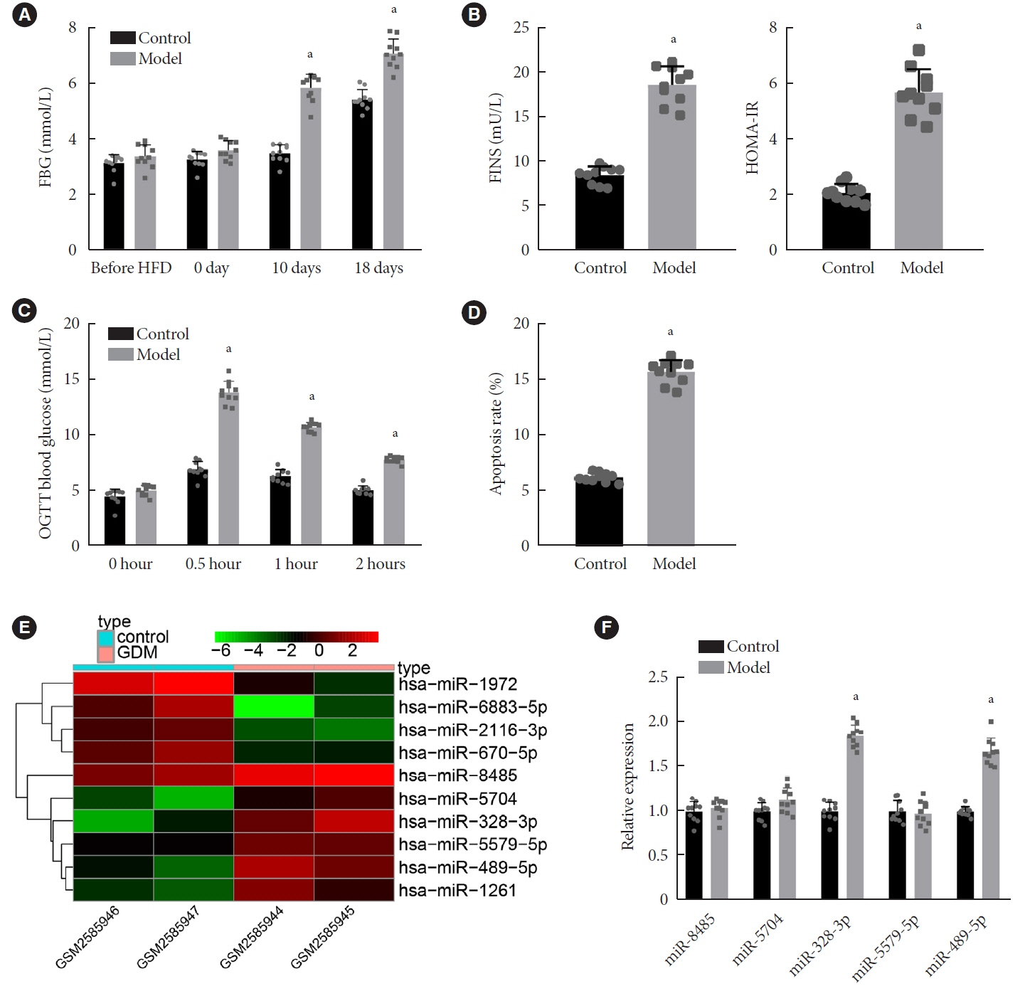
Fig. 1. Characterization of the gestational diabetes mellitus (GDM) mouse model and miRNA expression profiles in GDM. (A) Fasting blood glucose (FBG) levels (mmol/L) at different periods of gestation in control and GDM mice. (B) Levels of fasting insulin (FINS) and homeostasis model assessment of insulin resistance (HOMA-IR) at day 18 of gestation in control and GDM mice. (C) Glucose area under the curve at different time points in control and GDM mice. (D) Cell apoptosis in the islet tissues of control and GDM mice determined by terminal deoxynucleotidyl transferase-mediated dUTP-biotin nick-end labeling (TUNEL) assay. (E) A heat map of the top 10 differentially expressed miRNAs screened from the GSE98043 dataset. The abscissa indicates the sample number and the ordinate indicates the differentially expressed miRNAs; the upper right histogram is the color gradation; each rectangle corresponds to the value of a miRNA expression in one sample. (F) Expression of miR-8485, miR-5704, miR-328-3p, miR-5579-5p, and miR-489-5p determined by reverse transcription-quantitative polymerase chain reaction in islet tissues of control and GDM mice. OGTT, oral glucose tolerance test. aP<0.05 vs. control mice (n=10 for mice upon each treatment).
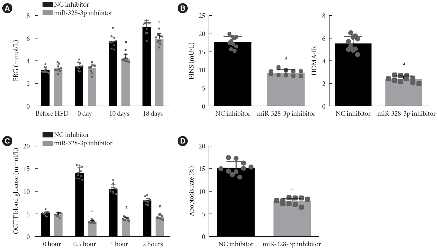
Fig. 2. Inhibition of miR-328-3p attenuates insulin resistance in gestational diabetes mellitus (GDM) mice. (A) Fasting blood glucose (FBG) levels (mmol/L) at different time points of gestation in GDM mice injected with nanoparticles (NPs) expressing negative control (NC) inhibitor or miR-328-3p inhibitor. (B) Levels of fasting insulin (FINS) and homeostasis model assessment of insulin resistance (HOMA-IR) at day 18 of gestation in GDM mice injected with NPs expressing NC inhibitor or miR-328-3p inhibitor. (C) Glucose area under the curve at different time points of gestation in GDM mice injected with NPs expressing NC inhibitor or miR-328-3p inhibitor. (D) Apoptosis of cells in islet tissues of GDM mice injected with NPs expressing NC inhibitor or miR-328-3p inhibitor detected using terminal deoxynucleotidyl transferase-mediated dUTP-biotin nick-end labeling (TUNEL) staining. OGTT, oral glucose tolerance test. aP<0.05 vs. GDM mice injected with NPs expressing NC inhibitor (n=10 for mice following each treatment).

Fig. 3. Sterol regulatory element binding protein 2 (SREBP-2) is a target gene of miR-328-3p. (A) Target genes of miR-328-3p predicted by the TargetScan, miRDB, starBase, DIANA, and RNA22 databases intersected with differentially expressed genes (DEGs) from the GSE41095 dataset. Red refers to the target genes of miR-328-3p simultaneously predicted by at least three databases and also differentially expressed in gestational diabetes mellitus (GDM) samples from the GSE41095 dataset. (B) An interaction network between candidate miR-328-3p target genes and GDM-related genes analyzed using the DisGeNET database; the circle indicates the disease gene, and the arrow indicates the DEG; genes with correlation coefficient of 0 are not shown in the figure. (C) Kyoto Encyclopedia of Genes and Genomes (KEGG) enrichment analysis of candidate miR-328-3p target genes and GDM-related genes obtained from the DisGeNET database. (D) Expression of SREBP-2 in normal and GDM samples in the GSE41095 dataset; red box refers to GDM samples, and blue box represents normal samples. (E) Expression of SREBP-2 determined by reverse transcription-quantitative polymerase chain reaction (RT-qPCR) in islet tissues of control and GDM mice. (F) Western blot analysis of SREBP-2 protein in islet tissues of control and GDM mice. (G) Immunohistochemistry staining of SREBP-2 protein in islet tissues of control and GDM mice. (H) Putative miR-328-3p binding sites in the 3’-untranslated region (3’-UTR) of SREBP-2 predicted by the starBase database (the left image) and miR-328-3p binding to SREBP-2 confirmed by dual-luciferase reporter assay in islet cells (the right image). (I) miR-328-3p expression and mRNA expression of SREBP-2 in islet tissues of GDM mice treated with nanoparticles (NPs) expressing negative control (NC) inhibitor or miR-328-3p inhibitor determined by RT-qPCR. (J) Western blot analysis of SREBP-2 protein in islet tissues of GDM mice treated with NPs expressing NC inhibitor or miR-328-3p inhibitor. aP<0.05 vs. GDM mice treated with NPs expressing NC mimic or NC inhibitor. Cell experiments were repeated three times.

Fig. 4. MiR-328-3p induces insulin resistance by disrupting the sterol regulatory element binding protein 2 (SREBP-2)-mediated extracellular signal-regulated kinase (ERK) signaling pathway inactivation in gestational diabetes mellitus (GDM) mice. GDM mice were injected with nanoparticles (NPs) expressing miR-328-3pmimic, overexpression (oe)-SREBP-2 or both. (A) Fasting blood glucose (FBG) levels (mmol/L) at different periods of gestation in GDM mice. (B) Levels of fasting insulin (FINS) and homeostasis model assessment of insulin resistance (HOMA-IR) at day 18 of gestation in GDM mice. (C) Glucose area under the curve at different gestational time points in GDM mice. (D) Apoptosis of cells in islet tissues of GDM mice detected using terminal deoxynucleotidyl transferase-mediated dUTP-biotin nick-end labeling (TUNEL) staining. (E) Western blot analysis of SREBP-2 and Ras proteins, and ERK phosphorylation level in islet tissues of GDM mice. (F) Immunohistochemistry analysis of SREBP-2- and p-ERK-positive cells in islet tissues of GDM mice. (G) The 3-hydroxy-3-methylglutaryl-CoA reductase (HMGCR) mRNA expression in pancreatic islet tissue of pregnant mice determined by reverse transcription-quantitative polymerase chain reaction. NC, negative control; OGTT, oral glucose tolerance test. aP<0.05 vs. GDM mice injected with NPs expressing NC inhibitor, bP<0.05 vs. GDM mice injected with NPs expressing NC mimic+oe-SREBP-2 (n=10 for mice following each treatment).
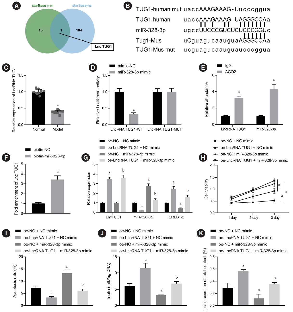
Fig. 5. Long non-coding RNAs (lncRNA) taurine upregulated gene 1 (TUG1) binds to miR-328-3p to upregulate sterol regulatory element binding protein 2 (SREBP-2) expression and enhance pancreatic β-cell viability and insulin secretion. (A) Upstream regulatory lncRNAs of miR-328-3p in human and mice predicted by the starBase database; the two circles in the figure represent the prediction results in human and mice respectively, and the central part represents the intersection of the two sets of data. (B) Binding sites between miR-328-3p and lncRNA TUG1 both in human and mice. (C) LncRNA TUG1 expression in the islet tissues of control and gestational diabetes mellitus (GDM) mice determined by reverse transcription-quantitative polymerase chain reaction (RT-qPCR). (D) Binding between miR-328-3p and lncRNA TUG1 confirmed by dual-luciferase reporter assay in islet cells transfected with miR-328-3p mimic or negative control (NC) mimic. (E) Binding of miR-328-3p and lncRNA TUG1 to the argonaute 2 (Ago2) antibody determined by RNA binding protein immunoprecipitation (RIP) assay. (F) Binding between miR-328-3p and lncRNA TUG1 determined by RNA pull-down assay; BETA-TC-6 cells were transfected with miR-328-3p mimic, overexpression (oe)-lncRNA TUG1 or both. (G) Expression of lncRNA TUG1, miR-328-3p and SREBP-2 in BETA-TC-6 cells determined by RT-qPCR. (H) BETA-TC-6 cell viability measured by cell counting kit-8 (CCK-8) assay. (I) Flow cytometric examination of BETA-TC-6 cell apoptosis. (J) Total insulin content in BETA-TC-6 cells measured by enzyme-linked immunosorbent assay (ELISA). (K) Insulin secretion in BETA-TC-6 cells measured by ELISA. IgG, immunoglobulin G. aP<0.05 vs. islet cells transfected with NC mimic or BETA-TC-6 cells transfected with oe-NC+NC mimic, bP<0.05 vs. BETA-TC-6 cells transfected with oe-NC+miR-328-3p mimic (n=10 for mice following each treatment). Cell experiments were repeated three times.

Fig. 6. Long non-coding RNAs (lncRNA) taurine upregulated gene 1 (TUG1) inhibits the insulin resistance in gestational diabetes mellitus (GDM) mice by regulating the miR-328-3p/sterol regulatory element binding protein 2 (SREBP-2)/extracellular signal-regulated kinase (ERK) signaling axis. (A) Expression of lncRNA TUG1, miR-328-3p and SREBP-2 in islet tissues of GDM mice overexpressing lncRNA TUG1 determined by reverse transcription-quantitative polymerase chain reaction. (B) Fasting blood glucose (FBG) levels (mM) at different periods of gestation in GDM mice overexpressing lncRNA TUG1. (C) Levels of fasting insulin (FINS) and homeostasis model assessment of insulin resistance (HOMA-IR) at day 18 of gestation in GDM mice overexpressing lncRNA TUG1. (D) Glucose area under the curve at different gestational days in GDM mice overexpressing lncRNA TUG1. (E) Apoptosis of cells in islet tissues of GDM mice overexpressing lncRNA TUG1 detected using terminal deoxynucleotidyl transferase-mediated dUTP-biotin nick-end labeling (TUNEL) staining. (F) Western blot analysis of Ras protein and ERK phosphorylation level in islet tissues of GDM mice overexpressing lncRNA TUG1. OGTT, oral glucose tolerance test. aP<0.05 vs. GDM mice injected with nanoparticles (NPs) expressing vector (n=10 for mice following each treatment).
Reference
-
1. Roglic G, Colagiuri S. Gestational diabetes mellitus: squaring the circle. Diabetes Care. 2014; 37:e143–4.2. Chiefari E, Arcidiacono B, Foti D, Brunetti A. Gestational diabetes mellitus: an updated overview. J Endocrinol Invest. 2017; 40:899–909.3. Kampmann U, Madsen LR, Skajaa GO, Iversen DS, Moeller N, Ovesen P. Gestational diabetes: a clinical update. World J Diabetes. 2015; 6:1065–72.4. Agha-Jaffar R, Oliver N, Johnston D, Robinson S. Gestational diabetes mellitus: does an effective prevention strategy exist? Nat Rev Endocrinol. 2016; 12:533–46.5. Voormolen DN, Abell SK, James R, Hague WM, Mol BW. Diagnostic criteria and treatment for gestational diabetes mellitus. Semin Reprod Med. 2016; 34:102–9.6. Nolan CJ. Lipotoxicity, β cell dysfunction, and gestational diabetes. Cell Metab. 2014; 19:553–4.7. Yang SJ, Kim TN, Baik SH, Kim TS, Lee KW, Nam M, et al. Insulin secretion and insulin resistance in Korean women with gestational diabetes mellitus and impaired glucose tolerance. Korean J Intern Med. 2013; 28:306–13.8. Boden G. Obesity, insulin resistance and free fatty acids. Curr Opin Endocrinol Diabetes Obes. 2011; 18:139–43.9. Johns EC, Denison FC, Norman JE, Reynolds RM. Gestational diabetes mellitus: mechanisms, treatment, and complications. Trends Endocrinol Metab. 2018; 29:743–54.10. Bhan A, Soleimani M, Mandal SS. Long noncoding RNA and cancer: a new paradigm. Cancer Res. 2017; 77:3965–81.11. Li Y, Zhi K, Han S, Li X, Li M, Lian W, et al. TUG1 enhances high glucose-impaired endothelial progenitor cell function via miR-29c-3p/PDGF-BB/Wnt signaling. Stem Cell Res Ther. 2020; 11:441.12. Wang S, Cao W, Gao S, Nie X, Zheng X, Xing Y, et al. TUG1 regulates pulmonary arterial smooth muscle cell proliferation in pulmonary arterial hypertension. Can J Cardiol. 2019; 35:1534–45.13. Poirier C, Desgagne V, Guerin R, Bouchard L. MicroRNAs in pregnancy and gestational diabetes mellitus: emerging role in maternal metabolic regulation. Curr Diab Rep. 2017; 17:35.14. Zheng Y, Wang Z, Zhou Z. miRNAs: novel regulators of autoimmunity-mediated pancreatic β-cell destruction in type 1 diabetes. Cell Mol Immunol. 2017; 14:488–96.15. Li XL, Subramanian M, Jones MF, Chaudhary R, Singh DK, Zong X, et al. Long noncoding RNA PURPL suppresses basal p53 levels and promotes tumorigenicity in colorectal cancer. Cell Rep. 2017; 20:2408–23.16. Prado M, de Goes TC, de Jesus ML, Mendonca L, Nascimento JS, Kaneto CM. Identification of miR-328-3p as an endogenous reference gene for the normalization of miRNA expression data from patients with diabetic retinopathy. Sci Rep. 2019; 9:19677.17. Shao W, Espenshade PJ. Expanding roles for SREBP in metabolism. Cell Metab. 2012; 16:414–9.18. Golic M, Stojanovska V, Bendix I, Wehner A, Herse F, Haase N, et al. Diabetes mellitus in pregnancy leads to growth restriction and epigenetic modification of the Srebf2 gene in rat fetuses. Hypertension. 2018; 71:911–20.19. Kotzka J, Lehr S, Roth G, Avci H, Knebel B, Muller-Wieland D. Insulin-activated Erk-mitogen-activated protein kinases phosphorylate sterol regulatory element-binding Protein-2 at serine residues 432 and 455 in vivo. J Biol Chem. 2004; 279:22404–11.20. Balachandiran M, Bobby Z, Dorairajan G, Gladwin V, Vinayagam V, Packirisamy RM. Decreased maternal serum adiponectin and increased insulin-like growth factor-1 levels along with increased placental glucose transporter-1 expression in gestational diabetes mellitus: possible role in fetal overgrowth. Placenta. 2021; 104:71–80.21. Roy R, Nguyen-Ngo C, Lappas M. Short-chain fatty acids as novel therapeutics for gestational diabetes. J Mol Endocrinol. 2020; 65:21–34.22. Jiang YK, Xin KY, Ge HW, Kong FJ, Zhao G. Upregulation Of renal GLUT2 and SGLT2 is involved in high-fat diet-induced gestational diabetes in mice. Diabetes Metab Syndr Obes. 2019; 12:2095–105.23. Liu Z, Yu X, Tong C, Qi H. Renal dysfunction in a mouse model of GDM is prevented by metformin through MAPKs. Mol Med Rep. 2019; 19:4491–9.24. Chen CP, Chang SC, Vivian Yang WC. High glucose alters proteoglycan expression and the glycosaminoglycan composition in placentas of women with gestational diabetes mellitus and in cultured trophoblasts. Placenta. 2007; 28:97–106.25. Fukui K, Ferris HA, Kahn CR. Effect of cholesterol reduction on receptor signaling in neurons. J Biol Chem. 2015; 290:26383–92.26. Zhang EB, Yin DD, Sun M, Kong R, Liu XH, You LH, et al. P53-regulated long non-coding RNA TUG1 affects cell proliferation in human non-small cell lung cancer, partly through epigenetically regulating HOXB7 expression. Cell Death Dis. 2014; 5:e1243.27. Young TL, Matsuda T, Cepko CL. The noncoding RNA taurine upregulated gene 1 is required for differentiation of the murine retina. Curr Biol. 2005; 15:501–12.28. Sweeting AN, Ross GP, Hyett J, Molyneaux L, Constantino M, Harding AJ, et al. Gestational diabetes mellitus in early pregnancy: evidence for poor pregnancy outcomes despite treatment. Diabetes Care. 2016; 39:75–81.29. Raghupathy P, Antonisamy B, Geethanjali FS, Saperia J, Leary SD, Priya G, et al. Glucose tolerance, insulin resistance and insulin secretion in young south Indian adults: relationships to parental size, neonatal size and childhood body mass index. Diabetes Res Clin Pract. 2010; 87:283–92.30. Zhang J, Chi H, Xiao H, Tian X, Wang Y, Yun X, et al. Interleukin 6 (IL-6) and tumor necrosis factor α (TNF-α) single nucleotide polymorphisms (SNPs), inflammation and metabolism in gestational diabetes mellitus in Inner Mongolia. Med Sci Monit. 2017; 23:4149–57.31. Zhu C, Yang H, Geng Q, Ma Q, Long Y, Zhou C, et al. Association of oxidative stress biomarkers with gestational diabetes mellitus in pregnant women: a case-control study. PLoS One. 2015; 10:e0126490.32. Pan J, Zhang F, Zhang L, Bao Y, Tao M, Jia W. Influence of insulin sensitivity and secretion on glycated albumin and hemoglobin A1c in pregnant women with gestational diabetes mellitus. Int J Gynaecol Obstet. 2013; 121:252–6.33. Lu J, Gu Y, Wang L, Li W, Zhang S, Liu H, et al. Glucose metabolism among obese and non-obese children of mothers with gestational diabetes. BMJ Open Diabetes Res Care. 2020; 8:e000822.34. Zhu Y, Tian F, Li H, Zhou Y, Lu J, Ge Q. Profiling maternal plasma microRNA expression in early pregnancy to predict gestational diabetes mellitus. Int J Gynaecol Obstet. 2015; 130:49–53.35. Huo W, Hou Y, Li Y, Li H. Downregulated lncRNA-MIAT confers protection against erectile dysfunction by downregulating lipoprotein lipase via activation of miR-328a-5p in diabetic rats. Biochim Biophys Acta Mol Basis Dis. 2019; 1865:1226–40.36. McManus DD, Rong J, Huan T, Lacey S, Tanriverdi K, Munson PJ, et al. Messenger RNA and MicroRNA transcriptomic signatures of cardiometabolic risk factors. BMC Genomics. 2017; 18:139.37. Guo L, Qiu Z, Wei L, Yu X, Gao X, Jiang S, et al. The microRNA-328 regulates hypoxic pulmonary hypertension by targeting at insulin growth factor 1 receptor and L-type calcium channel-α1C. Hypertension. 2012; 59:1006–13.38. Rayner KJ, Suarez Y, Davalos A, Parathath S, Fitzgerald ML, Tamehiro N, et al. MiR-33 contributes to the regulation of cholesterol homeostasis. Science. 2010; 328:1570–3.39. Rottiers V, Najafi-Shoushtari SH, Kristo F, Gurumurthy S, Zhong L, Li Y, et al. MicroRNAs in metabolism and metabolic diseases. Cold Spring Harb Symp Quant Biol. 2011; 76:225–33.40. Bommer GT, MacDougald OA. Regulation of lipid homeostasis by the bifunctional SREBF2-miR33a locus. Cell Metab. 2011; 13:241–7.41. Andres-Blasco I, Blesa S, Vinue A, Gonzalez-Navarro H, Real JT, Martinez-Hervas S, et al. Srebf2 locus overexpression reduces body weight, total cholesterol and glucose levels in mice fed with two different diets. Nutrients. 2020; 12:3130.42. Chen Y, Yang Q, Zhan Y, Ke J, Lv P, Huang J. The role of miR-328 in high glucose-induced endothelial-to-mesenchymal transition in human umbilical vein endothelial cells. Life Sci. 2018; 207:110–6.43. Ozaki KI, Awazu M, Tamiya M, Iwasaki Y, Harada A, Kugisaki S, et al. Targeting the ERK signaling pathway as a potential treatment for insulin resistance and type 2 diabetes. Am J Physiol Endocrinol Metab. 2016; 310:E643–51.44. Wang PY, Weng J, Anderson RG. OSBP is a cholesterol-regulated scaffolding protein in control of ERK 1/2 activation. Science. 2005; 307:1472–6.45. Fukui K, Ferris HA, Kahn CR. Effect of cholesterol reduction on receptor signaling in neurons. J Biol Chem. 2016; 291:15910.46. Chen K, Ma Y, Wu S, Zhuang Y, Liu X, Lv L, et al. Construction and analysis of a lncRNA-miRNA-mRNA network based on competitive endogenous RNA reveals functional lncRNAs in diabetic cardiomyopathy. Mol Med Rep. 2019; 20:1393–403.47. Zhao L, Sun H, Kong H, Chen Z, Chen B, Zhou M. The Lncrna-TUG1/EZH2 axis promotes pancreatic cancer cell proliferation, migration and EMT phenotype formation through sponging mir-382. Cell Physiol Biochem. 2017; 42:2145–58.48. Zang XJ, Li L, Du X, Yang B, Mei CL. LncRNA TUG1 inhibits the proliferation and fibrosis of mesangial cells in diabetic nephropathy via inhibiting the PI3K/AKT pathway. Eur Rev Med Pharmacol Sci. 2019; 23:7519–25.49. Zhang Y, Ma Y, Gu M, Peng Y. lncRNA TUG1 promotes the brown remodeling of white adipose tissue by regulating miR-204-targeted SIRT1 in diabetic mice. Int J Mol Med. 2020; 46:2225–34.50. Wei X, Zhou Y, Qiu J, Wang X, Xia Y, Sui L. Low expression of TUG1 promotes cisplatin sensitivity in cervical cancer by activating the MAPK pathway. J BUON. 2019; 24:1020–6.
- Full Text Links
-
- Actions
-
Cited
- CITED
-
- Close
- Share
-
- Similar articles
-
- Long Non-Coding RNA TUG1 Promotes Proliferation and Inhibits Apoptosis of Osteosarcoma Cells by Sponging miR-132-3p and Upregulating SOX4 Expression
- The Long Noncoding RNA DUXAP8 Facilitates the Malignant Progression of Colon Cancer via the microRNA-378a-3p/FOXQ1 Axis
- N6-Methyladenosine Methyltransferase METTL3 Alleviates Diabetes-Induced Testicular Damage through Modulating TUG1/Clusterin Axis
- LncRNA Taurine-Upregulated Gene 1 Promotes Cell Proliferation by Inhibiting MicroRNA-9 in MCF-7 Cells
- Response: Adipokines and Insulin Resistance According to Characteristics of Pregnant Women with Gestational Diabetes Mellitus (Diabetes Metab J 2017;41:457-65)
