Mixed adenoneuroendocrine carcinoma of the ampulla of Vater: Three case reports and a literature review
- Affiliations
-
- 1Division of Hepatobiliary and Pancreatic Surgery, Department of Surgery, Asan Medical Center, University of Ulsan College of Medicine, Seoul, Korea
- KMID: 2539521
- DOI: http://doi.org/10.14701/ahbps.22-054
Abstract
- Mixed adenoneuroendocrine carcinoma is defined as a tumor with a mixture of adenocarcinoma components and neuroendocrine neoplasm components. Each of these two components of mixed adenoneuroendocrine carcinoma accounts for at least 30% of all tumors. Mixed adenoneuroendocrine carcinoma might be located in the ampulla of Vater, a very rare location compared to other organs. Thus, its treatment and prognosis plans have not been established yet. We report three cases of mixed adenoneuroendocrine carcinoma occurring in the ampulla of Vater. Each patient had a different clinical course. In general, difficulty in preoperative diagnosis, risk of early recurrence, and poor disease course were main hallmarks of mixed adenoneuroendocrine carcinoma arising from the ampulla of Vater. However, one patient in this case report survived although she did not receive adjuvant chemotherapy due to her old age. Therefore, it is important to establish a careful treatment strategy for mixed adenoneuroendocrine carcinoma arising from the ampulla of Vater.
Figure
-
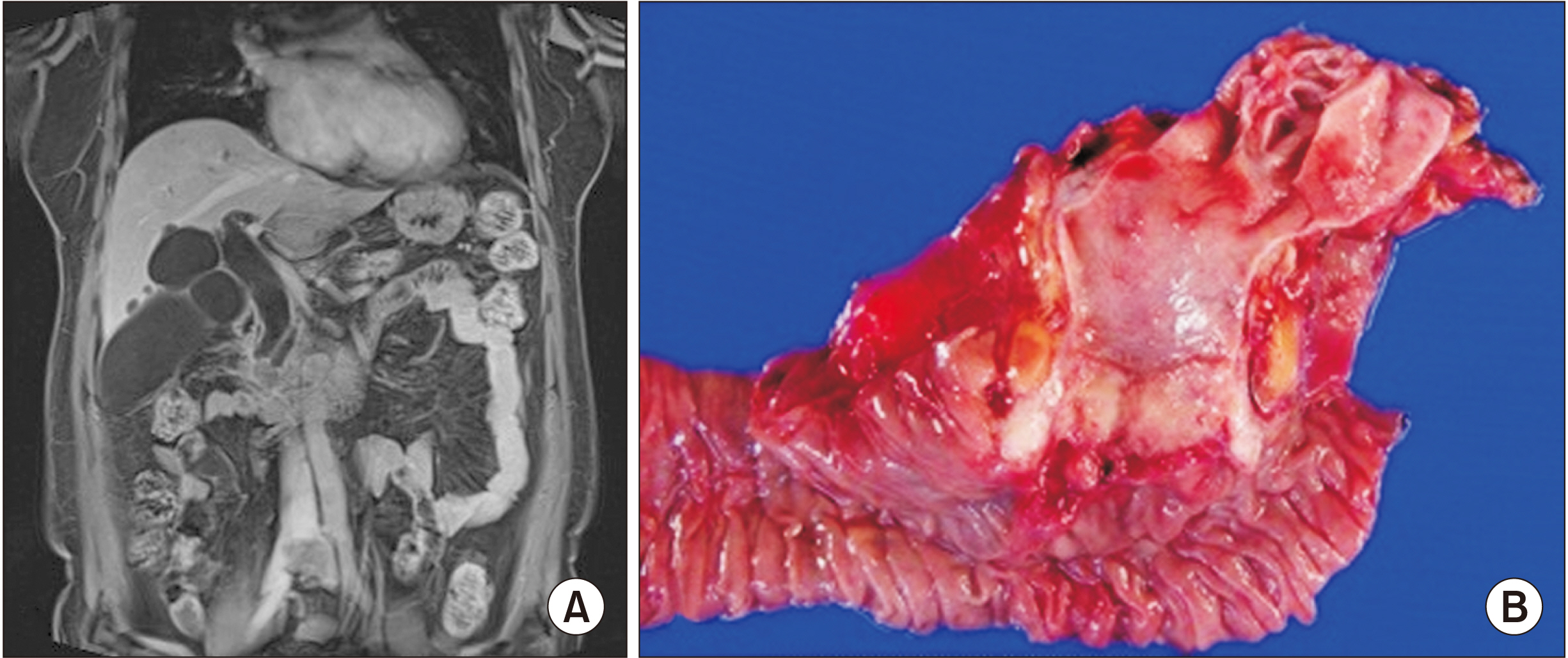
Fig. 1 (A) Case 1 magnetic resonance cholangiopancreatography (MRCP) findings of the neoplastic lesion showing bile duct dilatation, obstruction in the ampullary area of the stretched bile duct, a 2.3 cm long mass involving the ampulla of Vater, and lymph nodes less than 1 cm in the retropancreatic area. (B) Case 1 surgical specimen of the resected ampullary tumor.
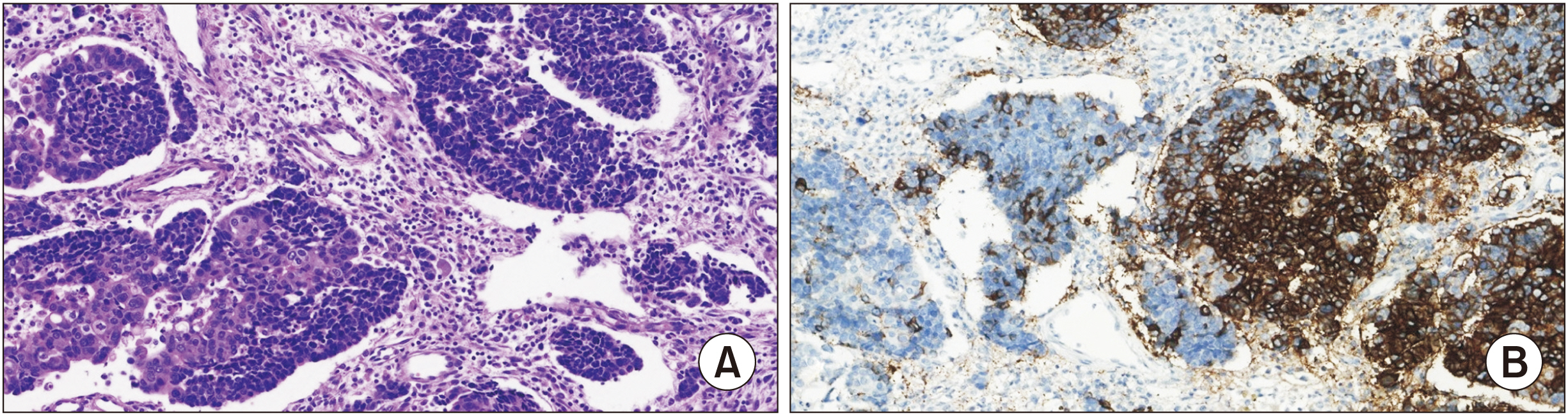
Fig. 2 (A) Case 1 H&E staining showing a mixed adenoneuroendocrine tumor (x200). (B) Case 1 immunohistochemical analysis showing neuroendocrine tumor cells being positive for CD56 (x200).
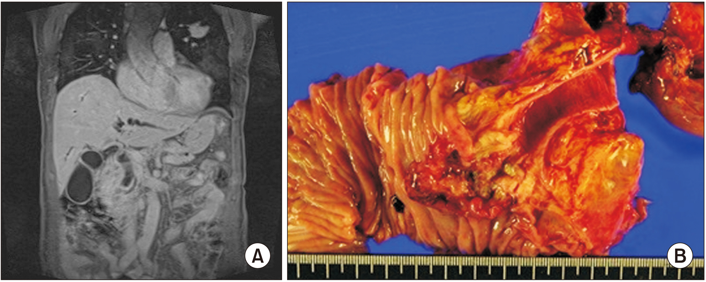
Fig. 3 (A) Case 2 magnetic resonance cholangiopancreatography (MRCP) findings of the neoplastic lesion showing bile duct dilatation, obstruction in the ampullary area of the stretched bile duct, and a 3.0 cm long mass involving the ampulla of Vater without enlarged lymph nodes. (B) Case 2 surgical specimen of the resected ampullary tumor.
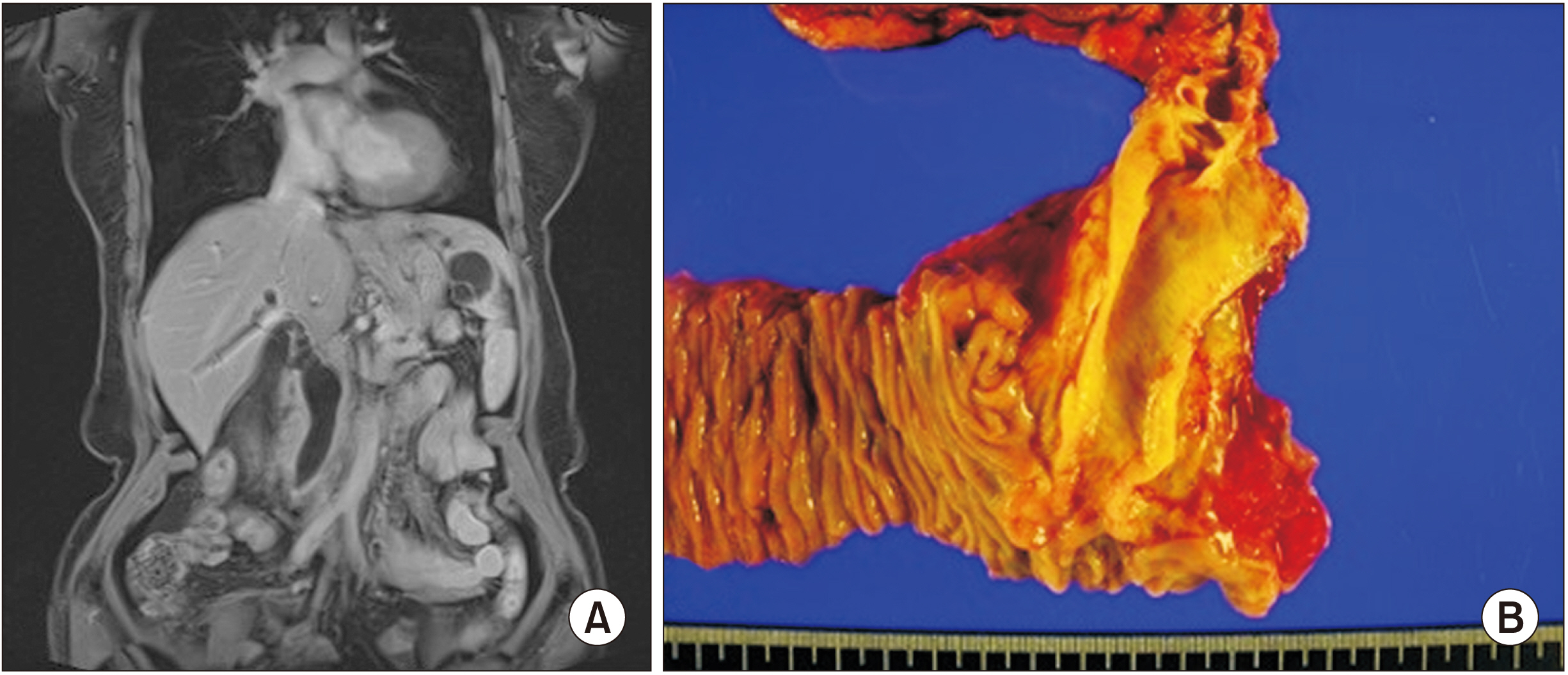
Fig. 4 (A) Case 3 magnetic resonance cholangiopancreatography (MRCP) findings of the neoplastic lesion showing bile duct dilatation, obstruction in the ampullary area of the stretched bile duct and main pancreatic duct dilatation, and a 1.5 cm long mass involving the ampulla of Vater without enlarged lymph nodes. (B) Case 3 surgical specimen of the resected ampullary tumor.
Reference
-
1. De Mestier L, Cros J, Neuzillet C, Hentic O, Egal A, Muller N, et al. 2017; Digestive system mixed neuroendocrine-non-neuroendocrine neoplasms. Neuroendocrinology. 105:412–425. DOI: 10.1159/000475527. PMID: 28803232.
Article2. Vilardell F, Velasco A, Cuevas D, Olsina JJ, Matias-Guiu X. 2011; Composite papillary intestinal-type adenocarcinoma/poorly differentiated neuroendocrine carcinoma of the ampulla of Vater. J Clin Pathol. 64:174–177. DOI: 10.1136/jcp.2010.084954. PMID: 21097790.
Article3. Yoshimachi S, Ohtsuka H, Aoki T, Miura T, Ariake K, Masuda K, et al. 2020; Mixed adenoneuroendocrine carcinoma of the ampulla of Vater: a case report and literature review. Clin J Gastroenterol. 13:37–45. DOI: 10.1007/s12328-019-01009-2. PMID: 31342462.
Article4. Jones MA, Griffith LM, West AB. 1989; Adenocarcinoid tumor of the periampullary region: a novel duodenal neoplasm presenting as biliary tract obstruction. Hum Pathol. 20:198–200. DOI: 10.1016/0046-8177(89)90187-1. PMID: 2914703.
Article5. Misonou J, Kanda M, Kitagawa T, Ota T, Muto E, Nenohi M, et al. 1990; A case of coexisting malignant carcinoid tumor and adenocarcinoma in the papilla of Vater. Gastroenterol Jpn. 25:630–635. DOI: 10.1007/BF02779365. PMID: 2227254.
Article6. Burke A, Lee YK. 1990; Adenocarcinoid (goblet cell carcinoid) of the duodenum presenting as gastric outlet obstruction. Hum Pathol. 21:238–239. DOI: 10.1016/0046-8177(90)90137-T. PMID: 2307453.
Article7. Shah IA, Schlageter MO, Boehm N. 1990; Composite carcinoid-adenocarcinoma of ampulla of Vater. Hum Pathol. 21:1188–1190. DOI: 10.1016/0046-8177(90)90158-2. PMID: 2227927.
Article8. Williams IM, Williams NW, Stock D, Foster ME. 1997; Collision tumour of the ampulla of Vater: carcinoid and adenocarcinoma. HPB Surg. 10:241–244. DOI: 10.1155/1997/46751. PMID: 9184878. PMCID: PMC2423872.
Article9. Alex WR, Auerbach HE, Pezzi CM. 1998; Adenocarcinoid tumor of the ampulla of Vater. Am Surg. 64:355–359.10. Moncur JT, Lacy BE, Longnecker DS. 2002; Mixed acinar-endocrine carcinoma arising in the ampulla of Vater. Hum Pathol. 33:449–451. DOI: 10.1053/hupa.2002.124040. PMID: 12055683.
Article11. Nassar H, Albores-Saavedra J, Klimstra DS. 2005; High-grade neuroendocrine carcinoma of the ampulla of vater: a clinicopathologic and immunohistochemical analysis of 14 cases. Am J Surg Pathol. 29:588–594. DOI: 10.1097/01.pas.0000157974.05397.4f. PMID: 15832081.12. Deschamps L, Dokmak S, Guedj N, Ruszniewski P, Sauvanet A, Couvelard A. 2010; Mixed endocrine somatostatinoma of the ampulla of Vater associated with a neurofibromatosis type 1: a case report and review of the literature. JOP. 11:64–68.13. Zhang L, DeMay RM. 2014; Cytological features of mixed adenoneuroendocrine carcinoma of the ampulla: two case reports with review of literature. Diagn Cytopathol. 42:1075–1084. DOI: 10.1002/dc.23107. PMID: 24554593.
Article14. Huang Z, Xiao WD, Li Y, Huang S, Cai J, Ao J. 2015; Mixed adenoneuroendocrine carcinoma of the ampulla: two case reports. World J Gastroenterol. 21:2254–2259. DOI: 10.3748/wjg.v21.i7.2254. PMID: 25717267. PMCID: PMC4326169.
Article15. Ginori A, Lo Bello G, Vassallo L, Tripodi SA. 2015; Amphicrine carcinoma of the ampullary region. Tumori. 101:e70–e72. DOI: 10.5301/tj.5000254. PMID: 25702653.
Article16. Li X, Li D, Sun X, Lv G. 2020; Mixed adenoneuroendocrine carcinoma (MANEC) of the ampulla of Vater in a Chinese patient: a case report. J Int Med Res. 48:300060520947918. DOI: 10.1177/0300060520947918. PMID: 32833541. PMCID: PMC7448144.
Article17. Doepker MP, Thompson ZJ, Centeno BA, Kim RD, Wong J, Hodul PJ. 2016; Clinicopathologic and survival analysis of resected ampullary adenocarcinoma. J Surg Oncol. 114:170–175. DOI: 10.1002/jso.24281. PMID: 27158031. PMCID: PMC7771532.
Article18. La Rosa S, Marando A, Sessa F, Capella C. 2012; Mixed adenoneuroendocrine carcinomas (MANECs) of the gastrointestinal tract: an update. Cancers (Basel). 4:11–30. DOI: 10.3390/cancers4010011. PMID: 24213223. PMCID: PMC3712682.
Article19. Acosta AM, Wiley EL. 2016; Primary biliary mixed adenoneuroendocrine carcinoma (MANEC): a short review. Arch Pathol Lab Med. 140:1157–1162. DOI: 10.5858/arpa.2015-0102-RS. PMID: 27684986.
Article20. Hatano H, Yoneyama C, Noda T, Tomimaru Y, Hirota M, Takata A, et al. 2015; [A case of surgical treatment for mixed adenoneuroendocrine carcinoma of the ampulla of Vater]. Gan To Kagaku Ryoho. 42:1755–1757. Japanese.
- Full Text Links
-
- Actions
-
Cited
- CITED
-
- Close
- Share
-
- Similar articles
-
- A Case of Early Carcinoma of the Ampulla of Vater Combined with Adenoma of Colon
- A Case of a Collision Tumor in the Ampulla of Vater with an Adenocarcinoma and a Large Cell Neuroendocrine Carcinoma
- Collision tumor of the ampulla of Vater - Coexistence of neuroendocrine carcinoma and adenocarcinoma: report of a case
- Concurrent occurrence of adenocarcinoma and neuroendocrine type small cell carcinoma in the ampulla of Vater
- A case of adenofibromatous hyperplasia of the ampulla of vater hardly differenciated from carcinoma
