Ann Rehabil Med.
2022 Dec;46(6):284-291. 10.5535/arm.22123.
Ultrasonographic Assessment of the Safe Zone for Carpal Tunnel Intervention: A Comparison Between Healthy Individuals and Patients With Carpal Tunnel Syndrome
- Affiliations
-
- 1Department of Physical Medicine and Rehabilitation, Korea University Guro Hospital, Seoul, Korea
- 2Goodbone Physical Medicine and Rehabilitation Clinic, Seoul, Korea
- 3Department of Plastic and Reconstructive Surgery, Korea University Guro Hospital, Seoul, Korea
- KMID: 2537159
- DOI: http://doi.org/10.5535/arm.22123
Abstract
Objective
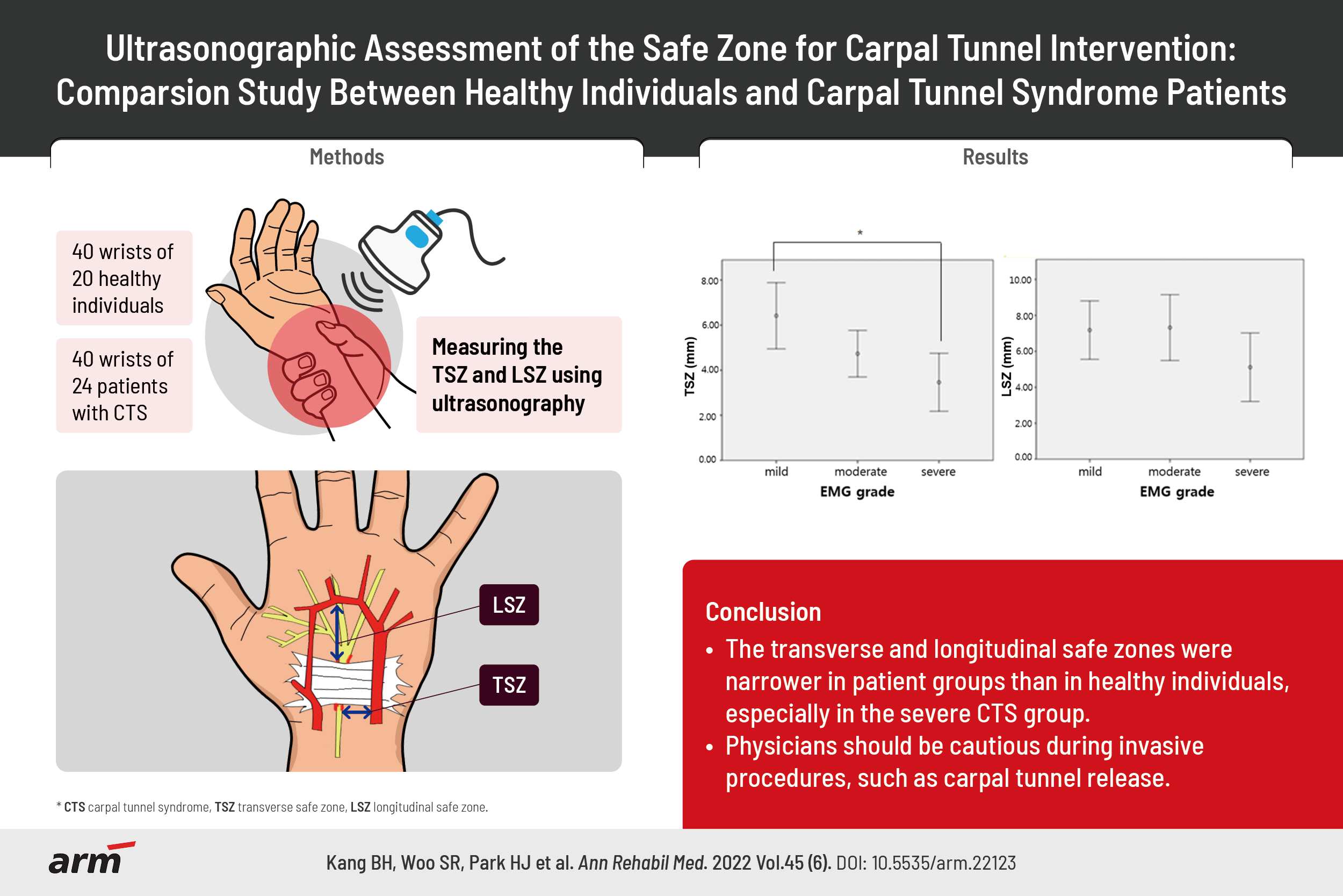
To compare transverse and longitudinal safe zones using ultrasonography between healthy individuals and patients with carpal tunnel syndrome (CTS).
Methods
This was a prospective observational case-control study. Forty wrists from 20 healthy individuals and 40 wrists from 24 patients with CTS were examined. Patients with CTS were classified into three groups (mild, moderate, and severe CTS) based on electrodiagnostic findings. Using ultrasonography, we measured the distance between the median nerve and ulnar vessels to identify the transverse safe zone, and between the distal flexor retinaculum and superficial palmar artery arch to identify the longitudinal safe zone.
Results
The transverse and longitudinal safe zones were significantly different between participants with CTS and those without CTS. The transverse safe zone significantly differed between the mild and severe CTS groups, while the longitudinal safe zone was not significantly different between the groups. The cross-sectional area of the median nerve negatively correlated with the transverse and longitudinal safe zones.
Conclusion
Transverse and longitudinal safe zones were narrower in patients with CTS than in the healthy group. A significant difference was observed between patients with mild CTS and those with severe CTS. Furthermore, the cross-sectional area of the median nerve was directly proportional to the degree of narrowing of the transverse and longitudinal safe zones.
Figure
-
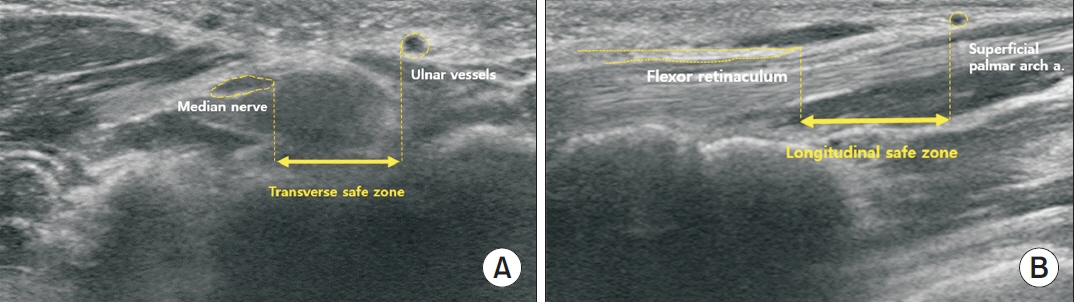
Fig. 1. Ultrasonographic measurement of (A) the transverse safe zone as the distance between the medial margin of the median nerve and lateral margin of the ulnar vessels; (B) the longitudinal safe zone as the distance between the distal end of the flexor retinaculum and proximal end of the superficial palmar arch artery.
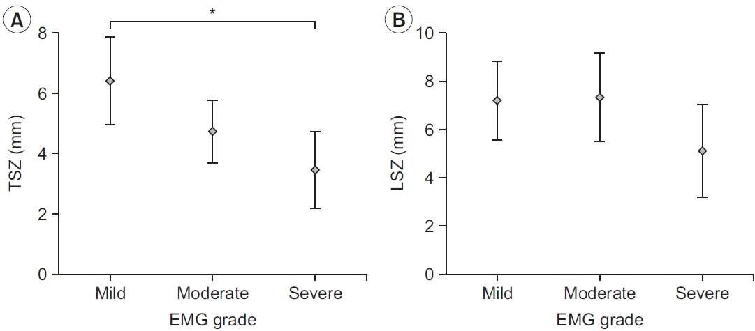
Fig. 2. Comparison of safe zones based on the severity of electromyographic (EMG) findings: (A) transverse safe zone (TSZ) and (B) longitudinal safe zone (LSZ). *p<0.05 using the Bonferroni test.
Reference
-
1. Demircay E, Civelek E, Cansever T, Kabatas S, Yilmaz C. Anatomic variations of the median nerve in the carpal tunnel: a brief review of the literature. Turk Neurosurg. 2011; 21:388–96.2. Gelberman RH, Hergenroeder PT, Hargens AR, Lundborg GN, Akeson WH. The carpal tunnel syndrome: a study of carpal canal pressures. J Bone Joint Surg Am. 1981; 63:380–3.3. Atroshi I, Gummesson C, Johnsson R, Ornstein E, Ranstam J, Rosen I. Prevalence of carpal tunnel syndrome in a general population. JAMA. 1999; 282:153–8.4. Skie M, Zeiss J, Ebraheim NA, Jackson WT. Carpal tunnel changes and median nerve compression during wrist flexion and extension seen by magnetic resonance imaging. J Hand Surg Am. 1990; 15:934–9.5. Uchiyama S, Itsubo T, Nakamura K, Kato H, Yasutomi T, Momose T. Current concepts of carpal tunnel syndrome: pathophysiology, treatment, and evaluation. J Orthop Sci. 2010; 15:1–13.6. Boya H, Ozcan O, Ozteki N HH. Long-term complications of open carpal tunnel release. Muscle Nerve. 2008; 38:1443–6.7. Kluge W, Simpson RG, Nicol AC. Late complications after open carpal tunnel decompression. J Hand Surg Br. 1996; 21:205–7.8. Hui AC, Wong S, Leung CH, Tong P, Mok V, Poon D, et al. A randomized controlled trial of surgery vs steroid injection for carpal tunnel syndrome. Neurology. 2005; 64:2074–8.9. Marshall S, Tardif G, Ashworth N. Local corticosteroid injection for carpal tunnel syndrome. Cochrane Database Syst Rev. 2007; (2):CD001554.10. Chern TC, Wu KC, Huang LW, Shao CJ, Wu TT, Kuo LC, et al. A cadaveric and preliminary clinical study of ultrasonographically assisted percutaneous carpal tunnel release. Ultrasound Med Biol. 2014; 40:1819–26.11. McShane JM, Slaff S, Gold JE, Nazarian LN. Sonographically guided percutaneous needle release of the carpal tunnel for treatment of carpal tunnel syndrome: preliminary report. J Ultrasound Med. 2012; 31:1341–9.12. Rojo-Manaute JM, Capa-Grasa A, Chana-Rodriguez F, Perez-Mananes R, Rodriguez-Maruri G, Sanz-Ruiz P, et al. Ultra-minimally invasive ultrasound-guided carpal tunnel release: a randomized clinical trial. J Ultrasound Med. 2016; 35:1149–57.13. Nakamichi K, Tachibana S. Distance between the median nerve and ulnar neurovascular bundle: clinical significance with ultrasonographically assisted carpal tunnel release. J Hand Surg Am. 1998; 23:870–4.14. Buncke G, McCormack B, Bodor M. Ultrasound-guided carpal tunnel release using the manos CTR system. Microsurgery. 2013; 33:362–6.15. Stevens JC. AAEM Minimonograph #26: the electrodiagnosis of carpal tunnel syndrome. Muscle Nerve. 1997; 20:1477–86.16. Lee HJ, Kwon HK. Electrophysiologic classification of severity of carpal tunnel syndrome. J Korean Assoc EMG-Electrodiagn Med. 2004; 6:1–3.17. Ajayi NO, Naidoo N, Lazarus L, Satyapal KS. Determination of the median nerve safe-zone in the carpal tunnel using the distal forearm bony prominences. Folia Morphol (Warsz). 2014; 73:409–13.18. Omokawa S, Tanaka Y, Ryu J, Suzuki J, Kish VL. Anatomy of the ulnar artery as it relates to the transverse carpal ligament. J Hand Surg Am. 2002; 27:101–4.19. Rotman MB, Manske PR. Anatomic relationships of an endoscopic carpal tunnel device to surrounding structures. J Hand Surg Am. 1993; 18:442–50.20. Olave E, Del Sol M, Gabriellp C, Mandiola E, Rodrigues CF. Biometric study of the relationships between palmar neurovas cular str uctures, the flexor retinaculum and the distal wrist crease. J Anat. 2001; 198(Pt 6):737–41.21. Bayrak IK, Bayrak AO, Tilki HE, Nural MS, Sunter T. Ultrasonography in carpal tunnel syndrome: comparison with electrophysiological stage and motor unit number estimate. Muscle Nerve. 2007; 35:344–8.22. Lee CH, Kim TK, Yoon ES, Dhong ES. Correlation of high-resolution ultrasonographic findings with the clinical symptoms and electrodiagnostic data in carpal tunnel syndrome. Ann Plast Surg. 2005; 54:20–3.23. Padua L, Pazzaglia C, Caliandro P, Granata G, Foschini M, Briani C, et al. Carpal tunnel syndrome: ultrasound, neurophysiology, clinical and patient-oriented assessment. Clin Neurophysiol. 2008; 119:2064–9.24. Ustun N, Tok F, Yagz AE, Kizil N, Korkmaz I, Karazincir S, et al. Ultrasound-guided vs. blind steroid injections in carpal tunnel syndrome: a single-blind randomized prospective study. Am J Phys Med Rehabil. 2013; 92:999–1004.25. Kim PT, Lee HJ, Kim TG, Jeon IH. Current approaches for carpal tunnel syndrome. Clin Orthop Surg. 2014; 6:253–7.
- Full Text Links
-
- Actions
-
Cited
- CITED
-
- Close
- Share
-
- Similar articles
-
- Ultrasound-Guided Nerve Hydrodissection for Carpal Tunnel Syndrome
- Analysis of Sonographic Measurement by Anatomical Area in Carpal Tunnel Syndrome and Correlation the Measurement with Electrodiagnostic Study
- Carpal Tunnel Syndrome in Children with Hypogammaglobulinemia: Case Report
- The Current Concepts for the Pathophysiology of Idiopathic Carpal Tunnel Syndrome
- The Carpal compression Test for Diagnosing Carpal Tunnel Syndrome
