Analysis of Sonographic Measurement by Anatomical Area in Carpal Tunnel Syndrome and Correlation the Measurement with Electrodiagnostic Study
- Affiliations
-
- 1Department of Orthopedic Surgery, Saeson Hospital, Daejeon, Korea
- KMID: 2501102
- DOI: http://doi.org/10.12790/ahm.19.0062
Abstract
- Purpose
To evaluate effectiveness of ultrasonographic measurement of carpal tunnel by anatomical area and correlation with electrodiagnostic study in diagnosis of carpal tunnel syndrome.
Methods
From September 2018 to March 2019, we performed the ultrasonography for 30 cases with carpal tunnel syndrome diagnosed with electrodiagnosis and 30 cases as control group. We measured median nerve diameter, cross-sectional area (CSA), and flattening ratio (FR) by area of carpal tunnel. We analyzed the difference of measurement between two groups and correlate the measurement with electrodiagnosis findings.
Results
There was significant statistically differences in sonographic measurement between two groups by independent t-test (CSA zone 1, p=0.01; FR zone 2, p=0.000; FR zone 3; p=0.001). With Pearson correlation test, there was correlation between sonographic measurement and electrodiagnostic findings (terminal latency and nerve conduction velocity) statistically, but the Pearson coefficient was low (r<0.4).
Conclusion
By anatomical area, the available value of sonographic measurement was different. But, as the values were has low power to diagnose the carpal tunnel syndrome, ultrasonography is proper to use as a complementary tool in diagnosis of carpal tunnel syndrome.
Figure
-
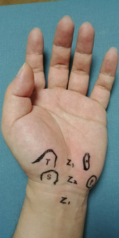
Fig. 1. Demonstration of zone of carpal tunnel. Zone 1 is proximal area to true carpal tunnel. Zone 2 is proximal carpal tunnel between scaphoid distal pole (S) and pisiform (P). Zone 3 is distal carpal tunnel between hamate (H) and trapezium (T).
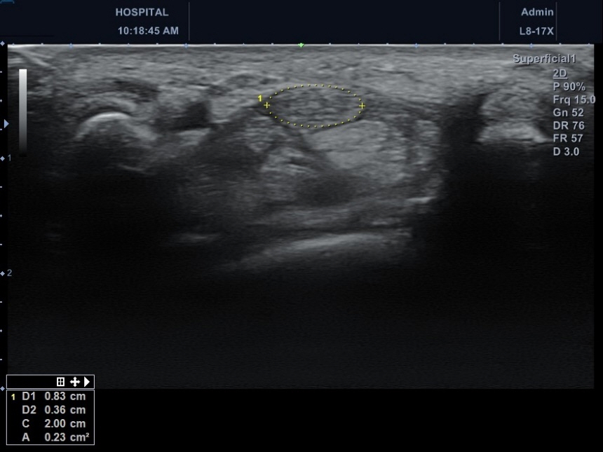
Fig. 2. The measurement of median nerve. D1 and D2 are horizontal and vertical diameter of median nerve. C is circumference and A is cross-sectional area.

Fig. 3. There was significant statistical difference between two groups in cross-sectional area of Zone 1 (A), flattening ratio of Zone 2 (B) and Zone 3 (C).
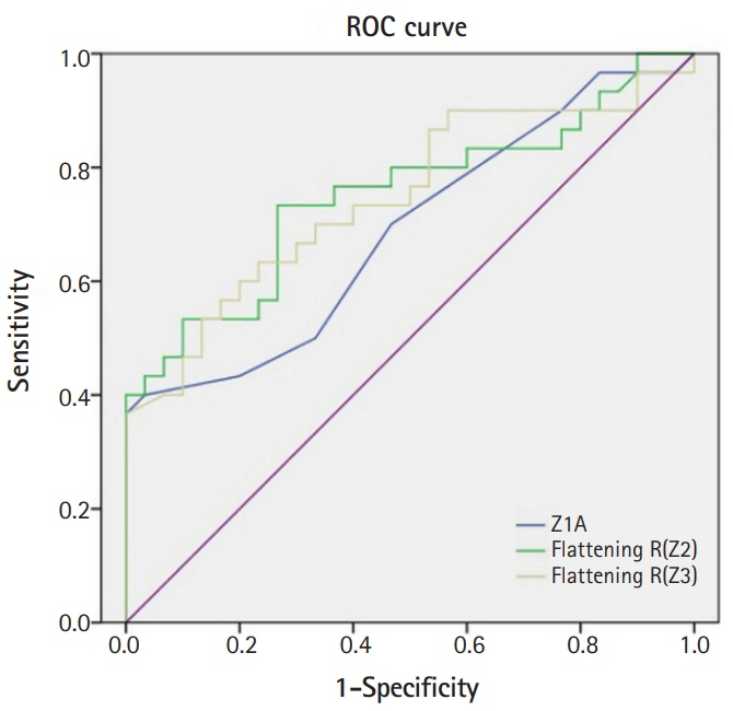
Fig. 4. Receiver operating characteristic (ROC) curve of cross-sectional area of Zone 1 (Z1A) and flattening ratio of Zone 2, Zone 3 (Flattening R(Z2), Flattening R(Z3)). Each area under the curve (AUC) was 0.69, 0.75, and 0.75.
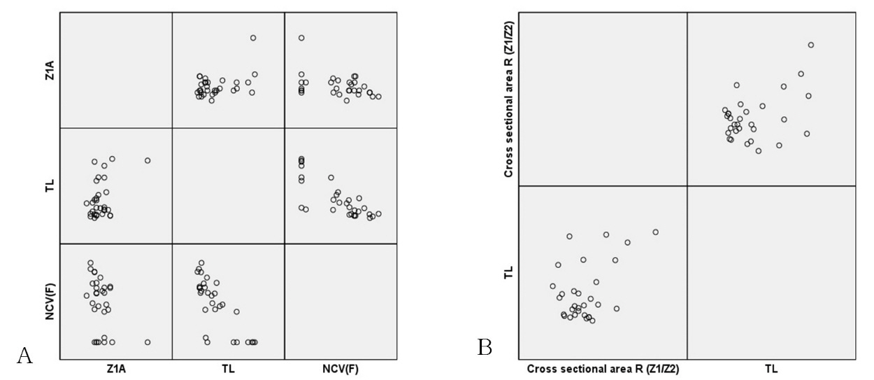
Fig. 5. There was statistical difference between ultrasonography measurement and electrodiagnostic study. But they had low coefficient of correlation and ultrasonography measurement could not represent electrodiagnostic severity. (A) Pearson correlation test between CSA of Zone 1 and EDS (terminal latency [TL], nerve conduction velocity [NCV]). (B) Pearson correlation test between cross-sectional area ratio and TL.
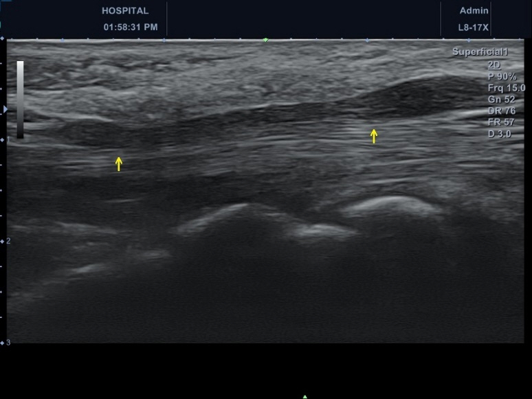
Fig. 6. Swelling of median nerve proximal to the carpal tunnel (arrow demonstrate the compression of median nerve and the right side of figure is proximal area). So-called by "notching sign". Generally, the median nerve in carpal tunnel syndrome is thickened, especially in proximal entrance of carpal tunnel.
Reference
-
References
1. Werner RA, Andary M. Carpal tunnel syndrome: pathophysiology and clinical neurophysiology. Clin Neurophysiol. 2002; 113:1373–81.
Article2. Phalen GS. The carpal-tunnel syndrome. Seventeen years' experience in diagnosis and treatment of six hundred fifty-four hands. J Bone Joint Surg Am. 1966; 48:211–28.3. Buchberger W, Judmaier W, Birbamer G, Lener M, Schmidauer C. Carpal tunnel syndrome: diagnosis with high-resolution sonography. AJR Am J Roentgenol. 1992; 159:793–8.
Article4. Wong SM, Griffith JF, Hui AC, Lo SK, Fu M, Wong KS. Carpal tunnel syndrome: diagnostic usefulness of sonography. Radiology. 2004; 232:93–9.
Article5. Ziswiler HR, Reichenbach S, Vogelin E, Bachmann LM, Villiger PM, Juni P. Diagnostic value of sonography in patients with suspected carpal tunnel syndrome: a prospective study. Arthritis Rheum. 2005; 52:304–11.
Article6. Mhoon JT, Juel VC, Hobson-Webb LD. Median nerve ultrasound as a screening tool in carpal tunnel syndrome: correlation of cross-sectional area measures with electrodiagnostic abnormality. Muscle Nerve. 2012; 46:871–8.
Article7. Billakota S, Hobson-Webb LD. Standard median nerve ultrasound in carpal tunnel syndrome: A retrospective review of 1,021 cases. Clin Neurophysiol Pract. 2017; 2:188–91.
Article8. Kang S, Kwon HK, Kim KH, Yun HS. Ultrasonography of median nerve and electrophysiologic severity in carpal tunnel syndrome. Ann Rehabil Med. 2012; 36:72–9.
Article9. Bianchi S, Martinoli C. Ultrasound of the musculoskeletal system. Heidelberg, Germany: Springer;2007.
Article10. Oh SJ. Clinical electromyography: nerve conduction studies. 3rd ed. Philadelphia, PA: Lippincott Williams & Wilkins;2003.11. Stevens JC. AAEM minimonograph# 26: the electrodiagnosis of carpal tunnel syndrome. Muscle Nerve. 1997; 20:1477–86.
Article12. Bland JD. A neurophysiological grading scale for carpal tunnel syndrome. Muscle Nerve. 2000; 23:1280–3.
Article13. Chen P, Maklad N, Redwine M, Zelitt D. Dynamic high-resolution sonography of the carpal tunnel. AJR Am J Roentgenol. 1997; 168:533–7.
Article14. Watanabe T, Ito H, Morita A, et al. Sonographic evaluation of the median nerve in diabetic patients: comparison with nerve conduction studies. J Ultrasound Med. 2009; 28:727–34.15. Duncan I, Sullivan P, Lomas F. Sonography in the diagnosis of carpal tunnel syndrome. AJR Am J Roentgenol. 1999; 173:681–4.
Article16. do Amaral e Castro A, Skare TL, Sakuma AK, Barros WH. Ultrasonography as a tool in diagnosis of carpal tunnel syndrome]. Rev Bras Reumatol. 2015; 55:330–3.
Article17. Wiesler ER, Chloros GD, Cartwright MS, Smith BP, Rushing J, Walker FO. The use of diagnostic ultrasound in carpal tunnel syndrome. J Hand Surg Am. 2006; 31:726–32.
Article18. Pastare D, Therimadasamy AK, Lee E, Wilder-Smith EP. Sonography versus nerve conduction studies in patients referred with a clinical diagnosis of carpal tunnel syndrome. J Clin Ultrasound. 2009; 37:389–93.
Article19. El Miedany YM, Aty SA, Ashour S. Ultrasonography versus nerve conduction study in patients with carpal tunnel syndrome: substantive or complementary tests? Rheumatology (Oxford). 2004; 43:887–95.
Article20. Fowler JR, Gaughan JP, Ilyas AM. The sensitivity and specificity of ultrasound for the diagnosis of carpal tunnel syndrome: a meta-analysis. Clin Orthop Relat Res. 2011; 469:1089–94.
Article
- Full Text Links
-
- Actions
-
Cited
- CITED
-
- Close
- Share
-
- Similar articles
-
- The Correlation Between Electrodiagnostic Results and Ultrasonographic Findings in the Severity of Carpal Tunnel Syndrome in Females
- Pressure Measurement in Carpal Tunnel Syndrome : Correlation with Electrodiagnostic and Ultrasonographic Findings
- Usefulness of Ultrasound for Carpal Tunnel Syndrome Proven in Meta-Analysis Studies
- Carpal Tunnel Configuration Measured by Ultrasonography as a Risk Factor of Carpal Tunnel Syndrome in Motor Part Manufacturing Workers
- Ulnar Neuropathy at the Wrist in a Patient with Carpal Tunnel Syndrome after Open Carpal Tunnel Release
