Abnormal Development of Neural Stem Cell Niche in the Dentate Gyrus of Menkes Disease
- Affiliations
-
- 1Cell Therapy Research Center, GC Cell, Yongin, Korea
- 2Department of Neurology, Dongguk University Ilsan Hospital, Goyang, Korea
- 3Department of Pediatrics, Seoul National University Children’s Hospital, Seoul National University College of Medicine, Seoul, Korea
- KMID: 2532401
- DOI: http://doi.org/10.15283/ijsc21088
Abstract
- Background and Objectives
Menkes disease (MNK) is a rare X-linked recessive disease, caused by mutations in the copper transporting ATP7A gene that is required for copper homeostasis. MNK patients experience various clinical symptoms including neurological defects that are closely related to the prognosis of MNK patients. Neural stem cells (NSCs) in the hippocampal dentate gyrus (DG) produce new neurons throughout life, and defects in DG neurogenesis are often correlated with cognitive and behavioral problems. However, neurodevelopmental defects in the DG during postnatal period in MNK have not been understood yet.
Methods and Results
Mottled-brindled (Mo Br/y ) mice (MNK mice) and littermate controls were used in this study. In vivo microCT imaging and immunohistochemistry results demonstrate that blood vasculatures in hippocampus are abnormally decreased in MNK mice. Furthermore, postnatal establishment of NSC population and their neurogenesis are severely compromised in the DG of MNK mice. In addition, in vitro analyses using hippocampal neurosphere culture followed by immunocytochemistry and immunoblotting suggest that neurogenesis from MNK NSCs is also significantly compromised, corresponding to defective neurogenic gene expression in MNK derived neurons.
Conclusions
Our study is the first reports demonstrating that improper expansion of the postnatal NSC population followed by significant reduction of neurogenesis may contribute to neurodevelopmental symptoms in MNK. In conclusion, our results provide new insight into early neurodevelopmental defects in MNK and emphasize the needs for early diagnosis and new therapeutic strategies in the postnatal central nerve system damage of MNK patients.
Figure
-
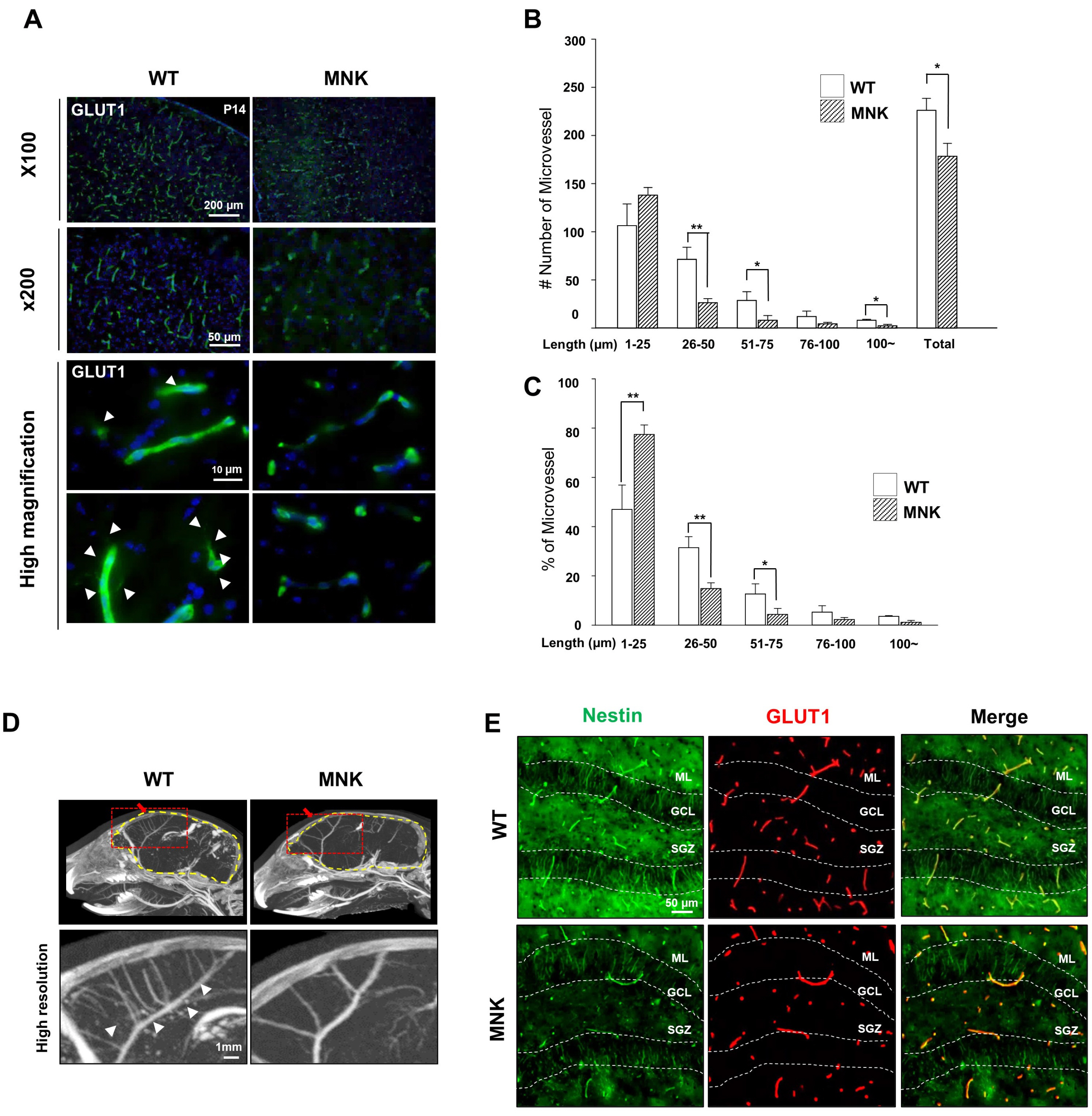
Fig. 1 Abnormal blood vessel development in P14 cerebral cortex and hippocampal dentate gyrus of MNK mice. (A) GLUT1 (marker of brain microvessel) was used to analyze brain vessel pattern in cerebral cortex of wild type and MNK mice. Expression of GLUT1 was decreased in P14 cerebral cortex of MNK mice compared with that of wild type. Scale bar 100 μm (upper), 50 μm (lower). Arrowheads in high magnification image show that sprouting formation in neovascular endothelial tip was reduced in P14 cerebral cortex of MNK mouse. DAPI was used to counterstain nuclei (Blue). (B, C) Quantitative data of blood vessels length show that number and proportion of blood vessels less than 25 μm length was increased in the P13 MNK cerebral cortex. *p<0.05, **p<0.01 by t-test. (D) Reduction of brain vessels revealed by microCT scan indicates the defect of cerebral vascular structure in the P14 of MNK mice. The red box indicates regions shown in below. (E) P13 hippocampal dentate gyrus of MNK mice were stained with neural stem cell marker NESTIN and cerebral vessel marker GLUT1. Decreased NESTIN+ radial processes and blood vessels in the P13 hippocampal dentate gyrus of MNK mice were observed compared to wild type littermate. Scale bar 50 μm. SGZ, subgranular zone; GCL, granule cell layer; ML, molecular layer.

Fig. 2 Neural stem cells and neural progenitors are severely compromised in the dentate gyrus of MNK mice. (A) NESTIN and SOX2 immunostainings were used to identify neural stem cells in the brain of P13 wild type and MNK mice. Neural stem cells in the dentate gyrus were significantly reduced in the brain of P13 MNK mice compared with wild type. White arrowheads of high magnification image indicate neural stem cells stained by NESTIN or SOX2 respectively. Number of NESTIN+, SOX2+ NSCs per 100 μm of SGZ were significantly reduced in the MNK mice compared to control. Scale bar 50 μm and 10 μm (high magnification). (B) Coronal sections of P13 mouse brains were stained with neural progenitor cell marker doublecortin (DCX, Green) and Prospero homeobox protein 1 (PROX1, Red). DCX+ and PROX1+ neural progenitor cells were reduced respectively in GCL. Furthermore, number of DCX+, PROX1+ cells were also significantly reduced by 63% per 100 μm of the GCL and SGZ of MNK mice compared to control. **p<0.001, n=4 each, by t-test. (C) Coronal sections of P13 mouse brains were stained with neuronal marker NeuN and mature neuron maker Calbindin. Number of NeuN+, Calbindin+ mature neurons were significantly decreased by 55% in MNK mice compared with wild type per 100 μm of the upper GCL region of the dentate gyrus. The White box indicates regions shown in high magnification. Scale bar 50 μm. SGZ, subgranular zone; GCL, granule cell layer; ML, molecular layer. *p<0.05, n=4 each, by t-test. **p<0.001, n=4 each, by t-test.
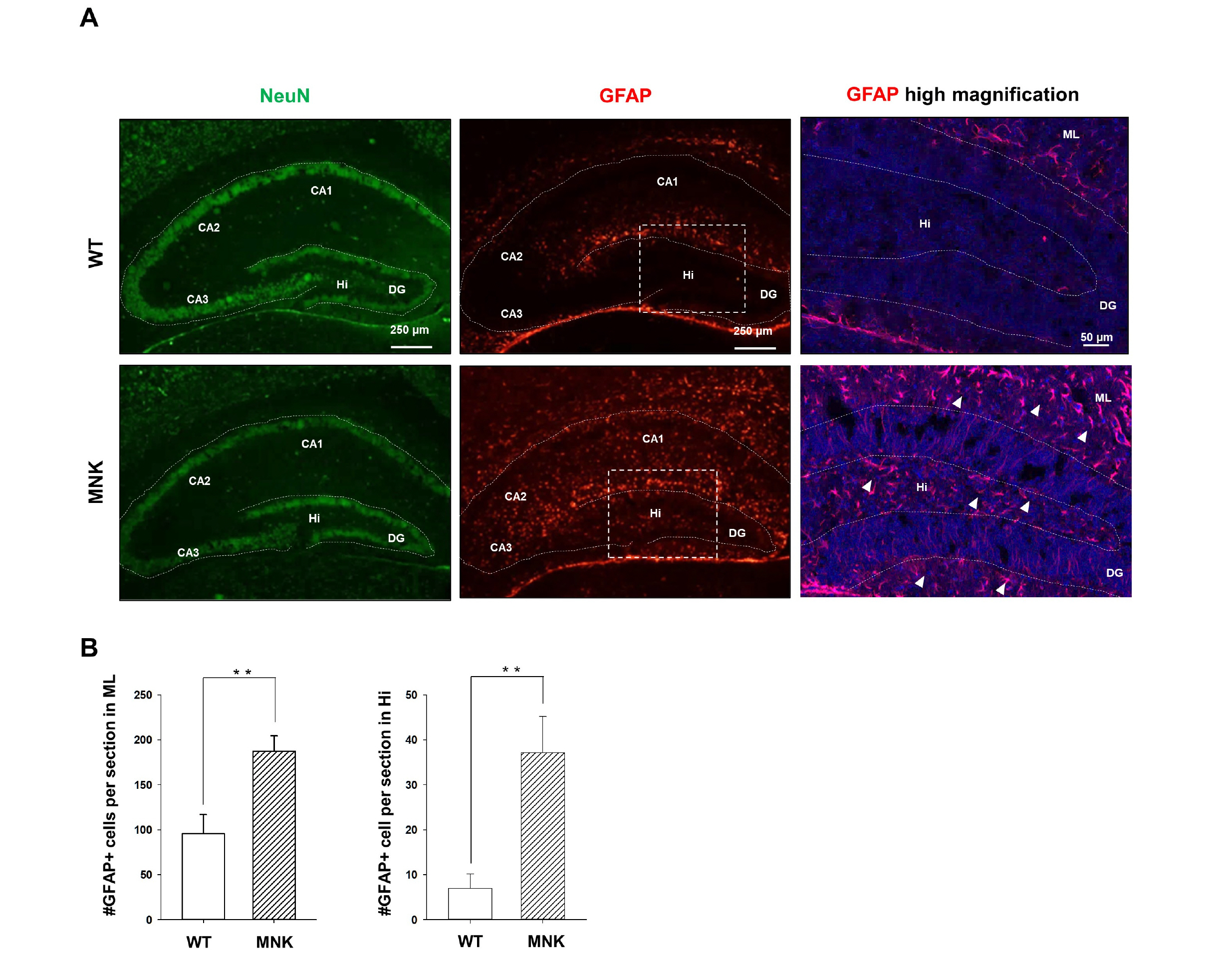
Fig. 3 Astrogenesis is promoted in the hippocampus of MNK mice. (A) Hippocampal sections were immunostained with neuronal marker NeuN and astrocyte marker GFAP at P13. The white box indicates regions shown in high magnification. DAPI was used to counterstain nuclei (Blue). (B) GFAP+ (Red, arrowhead) astrocytes in the highly magnified image were dramatically increased in the hillus and molecular layer of P13 of MNK mice compared with wild type. Scale bar 50 μm and 250 μm (high magnification). CA, Cornu ammonis; Hi, Hilus; DG, dentate gyrus; ML, molecular layer. **p<0.001, n=4 each, by t-test.

Fig. 4 Postnatal development of neural stem cell population in the dentate gyrus of MNK mice. (A) Dentate gyrus sections from MNK and wild type mice were stained with NESTIN and SOX2 during postnatal brain development (P3∼P10). The white box indicates regions shown below (high magnification). Arrowheads indicate NESTIN+, SOX2+ neural stem cells. (B) Quantitative analysis of NESTIN+, SOX2+ cells during postnatal development of the DG. At P3, distribution of NESTIN+, SOX2+ cells in the dentate gyrus of MNK mice were not different from that of wild type. However, at P7 and P10, the number of NESTIN+, SOX2+ neural stem cells was significantly decreased in the SGZ of MNK mice. Scale bar 50 μm and 20 μm (high magnification). SGZ, subgranular zone; GCL, granule cell layer; ML, molecular layer. **p<0.001, n=4 each, by t-test.
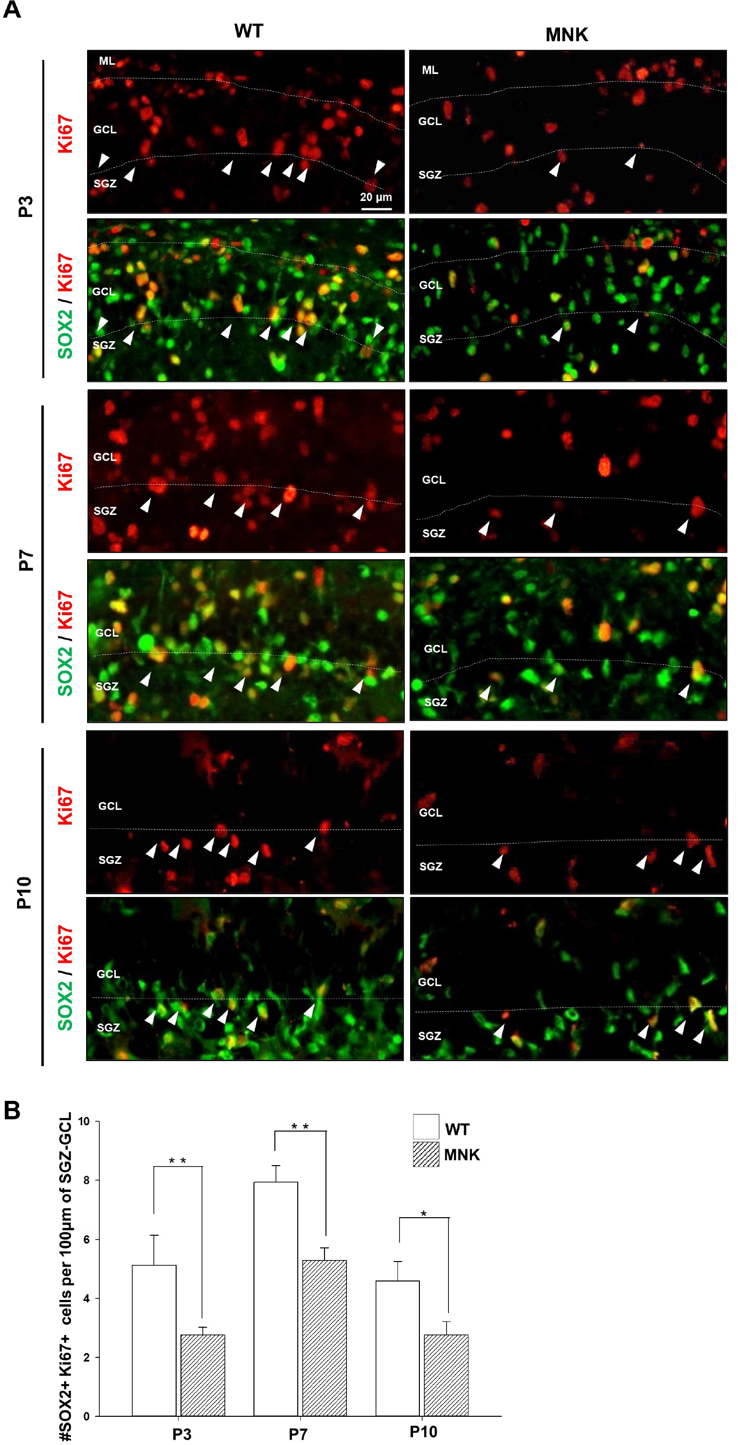
Fig. 5 Decrease of proliferating neural progenitor cells during DG development in MNK mice. (A) Dentate gyrus sections from MNK and wild type mice were stained with SOX2 and proliferation marker Ki67 during postnatal brain development (P3∼P10). White arrowheads indicate SOX2+, Ki67+ proliferating neural progenitor cells. (B) Quantitative analysis of SOX2+, Ki67+ cells during postnatal development of the DG. SOX2+, Ki67+ proliferating neural progenitor cells in SGZ were significantly reduced in the MNK mice. Scale bar 20 μm. SGZ, subgranular zone; GCL, granule cell layer; ML, molecular layer. *p< 0.05, **p<0.001, n=4 each, by t-test.
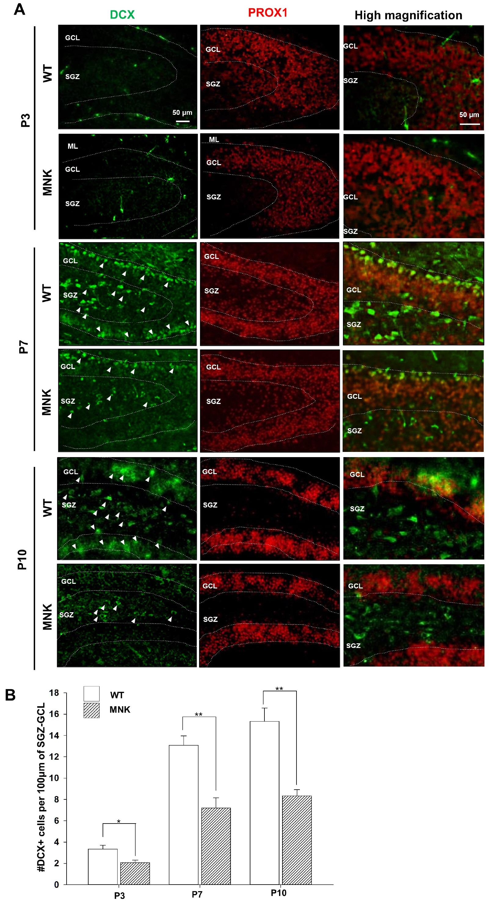
Fig. 6 Abnormal postnatal neurogenesis in the dentate gyrus of MNK mice. (A) Dentate gyrus sections from MNK and wild type mice were stained with neuroblast marker DCX and PROX1 during postnatal brain development (P3∼P10). The number of DCX+ neuroblasts in the dentate gyrus of MNK mice was not different from that of wild type at P3. (B) Quantitative analysis of DCX+, PROX1+ cells during the postnatal development of the DG. More than 40% of DCX+ neuroblasts (arrowheads) were decreased at P7 and P10 in the DG of MNK mice. Scale bar 50 μm. SGZ, subgranular zone; GCL, granule cell layer; ML, molecular layer. *p<0.05, **p<0.001, n=4 each, by t-test.
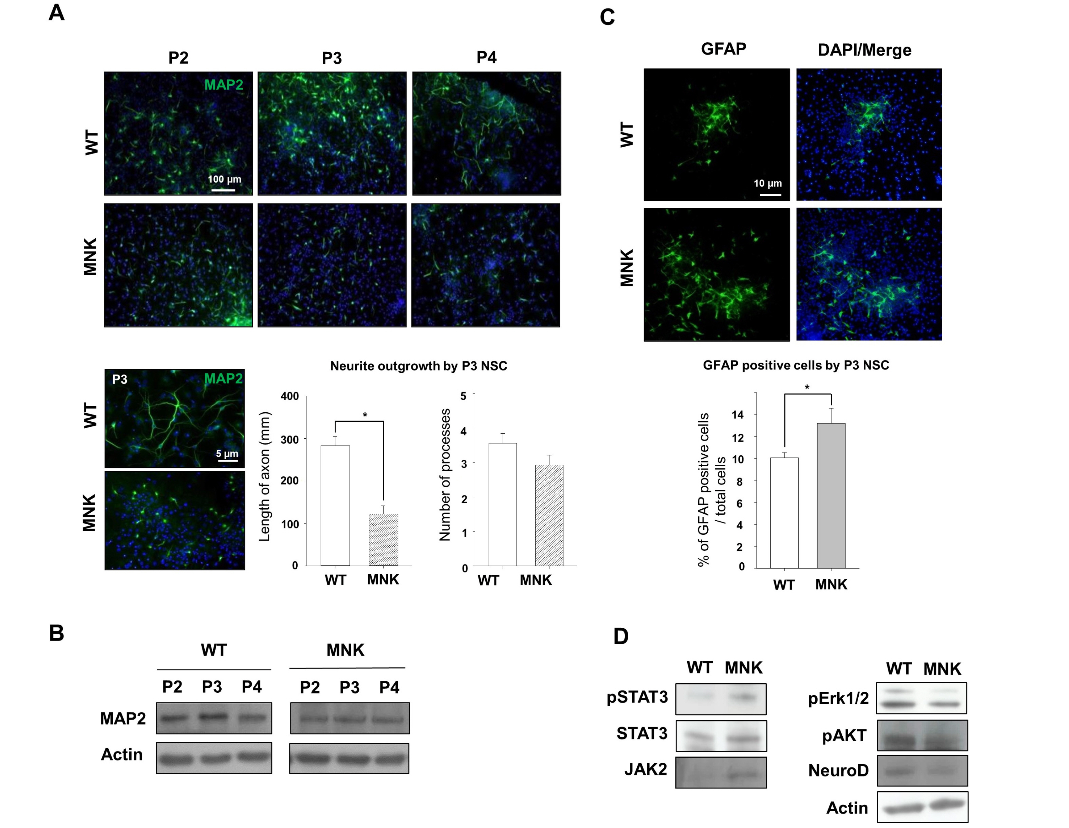
Fig. 7 Neurogenesis and neuronal maturation are severely compromised in MNK derived NSCs. Hippocampal neurosphere cultures from the brain of E18.5 wild type and MNK mice were established and induced neuronal differentiation for 4 days. (A) Neuronal processes marker MAP2 and DAPI (blue) were stained after neuronal differentiation. Neurite outgrowth of neurons derived from the hippocampal NSCs of MNK mice was significantly decreased through 4 passages in differentiation condition. Scale bar 100 μm (upper), scale bar 5 μm (lower). Metamorph analysis indicated more than 50% shorter neurites in the MNK derived neurons were observed relative to that of wild type. Scale bar 5 μm. *p<0.05 by t-test. (B) MAP2 expression was reduced in the MNK derived neurons compared with that of wild type through 4 passages. (C) Astrocyte marker GFAP+ cells were significantly increased in the MNK derived NSCs during neuronal differentiation. Scale bar 10 μm. *p<0.05 by t-test. (D) Western blot analysis was performed for neurogenesis makers (anti-NeuroD, anti-pAKT, and anti-pErk1/2) and astrogenesis makers (anti-pSTAT3 and anti-JAK2).
Reference
-
References
1. Vulpe C, Levinson B, Whitney S, Packman S, Gitschier J. 1993; Isolation of a candidate gene for Menkes disease and evidence that it encodes a copper-transporting ATPase. Nat Genet. 7–13. Erratum in: Nat Genet 1993;3:273. DOI: 10.1038/ng0193-7. PMID: 8490659. PMID: https://www.scopus.com/inward/record.uri?partnerID=HzOxMe3b&scp=0027446365&origin=inward.2. Mercer JF, Livingston J, Hall B, Paynter JA, Begy C, Chandrasekharappa S, Lockhart P, Grimes A, Bhave M, Siemieniak D, Glover TW. 1993; Isolation of a partial candidate gene for Menkes disease by positional cloning. Nat Genet. 3:20–25. DOI: 10.1038/ng0193-20. PMID: 8490647. PMID: https://www.scopus.com/inward/record.uri?partnerID=HzOxMe3b&scp=0027475976&origin=inward.3. Menkes JH, Alter M, Steigleder GK, Weakley DR, Sung JH. 1962; A sex-linked recessive disorder with retardation of growth, peculiar hair, and focal cerebral and cerebellar degeneration. Pediatrics. 29:764–779. PMID: 14472668. PMID: https://www.scopus.com/inward/record.uri?partnerID=HzOxMe3b&scp=78651124591&origin=inward.4. Kaler SG. 2011; ATP7A-related copper transport diseases-emerging concepts and future trends. Nat Rev Neurol. 7:15–29. DOI: 10.1038/nrneurol.2010.180. PMID: 21221114. PMCID: PMC4214867. PMID: https://www.scopus.com/inward/record.uri?partnerID=HzOxMe3b&scp=78651355486&origin=inward.5. Paynter JA, Grimes A, Lockhart P, Mercer JF. 1994; Expression of the Menkes gene homologue in mouse tissues lack of effect of copper on the mRNA levels. FEBS Lett. 351:186–190. DOI: 10.1016/0014-5793(94)00868-X. PMID: 8082762. PMID: https://www.scopus.com/inward/record.uri?partnerID=HzOxMe3b&scp=0027968802&origin=inward.6. Madsen E, Gitlin JD. 2007; Copper and iron disorders of the brain. Annu Rev Neurosci. 30:317–337. DOI: 10.1146/annurev.neuro.30.051606.094232. PMID: 17367269. PMID: https://www.scopus.com/inward/record.uri?partnerID=HzOxMe3b&scp=34547782810&origin=inward.7. Kodama H, Fujisawa C, Bhadhprasit W. 2012; Inherited copper transport disorders: biochemical mechanisms, diagnosis, and treatment. Curr Drug Metab. 13:237–250. DOI: 10.2174/138920012799320455. PMID: 21838703. PMCID: PMC3290776. PMID: https://www.scopus.com/inward/record.uri?partnerID=HzOxMe3b&scp=84860505334&origin=inward.8. Qian Y, Tiffany-Castiglioni E, Welsh J, Harris ED. 1998; Copper efflux from murine microvascular cells requires expression of the menkes disease Cu-ATPase. J Nutr. 128:1276–1282. DOI: 10.1093/jn/128.8.1276. PMID: 9687544. PMID: https://www.scopus.com/inward/record.uri?partnerID=HzOxMe3b&scp=0031829282&origin=inward.9. Kaler SG, DiStasio AT. Adam MP, Ardinger HH, Pagon RA, Wallace SE, Bean LJH, Gripp KW, Mirzaa GM, Amemiya A, editors. 1993. ATP7A-related copper transport disorders. GeneReviews®. University of Washington;Seattle: PMID: https://www.scopus.com/inward/record.uri?partnerID=HzOxMe3b&scp=0027968802&origin=inward.10. Donsante A, Yi L, Zerfas PM, Brinster LR, Sullivan P, Goldstein DS, Prohaska J, Centeno JA, Rushing E, Kaler SG. 2011; ATP7A gene addition to the choroid plexus results in long-term rescue of the lethal copper transport defect in a Menkes disease mouse model. Mol Ther. 19:2114–2123. DOI: 10.1038/mt.2011.143. PMID: 21878905. PMCID: PMC3242653. PMID: https://www.scopus.com/inward/record.uri?partnerID=HzOxMe3b&scp=82955232408&origin=inward.11. Deng W, Aimone JB, Gage FH. 2010; New neurons and new memories: how does adult hippocampal neurogenesis affect learning and memory? Nat Rev Neurosci. 11:339–350. DOI: 10.1038/nrn2822. PMID: 20354534. PMCID: PMC2886712. PMID: https://www.scopus.com/inward/record.uri?partnerID=HzOxMe3b&scp=77951498581&origin=inward.12. Ming GL, Song H. 2011; Adult neurogenesis in the mammalian brain: significant answers and significant questions. Neu-ron. 70:687–702. DOI: 10.1016/j.neuron.2011.05.001. PMID: 21609825. PMCID: PMC3106107. PMID: https://www.scopus.com/inward/record.uri?partnerID=HzOxMe3b&scp=79956209852&origin=inward.13. Jacob FD, Habas PA, Kim K, Corbett-Detig J, Xu D, Studholme C, Glenn OA. 2011; Fetal hippocampal development: analysis by magnetic resonance imaging volumetry. Pediatr Res. 69(5 Pt 1):425–429. DOI: 10.1203/PDR.0b013e318211dd7f. PMID: 21270675. PMCID: PMC3132078. PMID: https://www.scopus.com/inward/record.uri?partnerID=HzOxMe3b&scp=79954588964&origin=inward.14. Yu DX, Marchetto MC, Gage FH. 2014; How to make a hippocampal dentate gyrus granule neuron. Development. 141:2366–2375. DOI: 10.1242/dev.096776. PMID: 24917496. PMID: https://www.scopus.com/inward/record.uri?partnerID=HzOxMe3b&scp=84902189439&origin=inward.15. Wegiel J, Kuchna I, Nowicki K, Imaki H, Wegiel J, Marchi E, Ma SY, Chauhan A, Chauhan V, Bobrowicz TW, de Leon M, Louis LA, Cohen IL, London E, Brown WT, Wisniewski T. 2010; The neuropathology of autism: defects of neurogenesis and neuronal migration, and dysplastic changes. Acta Neuropathol. 119:755–770. DOI: 10.1007/s00401-010-0655-4. PMID: 20198484. PMCID: PMC2869041. PMID: https://www.scopus.com/inward/record.uri?partnerID=HzOxMe3b&scp=77953028667&origin=inward.16. Mann JR, Camakaris J, Danks DM. 1979; Copper metabolism in mottled mouse mutants: distribution of 64Cu in brindled (Mobr) mice. Biochem J. 180:613–619. DOI: 10.1042/bj1800613. PMID: 573619. PMCID: PMC1161101. PMID: https://www.scopus.com/inward/record.uri?partnerID=HzOxMe3b&scp=0018791656&origin=inward.17. Emanuele P, Goodman ZD. 1998; A simple and rapid stain for copper in liver tissue. Ann Diagn Pathol. 2:125–126. DOI: 10.1016/S1092-9134(98)80049-X. PMID: 9845729. PMID: https://www.scopus.com/inward/record.uri?partnerID=HzOxMe3b&scp=0032056045&origin=inward.18. Weiss S, Dunne C, Hewson J, Wohl C, Wheatley M, Peterson AC, Reynolds BA. 1996; Multipotent CNS stem cells are present in the adult mammalian spinal cord and ventricular neuroaxis. J Neurosci. 16:7599–7609. DOI: 10.1523/JNEUROSCI.16-23-07599.1996. PMID: 8922416. PMCID: PMC6579089. PMID: https://www.scopus.com/inward/record.uri?partnerID=HzOxMe3b&scp=0029860591&origin=inward.19. Ding Y, Zhang Z, Ma J, Xia H, Wang Y, Liu Y, Ma Q, Sun T, Liu J. 2016; Directed differentiation of postnatal hippocampal neural stem cells generates nuclear receptor related-1 protein- and tyrosine hydroxylase‑expressing cells. Mol Med Rep. 14:1993–1999. DOI: 10.3892/mmr.2016.5489. PMID: 27432537. PMCID: PMC4991738. PMID: https://www.scopus.com/inward/record.uri?partnerID=HzOxMe3b&scp=84989201357&origin=inward.20. Wachs FP, Couillard-Despres S, Engelhardt M, Wilhelm D, Ploetz S, Vroemen M, Kaesbauer J, Uyanik G, Klucken J, Karl C, Tebbing J, Svendsen C, Weidner N, Kuhn HG, Winkler J, Aigner L. 2003; High efficacy of clonal growth and expansion of adult neural stem cells. Lab Invest. 83:949–962. DOI: 10.1097/01.LAB.0000075556.74231.A5. PMID: 12861035. PMID: https://www.scopus.com/inward/record.uri?partnerID=HzOxMe3b&scp=0041364517&origin=inward.21. Doetsch F, Caillé I, Lim DA, García-Verdugo JM, Alvarez-Buylla A. 1999; Subventricular zone astrocytes are neural stem cells in the adult mammalian brain. Cell. 97:703–716. DOI: 10.1016/S0092-8674(00)80783-7. PMID: https://www.scopus.com/inward/record.uri?partnerID=HzOxMe3b&scp=0033040497&origin=inward.22. Kodama H. 1993; Recent developments in Menkes disease. J Inherit Metab Dis. 16:791–799. DOI: 10.1007/BF00711911. PMID: 8412022. PMID: https://www.scopus.com/inward/record.uri?partnerID=HzOxMe3b&scp=0027236562&origin=inward.23. Tiffany-Castiglion E, Qian Y. 2001; Astroglia as metal depots: molecular mechanisms for metal accumulation, storage and release. Neurotoxicology. 22:577–592. DOI: 10.1016/S0161-813X(01)00050-X. PMID: 11770879. PMID: https://www.scopus.com/inward/record.uri?partnerID=HzOxMe3b&scp=0035211681&origin=inward.24. Gonzalez-Perez O, Quiñones-Hinojosa A. 2012; Astrocytes as neural stem cells in the adult brain. J Stem Cells. 7:181–188. DOI: 10.1155/2012/378356. PMID: 23213339. PMCID: PMC3504454. PMID: https://www.scopus.com/inward/record.uri?partnerID=HzOxMe3b&scp=84870185940&origin=inward.25. Suzuki J, Takaku A. 1969; Cerebrovascular "moyamoya" disease. Disease showing abnormal net-like vessels in base of brain. Arch Neurol. 20:288–299. DOI: 10.1001/archneur.1969.00480090076012. PMID: 5775283. PMID: https://www.scopus.com/inward/record.uri?partnerID=HzOxMe3b&scp=0014477593&origin=inward.26. Palmer TD, Willhoite AR, Gage FH. 2000; Vascular niche for adult hippocampal neurogenesis. J Comp Neurol. 425:479–494. DOI: 10.1002/1096-9861(20001002)425:4<479::AID-CNE2>3.0.CO;2-3. PMID: 10975875.27. Patrício P, Mateus-Pinheiro A, Sousa N, Pinto L. 2013; Re-cycling paradigms: cell cycle regulation in adult hippocampal neurogenesis and implications for depression. Mol Neuro-biol. 48:84–96. DOI: 10.1007/s12035-013-8422-x. PMID: 23471746. PMCID: PMC3718990. PMID: https://www.scopus.com/inward/record.uri?partnerID=HzOxMe3b&scp=84880924582&origin=inward.28. Nicola Z, Fabel K, Kempermann G. 2015; Development of the adult neurogenic niche in the hippocampus of mice. Front Neuroanat. 9:53. DOI: 10.3389/fnana.2015.00053. PMID: 25999820. PMCID: PMC4423450. PMID: https://www.scopus.com/inward/record.uri?partnerID=HzOxMe3b&scp=84930615910&origin=inward.29. Negishi T, Ishii Y, Kyuwa S, Kuroda Y, Yoshikawa Y. 2003; Primary culture of cortical neurons, type-1 astrocytes, and microglial cells from cynomolgus monkey (Macaca fascicularis) fetuses. J Neurosci Methods. 131:133–140. DOI: 10.1016/j.jneumeth.2003.08.006. PMID: 14659833. PMID: https://www.scopus.com/inward/record.uri?partnerID=HzOxMe3b&scp=0344945396&origin=inward.30. Rajan P, McKay RD. 1998; Multiple routes to astrocytic differentiation in the CNS. J Neurosci. 18:3620–3629. DOI: 10.1523/JNEUROSCI.18-10-03620.1998. PMID: 9570793. PMCID: PMC6793143. PMID: https://www.scopus.com/inward/record.uri?partnerID=HzOxMe3b&scp=0032525155&origin=inward.31. Rhim JH, Luo X, Gao D, Xu X, Zhou T, Li F, Wang P, Wong ST, Xia X. 2016; Cell type-dependent Erk-Akt pathway crosstalk regulates the proliferation of fetal neural progenitor cells. Sci Rep. 6:26547. DOI: 10.1038/srep26547. PMID: 27211495. PMCID: PMC4876380. PMID: https://www.scopus.com/inward/record.uri?partnerID=HzOxMe3b&scp=84970005266&origin=inward.32. Peltier J, O'Neill A, Schaffer DV. 2007; PI3K/Akt and CREB regulate adult neural hippocampal progenitor proliferation and differentiation. Dev Neurobiol. 67:1348–1361. DOI: 10.1002/dneu.20506. PMID: 17638387. PMID: https://www.scopus.com/inward/record.uri?partnerID=HzOxMe3b&scp=34547793039&origin=inward.33. Shioda N, Han F, Fukunaga K. 2009; Role of Akt and ERK signaling in the neurogenesis following brain ischemia. Int Rev Neurobiol. 85:375–387. DOI: 10.1016/S0074-7742(09)85026-5. PMID: 19607982. PMID: https://www.scopus.com/inward/record.uri?partnerID=HzOxMe3b&scp=67650079547&origin=inward.34. Cosimo QC, Daniela L, Elsa B, Carlo DV, Giuseppe F. 2011; Kinky hair, kinky vessels, and bladder diverticula in Menkes disease. J Neuroimaging. 21:e114–e116. DOI: 10.1111/j.1552-6569.2010.00476.x. PMID: 20412396. PMID: https://www.scopus.com/inward/record.uri?partnerID=HzOxMe3b&scp=79953040569&origin=inward.35. Kishimoto T, Fukuzawa Y, Abe M, Hashimoto M, Ohno M, Tada M. 1992; Injury to cultured human vascular endothelial cells by copper (CuSO4). Nihon Eiseigaku Zasshi. 47:965–970. DOI: 10.1265/jjh.47.965. PMID: 1287265. PMID: https://www.scopus.com/inward/record.uri?partnerID=HzOxMe3b&scp=0027090048&origin=inward.36. Goldberg JS, Hirschi KK. 2009; Diverse roles of the vasculature within the neural stem cell niche. Regen Med. 4:879–897. DOI: 10.2217/rme.09.61. PMID: 19903006. PMCID: PMC2836203. PMID: https://www.scopus.com/inward/record.uri?partnerID=HzOxMe3b&scp=75649144081&origin=inward.37. Shen Q, Temple S. 2009; Fine control: microRNA regulation of adult neurogenesis. Nat Neurosci. 12:369–370. DOI: 10.1038/nn0409-369. PMID: 19322237. PMID: https://www.scopus.com/inward/record.uri?partnerID=HzOxMe3b&scp=63649088898&origin=inward.38. Suh JH, Kim D, Kim H, Helfman DM, Choi JH, Lee BH, Yoo HW, Han YM. 2014; Modeling of Menkes disease via human induced pluripotent stem cells. Biochem Biophys Res Co-mmun. 444:311–318. DOI: 10.1016/j.bbrc.2014.01.038. PMID: 24468087. PMID: https://www.scopus.com/inward/record.uri?partnerID=HzOxMe3b&scp=84894144906&origin=inward.39. Watanabe M, Tezuka M. 2006; Copper is required for retinoic acid receptor-dependent transcription and neuronal differentiation in P19 embryonal carcinoma cells. J Health Sci. 52:540–548. DOI: 10.1248/jhs.52.540. PMID: https://www.scopus.com/inward/record.uri?partnerID=HzOxMe3b&scp=33749435734&origin=inward.40. Birkaya B, Aletta JM. 2005; NGF promotes copper accumulation required for optimum neurite outgrowth and protein methylation. J Neurobiol. 63:49–61. DOI: 10.1002/neu.20114. PMID: 15627265. PMID: https://www.scopus.com/inward/record.uri?partnerID=HzOxMe3b&scp=14844337444&origin=inward.
- Full Text Links
-
- Actions
-
Cited
- CITED
-
- Close
- Share
-
- Similar articles
-
- Adult Neurogenesis in the Central and Peripheral Nervous Systems
- Endogenous Neurogenesis in Postnatal Brain
- Polarized and Stage-Dependent Distribution of Immunoreactivity for Novel PDZ-Binding Protein Preso1 in Adult Neurogenic Regions
- Toll-like receptor 2 promotes neurogenesis from the dentate gyrus after photothrombotic cerebral ischemia in mice
- Comparison of pharmacological and genetic inhibition of cyclooxygenase-2: effects on adult neurogenesis in the hippocampal dentate gyrus
