Occurrence of De Novo Dural Arteriovenous Fistula after Transvenous Embolization of Dural Arteriovenous Fistula : Case Reports of Two Patients
- Affiliations
-
- 1Department of Neurosurgery, Hamamatsu University School of Medicine, Hamamatsu, Japan
- KMID: 2531629
- DOI: http://doi.org/10.3340/jkns.2021.0144
Abstract
- Development of de novo dural arteriovenous fistula (DAVF) at a different site after resolution of an initial DAVF, is rare. Here we report two cases, which we encountered in our hospital. A 68-year-old woman presented with pulsatile tinnitus on the left side. Cerebral angiography demonstrated a left anterior condylar confluence (ACC) DVAF and she underwent transvenous embolization. Four years after this treatment, she presented with tinnitus on the left side, and cerebral angiography revealed a right DAVF around the sinus of the lesser sphenoid wing. Another 69-year-old woman presented with left-sided orbital bruits, chemosis, and conjunctival hyperemia. Cerebral angiography showed left cavernous sinus (CS) DAVF, for which she underwent transvenous embolization for CS DAVF. One year later, she developed a left ACC and transverse-sigmoid sinus (TSS) DAVF.
Figure
-

Fig. 1. Right ascending pharyngeal artery angiogram demonstrates a left anterior condylar confluence dural arteriovenous fistula (DAVF) draining to left internal jugular and vertebral venous plexus (A, arrow). Postoperative cerebral angiogram demonstrates obliteration of the DAVF (B). Three-dimensional digital subtraction angiogram demonstrates that the DAVF (asterisk) around the sinus of the lesser sphenoid wing (SLSW) is mainly fed by the accessory meningeal artery. The DAVF drains from the SLWS through the sphenopetrosal vein and superior petrosal sinus into the inferior petrosal sinus via the cavernous sinus (dotted arrow) (C). The maximum intensity projection image on cerebral angiogram shows that retrograde leptomeningeal venous drainage is refluxed from the SLSW (white arrowhead) to the superficial middle cerebral vein (double arrowhead) via a bridging vein (white arrow) (D). Postoperative cerebral angiogram shows complete remission of the DAVF (E). SMCV : superficial middle cerebral vein, CS : cavernous sinus, SPV : sphenopetrosal vein, SPS : superior petrosal sinus, AMA : accessory meningeal artery, IPS : inferior petrosal sinus.
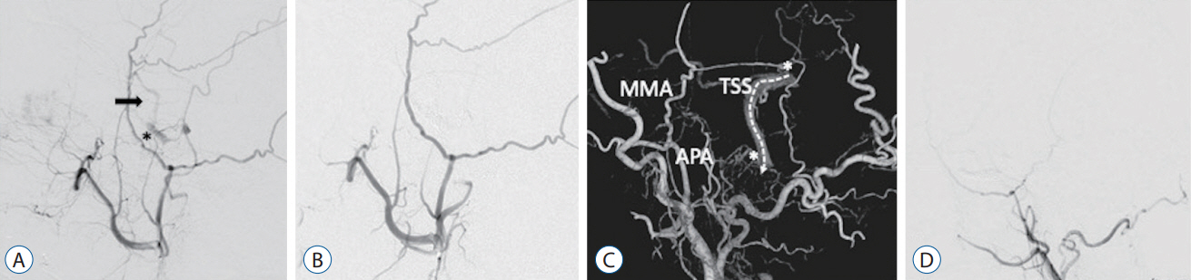
Fig. 2. Cerebral angiogram demonstrates that the left cavernous sinus dural arteriovenous fistula (DAVF) (asterisk) drains to the superficial middle cerebral vein (black arrow) (A). Postoperative cerebral angiogram shows that the fistula is occluded (B). Three-dimensional digital subtraction angiogram shows that the left anterior condylar confluence and transverse-sigmoid sinus (asterisks) DAVFs are mainly fed by the left ascending pharyngeal artery (APA) and middle meningeal artery (MMA) and drain to the left internal jugular vein without dangerous cortical venous reflux (dotted arrow) (C). Cerebral angiogram shows that the DAVFs are spontaneously are occluded (D). TSS : transverse-sigmoid sinus.
Reference
-
References
1. Gupta R, Horowitz M, Tayal A, Jovin T. De novo development of a remote arteriovenous fistula following transarterial embolization of a carotid cavernous fistula: case report and review of the literature. AJNR Am J Neuroradiol. 26:2587–2590. 2005.2. Ha SY, Kwon YS, Kim BM, Kim DI, Kim DJ. Clinical and angiographic characteristics of multiple dural arteriovenous shunts. AJNR Am J Neuroradiol. 33:1691–1695. 2012.
Article3. Hagiwara S, Miyazaki T, Tsuji M, Kambara M, Yoshikane T, Nagai H, et al. A case of de novo anterior condylar dural arteriovenous fistula long after curative transvenous embolization of contralateral anterior condylar arteriovenous fistula. Case Rep Med. 2016:6974526. 2016.
Article4. Kiyosue H, Tanoue S, Okahara M, Yamashita M, Nagatomi H, Mori H. Recurrence of dural arteriovenous fistula in another location after selective transvenous coil embolization: report of two cases. AJNR Am J Neuroradiol. 23:689–692. 2002.5. Kubota Y, Ueda T, Kaku Y, Sakai N. Development of a dural arteriovenous fistula around the jugular valve after transvenous embolization of cavernous dural arteriovenous fistula. Surg Neurol. 51:174–176. 1999.
Article6. Kurl S, Vanninen R, Saari T, Hernesniemi J. Development of right transverse sinus dural arteriovenous malformation after embolisation of a similar lesion on the left. Neuroradiology. 38:386–388. 1996.
Article7. Makiuchi T, Takasaki K, Yamagami M, Oda H, Todoroki K, Atsuchi M, et al. A case of sigmoid sinus dural arteriovenous fistula after treated cavernous dural arteriovenous fistula. Interv Neuroradiol 4 Suppl. 1:219–222. 1998.
Article8. Nakagawa H, Kubo S, Nakajima Y, Izumoto S, Fujita T. Shifting of dural arteriovenous malformation from the cavernous sinus to the sigmoid sinus to the transverse sinus after transvenous embolization. A case of left spontaneous carotid-cavernous sinus fistula. Surg Neurol. 37:30–38. 1992.
Article9. Newton TH, Cronqvist S. Involvement of dural arteries in intracranial arteriovenous malformations. Radiology. 93:1071–1078. 1969.
Article10. Oh JS, Yoon SM, Oh HJ, Shim JJ, Bae HG, Lee KS. Endovascular treatment of dural arteriovenous fistulas: single center experience. J Korean Neurosurg Soc. 59:17–25. 2016.
Article11. Yamashita K, Taki W, Nakahara I, Nishi S, Sadato A, Kikuchi H. Development of sigmoid dural arteriovenous fistulas after transvenous embolization of cavernous dural arteriovenous fistulas. AJNR Am J Neuroradiol. 14:1106–1108. 1993.
- Full Text Links
-
- Actions
-
Cited
- CITED
-
- Close
- Share
-
- Similar articles
-
- Transvenous coil embolization of hypoglossal canal dural arteriovenous fistula using detachable coils: A case report
- Dural Arteriovenous Fistula Involving Transverse Sinus: Successful Embolization Using Onyx(R)
- A Case of Dural Arteriovenous Fistula of the Anterior Condylar Vein
- Transvenous injection of n-butyl 2-cyanoacrylate to obliterate the pathologic cavernous sinus as a salvage technique for incompletely obliterated complex cavernous sinus dural arteriovenous fistula after transvenous coil embolization
- Occurrence of Metachronous Intracranial Dural Arteriovenous Fistula after Embolization of Intracranial Dural Arteriovenous Fistula: A Case Report
