Transvenous coil embolization of hypoglossal canal dural arteriovenous fistula using detachable coils: A case report
- Affiliations
-
- 1Department of Neurosurgery, Hallym University Sacred Heart Hospital, Anyang, Korea
- KMID: 2531660
- DOI: http://doi.org/10.7461/jcen.2021.E2021.08.004
Abstract
- The hypoglossal canal (HC) is an unusual location of the posterior fossa dural arteriovenous fistula (AVF), which usually occurs in the transverse or sigmoid sinus. Herein, we report a case of HC dural AVF successfully treated with transvenous coil embolization using detachable coils in a 68-year-old woman who presented with headache and left pulsatile tinnitus for 2 months. Brain magnetic resonance imaging (MRI) and cerebral angiography revealed left HC dural AVF. The pulsatile bruit disappeared immediately after the procedure. Follow-up MRI showed complete disappearance of the fistula. Precise localization of the fistula through careful consideration of the anatomy and transvenous coil embolization using a detachable coil can facilitate the treatment for HC dural AVF.
Figure
-
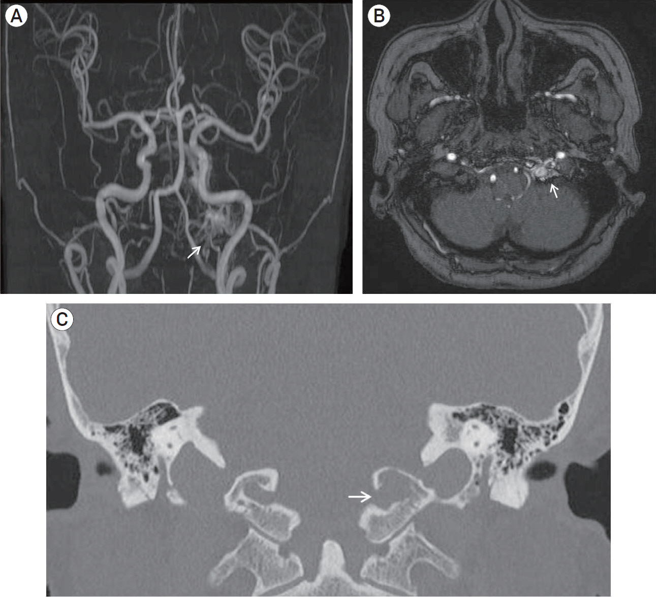
Fig. 1. Magnetic resonance angiography shows abnormal signal hyperintensity around the left hypoglossal canal (the “magic wand appearance”) (arrow) (A, B). A bone imaging computed tomography shows bone erosion and expansion of left hypoglossal canal (arrow) (C).

Fig. 2. Cerebral angiography reveals a dural arteriovenous fistula in the anterior condylar vein within the left hypoglossal canal (HC) fed by neuromeningeal branches of the bilatreral ascending pharyngeal artery (APA), occipital artery (A, B, C) and vertebral artery (D), which drained into internal jugular vein (IJV), cavernous sinus (CS) through inferior petrosal sinus (IPS), and vertebral venous plexus (VVP). Schematic illustration shows detailed anatomy of dural arteriovenous fistula (E).

Fig. 3. Microcatheter angiography demonstrated dilated venous pouch connected with vertebral venous plexus (arrow) and inferior petrosal sinus (arrowhead) (A). Final angiography showed complete occlusion of dural arteriovenous fistula and contrast stagnation around the hypoglossal canal (arrow) (B, C). CT shows coil mass in the hypoglossal canal (D). CT, computed tomography.
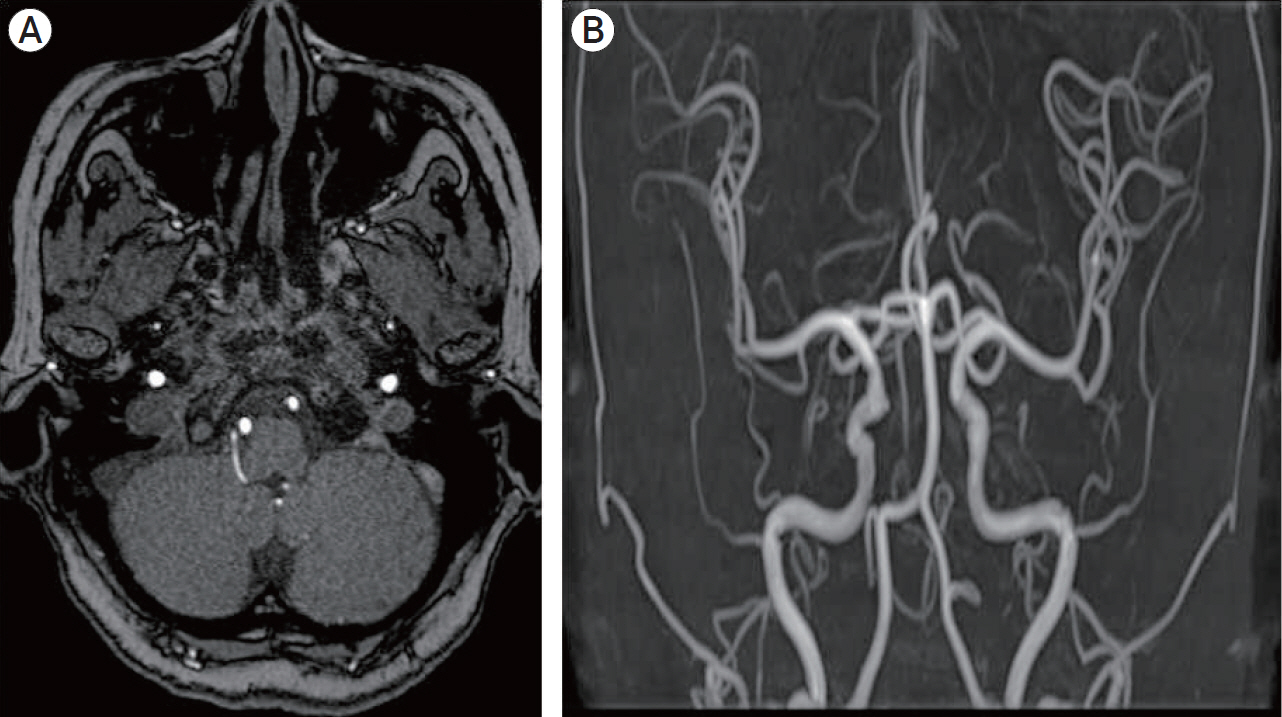
Fig. 4. Follow-up magnetic resonance image (A), and magnetic resonance angiography (B) showed complete disappearance of dural arteriovenous fisula.
Cited by 1 articles
-
In Vitro Head-to-Head Comparison of Flow Reduction between Fibered and Non-Fibered Pushable Coils
Jong-Tae Yoon, Boseong Kwon, Joon Ho Choi, Sun Moon Hwang, Mihyeon Kim, Sungbin Hwang, Yunsun Song, Deok Hee Lee
Neurointervention. 2024;19(1):31-38. doi: 10.5469/neuroint.2024.00031.
Reference
-
1. Barnwell SL, Halbach VV, Dowd CF, Higashida RT, Hieshima GB. Dural arteriovenous fistulas involving the inferior petrosal sinus: angiographic findings in six patients. AJNR Am J Neuroradiol. 1990; May. 11(3):511–6.2. Blomquist MH, Barr JD, Hurst RW. Isolated unilateral hypoglossal neuropathy caused by dural arteriovenous fistula. AJNR Am J Neuroradiol. 1998; May. 19(5):951–3.3. Choi JW, Kim BM, Kim DJ, Kim DI, Suh SH, Shin N-Y, et al. Hypoglossal canal dural arteriovenous fistula: incidence and the relationship between symptoms and drainage pattern. J Neurosurg. 2013; Oct. 119(4):955–60.
Article4. Combarros O, Alvarez de Arcaya A, Berciano J. Isolated unilateral hypoglossal nerve palsy: nine cases. J Neurol. 1998; Feb. 245(2):98–100.
Article5. Ernst R, Bulas R, Tomsick T, van Loveren H, Aziz KA. Three cases of dural arteriovenous fistula of the anterior condylar vein within the hypoglossal canal. AJNR Am J Neuroradiol. 1999; Nov-Dec. 20(10):2016–20.6. Manabe S, Satoh K, Matsubara S, Satomi J, Hanaoka M, Nagahiro S. Characteristics, diagnosis and treatment of hypoglossal canal dural arteriovenous fistula: report of nine cases. Neuroradiology. 2008; Aug. 50(8):715–21.
Article7. McDougall CG, Halbach VV, Dowd CF, Higashida RT, Larsen DW, Hieshima GB. Dural arteriovenous fistulas of the marginal sinus. AJNR Am J Neuroradiol. 1997; Sep. 18(8):1565–72.
Article8. San Millán Ruiz D, Gailloud P, Rüfenacht DA, Delavelle J, Henry F, Fasel JHD. The craniocervical venous system in relation to cerebral venous drainage. AJNR Am J Neuroradiol. 2002; Oct. 23(9):1500–8.9. Tanoue S, Goto K, Oota S. Endovascular treatment for dural arteriovenous fistula of the anterior condylar vein with unusual venous drainage: report of two cases. AJNR Am J Neuroradiol. 2005; Sep. 26(8):1955–9.
- Full Text Links
-
- Actions
-
Cited
- CITED
-
- Close
- Share
-
- Similar articles
-
- Transvenous injection of n-butyl 2-cyanoacrylate to obliterate the pathologic cavernous sinus as a salvage technique for incompletely obliterated complex cavernous sinus dural arteriovenous fistula after transvenous coil embolization
- A Case of Dural Arteriovenous Fistula of the Anterior Condylar Vein
- Transvenous Coil Embolization for Dural Arteriovenous Fistulas of the Ophthalmic Sheath: Report of Two Cases and Review of the Literature
- Dural Arteriovenous Fistula Involving Transverse Sinus: Successful Embolization Using Onyx(R)
- Transvenous Embolization in Patients with Dural Arteriovenous Fistula
