Clin Endosc.
2022 May;55(3):463-464. 10.5946/ce.2020.239.
Metastasis of breast cancer presenting as enlarged folds in the stomach
- Affiliations
-
- 1Department of Internal Medicine, Pusan National University College of Medicine, Busan, Korea
- 2Biomedical Research Institute, Pusan National University Hospital, Busan, Korea
- 3Department of Pathology, Pusan National University College of Medicine, Busan, Korea
- KMID: 2529971
- DOI: http://doi.org/10.5946/ce.2020.239
Figure
-
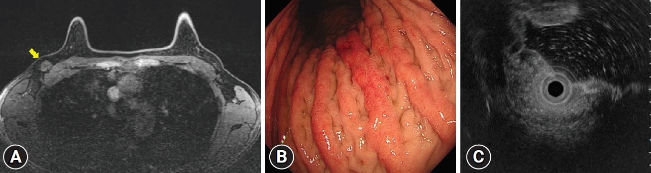
Fig. 1. (A) Breast magnetic resonance imaging shows a 2 cm-sized mass in the right breast (arrow). (B) On endoscopy, diffusely nodular, enlarged folds with hyperemia are seen on the greater curvature of the gastric body. On air inflation, the stomach maintains relatively good distensibility. (C) Endoscopic ultrasonography reveals the thickening of the submucosal layer.
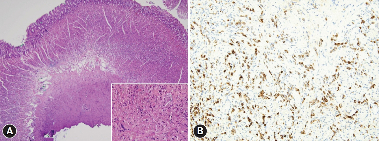
Fig. 2. (A) Strip biopsy shows that discohesive tumor cells infiltrate the submucosa of the stomach (hematoxylin & eosin, ×40; boxed area: hematoxylin & eosin, ×400). (B) The tumor cells are immunopositive for gross cystic disease fluid protein-15 (immunohistochemical stain, ×200).
Reference
-
1. McLemore EC, Pockaj BA, Reynolds C, et al. Breast cancer: presentation and intervention in women with gastrointestinal metastasis and carcinomatosis. Ann Surg Oncol. 2005; 12:886–894.2. Yim K, Ro SM, Lee J. Breast cancer metastasizing to the stomach mimicking primary gastric cancer: a case report. World J Gastroenterol. 2017; 23:2251–2257.3. Honma N, Horii R, Iwase T, et al. Clinical importance of estrogen receptor-beta evaluation in breast cancer patients treated with adjuvant tamoxifen therapy. J Clin Oncol. 2008; 26:3727–3734.
- Full Text Links
-
- Actions
-
Cited
- CITED
-
- Close
- Share
-
- Similar articles
-
- An Unusual Case of Gastric Cancer Presenting with Breast Metastasis with Pleomorphic Microcalcifications
- Bladder Cancer Metastasis to the Breast in a Male Patient: Imaging Findings on Mammography and Ultrasonography
- A case of stomach metastasis from breast cancer
- Breast Cancer Metastasis to the Stomach Resembling Early Gastric Cancer
- Occult Invasive Lobular Carcinoma of Breast Detected by Stomach Metastasis: A Case Report
