Generation of Cortical Brain Organoid with Vascularization by Assembling with Vascular Spheroid
- Affiliations
-
- 1Adult Stem Cell Research Center, College of Veterinary Medicine, Seoul National University, Seoul, Korea
- 2College of Veterinary Medicine and Research Institute for Veterinary Science, Seoul National University, Seoul, Korea
- KMID: 2526740
- DOI: http://doi.org/10.15283/ijsc21157
Abstract
- Background and Objectives
Brain organoids have the potential to improve our understanding of brain development and neurological disease. Despite the importance of brain organoids, the effect of vascularization on brain organoids is largely unknown. The objective of this study is to develop vascularized organoids by assembling vascular spheroids with cerebral organoids.
Methods and Results
In this study, vascularized spheroids were generated from non-adherent microwell culture system of human umbilical vein endothelial cells, human dermal fibroblasts and human umbilical cord blood derived mesenchymal stem cells. These vascular spheroids were used for fusion with iPSCs induced cerebral organoids. Immunostaining studies of vascularized organoids demonstrated well organized vascular structures and reduced apoptosis. We showed that the vascularization in cerebral organoids up-regulated the Wnt/β-catenin signaling.
Conclusions
We developed vascularized cerebral organoids through assembly of brain organoids with vascular spheroids. This method could not only provide a model to study human cortical development but also represent an opportunity to explore neurological disease.
Keyword
Figure
-
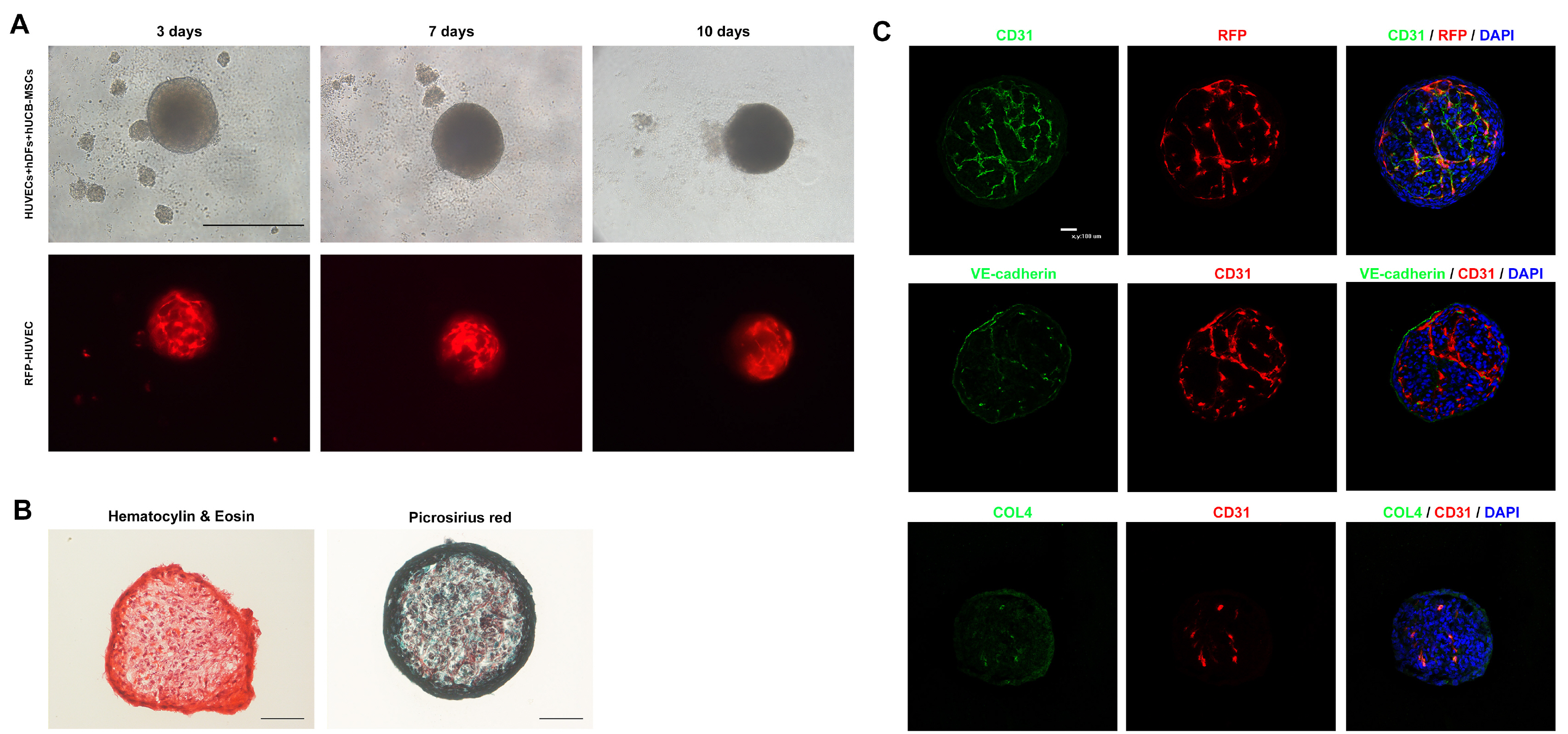
Fig. 1 Phenotypical characterization of vascular spheroids. (A) Representative images of vascular spheroids. HUVECs, hDFs and hUCB-MSCs were cultured directly for 10 days. Angiogenesis is shown using RFP fluorescence in the vascular spheroid (Scale bar: 500 μm). (B) Representative H&E and picrosirius staining images of vascular spheroids. Collagen deposition is shown in the vascular spheroid (Scale bar: 100 μm). (C) Representative images from day 10 spheroids immunostained with CD31, VE-cadherin, vWF and collagen IV. Nuclei are stained with DAPI (Scale bar: 100 μm).

Fig. 2 Characterization of vascular structure in the assembloid of vascular spheroids and cortical organoids. (A) Morphology and size of organoids on days 20, 30 and 43. Quantification of diameter and area from each group at different stages (Scale bar: 500 μm). (B) Representative CD31 immunostaining images of organoids on day 43 (Scale bar: 100 μm). (C) Immunostaining for CD31 and endothelial markers vWF and CDH5 in sectioned organoids at different time points (days 43 and 60). Expression of CD31 is shown in the vascular spheroid and cerebral organoid. The endothelial markers CDH5 and vWF are expressed in the vascular spheroid. White dash line indicates the location of the vascular spheroid (Scale bar: 100 μm). One-way ANOVA analysis. (D) Immunostaining images of whole-mount organoids at different time points (days 43 and 60) showing vascular-like structures in the organoids. Total vessel length was quantified by angiogenesis analysis (Scale bar: 1 mm). **p<0.01.

Fig. 3 Neuronal characterization of cerebral organoids. (A) Representative images of each organoid on day 43 stained with NeuN, CTIP2, TUJ1 and MAP2. Nuclei are stained with DAPI (Scale bar: 100 μm). (B) Immunostaining images of α-ZO1 in each group on day 60 (Scale bar: 100 μm). Student’s t-test, n=3, independent organoids from 2 different batches. *p<0.05; n.s. not significant.

Fig. 4 Vascularization of hCOs with vascular spheroids reduces apoptosis. (A) Representative images of the TUNEL assay for detecting apoptotic cells. Quantification showing the percentage of TUNEL+ cells that were positive for DAPI in each group (Scale bar: 100 μm). (B) Protein levels of rH2AX and cleaved caspase-3. (C) Immunostaining images and quantification of rH2AX and SOX2 in each group on day 60 (Scale bar: 100 μm). (D) Immunostaining images and quantification of cleaved caspase-3 and PAX6 in each group on day 60 (Scale bar: 100 μm). Student’s t-test, n=4, independent organoids from 2 different batches. *p<0.05, ***p<0.001; n.s. not significant.
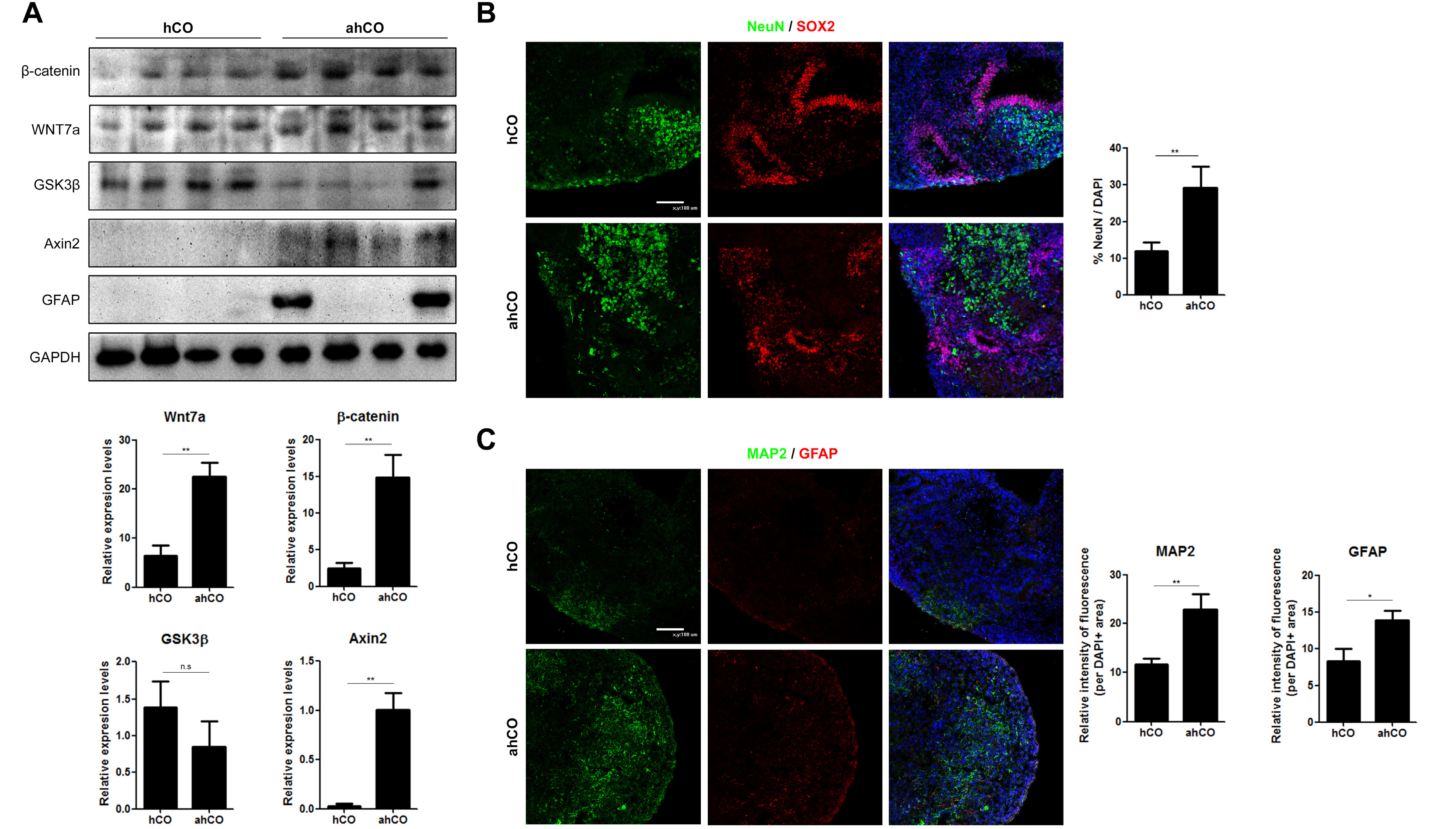
Fig. 5 Wnt/β-catenin signaling are upregulated in ahCOs. (A) Western blot of Wnt7a, β-catenin, GSK3β, AXIN2 and GFAP in each group of organoids. Western blot analysis revealed that decrease tendency of GSK3β and up-regulation of axin2 along with Wnt7a activation and that β-catenin was increased in the ahCO group compared to the hCO group. (B) Immunostaining images and quantification of SOX2 and NeuN in each group on day 60 (Scale bar: 100 μm). (C) Immunostaining images and quantification of MAP2 and GFAP in each group on day 60 (Scale bar: 100 μm). Student’s t-test, n=4, independent organoids from 2 different batches. **p<0.01; n.s. not significant.
Cited by 1 articles
-
Lo and Behold, the Lab-Grown Organs Have Arrived!
Jaesang Kim
Int J Stem Cells. 2022;15(1):1-2. doi: 10.15283/ijsc22026.
Reference
-
References
1. Marshall JJ, Mason JO. 2019; Mouse vs man: organoid models of brain development & disease. Brain Res. 1724:146427. DOI: 10.1016/j.brainres.2019.146427. PMID: 31473222.2. Lancaster MA, Renner M, Martin CA, Wenzel D, Bicknell LS, Hurles ME, Homfray T, Penninger JM, Jackson AP, Knoblich JA. 2013; Cerebral organoids model human brain development and microcephaly. Nature. 501:373–379. DOI: 10.1038/nature12517. PMID: 23995685. PMCID: PMC3817409.
Article3. Lancaster MA, Knoblich JA. 2014; Generation of cerebral organoids from human pluripotent stem cells. Nat Protoc. 9:2329–2340. DOI: 10.1038/nprot.2014.158. PMID: 25188634. PMCID: PMC4160653.
Article4. Qian X, Song H, Ming GL. 2019; Brain organoids: advances, applications and challenges. Development. 146:dev166074. DOI: 10.1242/dev.166074. PMID: 30992274. PMCID: PMC6503989.
Article5. Clevers H. 2016; Modeling development and disease with organoids. Cell. 165:1586–1597. DOI: 10.1016/j.cell.2016.05.082. PMID: 27315476.
Article6. Luo C, Lancaster MA, Castanon R, Nery JR, Knoblich JA, Ecker JR. 2016; Cerebral organoids recapitulate epigenomic signatures of the human fetal brain. Cell Rep. 17:3369–3384. DOI: 10.1016/j.celrep.2016.12.001. PMID: 28009303. PMCID: PMC5495578.
Article7. Sloan SA, Andersen J, Pașca AM, Birey F, Pașca SP. 2018; Generation and assembly of human brain region-specific three-dimensional cultures. Nat Protoc. 13:2062–2085. DOI: 10.1038/s41596-018-0032-7. PMID: 30202107. PMCID: PMC6597009.
Article8. Koo B, Choi B, Park H, Yoon KJ. 2019; Past, present, and future of brain organoid technology. Mol Cells. 42:617–627. DOI: 10.14348/molcells.2019.0162. PMID: 31564073. PMCID: PMC6776157.9. Matsui TK, Tsuru Y, Hasegawa K, Kuwako KI. 2021; Vasculari-zation of human brain organoids. Stem Cells. 39:1017–1024. DOI: 10.1002/stem.3368. PMID: 33754425.
Article10. Cakir B, Xiang Y, Tanaka Y, Kural MH, Parent M, Kang YJ, Chapeton K, Patterson B, Yuan Y, He CS, Raredon MSB, Dengelegi J, Kim KY, Sun P, Zhong M, Lee S, Patra P, Hyder F, Niklason LE, Lee SH, Yoon YS, Park IH. 2019; Engineering of human brain organoids with a functional vascular-like system. Nat Methods. 16:1169–1175. DOI: 10.1038/s41592-019-0586-5. PMID: 31591580. PMCID: PMC6918722.
Article11. Shi Y, Sun L, Wang M, Liu J, Zhong S, Li R, Li P, Guo L, Fang A, Chen R, Ge WP, Wu Q, Wang X. 2020; Vascularized human cortical organoids (vOrganoids) model cortical development in vivo. PLoS Biol. 18:e3000705. DOI: 10.1371/journal.pbio.3000705. PMID: 32401820. PMCID: PMC7250475. PMID: 66f6d290c2f84ede886185d58cd7368f.12. Licht T, Keshet E. 2015; The vascular niche in adult neurogenesis. Mech Dev. 138 Pt 1:56–62. DOI: 10.1016/j.mod.2015.06.001. PMID: 26103548.
Article13. Karakatsani A, Shah B, Ruiz de Almodovar C. 2019; Blood vessels as regulators of neural stem cell properties. Front Mol Neurosci. 12:85. DOI: 10.3389/fnmol.2019.00085. PMID: 31031591. PMCID: PMC6473036.
Article14. Daneman R, Agalliu D, Zhou L, Kuhnert F, Kuo CJ, Barres BA. 2009; Wnt/beta-catenin signaling is required for CNS, but not non-CNS, angiogenesis. Proc Natl Acad Sci U S A. 106:641–646. DOI: 10.1073/pnas.0805165106. PMID: 19129494. PMCID: PMC2626756.15. Chenn A. 2008; Wnt/beta-catenin signaling in cerebral cortical development. Organogenesis. 4:76–80. DOI: 10.4161/org.4.2.5852. PMID: 19279718. PMCID: PMC2634251.
Article16. De Moor L, Merovci I, Baetens S, Verstraeten J, Kowalska P, Krysko DV, De Vos WH, Declercq H. 2018; High-throughput fabrication of vascularized spheroids for bioprinting. Biofabrication. 10:035009. DOI: 10.1088/1758-5090/aac7e6. PMID: 29798932.
Article17. Mulligan KA, Cheyette BN. 2012; Wnt signaling in vertebrate neural development and function. J Neuroimmune Phar-macol. 7:774–787. DOI: 10.1007/s11481-012-9404-x. PMID: 23015196. PMCID: PMC3518582.
Article18. Muoio V, Persson PB, Sendeski MM. 2014; The neurovascular unit - concept review. Acta Physiol (Oxf). 210:790–798. DOI: 10.1111/apha.12250. PMID: 24629161.
Article19. Bell AH, Miller SL, Castillo-Melendez M, Malhotra A. 2020; The neurovascular unit: effects of brain insults during the perinatal period. Front Neurosci. 13:1452. DOI: 10.3389/fnins.2019.01452. PMID: 32038147. PMCID: PMC6987380. PMID: 469ce704fd7343508e166500e1437879.
Article20. Pham MT, Pollock KM, Rose MD, Cary WA, Stewart HR, Zhou P, Nolta JA, Waldau B. 2018; Generation of human vascularized brain organoids. Neuroreport. 29:588–593. DOI: 10.1097/WNR.0000000000001014. PMID: 29570159. PMCID: PMC6476536.
Article21. Zhao X, Xu Z, Xiao L, Shi T, Xiao H, Wang Y, Li Y, Xue F, Zeng W. 2021; Review on the vascularization of organoids and organoids-on-a-chip. Front Bioeng Biotechnol. 9:637048. DOI: 10.3389/fbioe.2021.637048. PMID: 33912545. PMCID: PMC8072266. PMID: d9603ef2f6584acfa5016be2d649b8c3.
Article22. Rademakers T, Horvath JM, van Blitterswijk CA, LaPointe VLS. 2019; Oxygen and nutrient delivery in tissue engineering: approaches to graft vascularization. J Tissue Eng Regen Med. 13:1815–1829. DOI: 10.1002/term.2932. PMID: 31310055. PMCID: PMC6852121.
Article23. Heiss M, Hellström M, Kalén M, May T, Weber H, Hecker M, Augustin HG, Korff T. 2015; Endothelial cell spheroids as a versatile tool to study angiogenesis in vitro. FASEB J. 29:3076–3084. DOI: 10.1096/fj.14-267633. PMID: 25857554.
Article24. Pfisterer L, Korff T. 2016; Spheroid-based in vitro angiogenesis model. Methods Mol Biol. 1430:167–177. DOI: 10.1007/978-1-4939-3628-1_11. PMID: 27172953.25. Serra J, Alves CPA, Brito L, Monteiro GA, Cabral JMS, Prazeres DMF, da Silva CL. 2019; Engineering of human mesenchymal stem/stromal cells with vascular endothelial growth factor-encoding minicircles for angiogenic ex vivo gene therapy. Hum Gene Ther. 30:316–329. DOI: 10.1089/hum.2018.154. PMID: 30200778.
Article26. Arutyunyan I, Fatkhudinov T, Kananykhina E, Usman N, Elchaninov A, Makarov A, Bolshakova G, Goldshtein D, Sukhikh G. 2016; Role of VEGF-A in angiogenesis promoted by umbilical cord-derived mesenchymal stromal/stem cells: in vitro study. Stem Cell Res Ther. 7:46. DOI: 10.1186/s13287-016-0305-4. PMID: 27001300. PMCID: PMC4802928.27. Maacha S, Sidahmed H, Jacob S, Gentilcore G, Calzone R, Grivel JC, Cugno C. 2020; Paracrine mechanisms of mesenchymal stromal cells in angiogenesis. Stem Cells Int. 2020:4356359. DOI: 10.1155/2020/4356359. PMID: 32215017. PMCID: PMC7085399.
Article28. Mansour AA, Gonçalves JT, Bloyd CW, Li H, Fernandes S, Quang D, Johnston S, Parylak SL, Jin X, Gage FH. 2018; An in vivo model of functional and vascularized human brain organoids. Nat Biotechnol. 36:432–441. DOI: 10.1038/nbt.4127. PMID: 29658944. PMCID: PMC6331203.
Article29. Lau M, Li J, Cline HT. 2017; In vivo analysis of the neurovascular niche in the developing Xenopus brain. eNeuro. 4:ENEURO.0030-17.2017. DOI: 10.1523/ENEURO.0030-17.2017. PMID: 28795134. PMCID: PMC5548361.30. Tavazoie M, Van der Veken L, Silva-Vargas V, Louissaint M, Colonna L, Zaidi B, Garcia-Verdugo JM, Doetsch F. 2008; A specialized vascular niche for adult neural stem cells. Cell Stem Cell. 3:279–288. DOI: 10.1016/j.stem.2008.07.025. PMID: 18786415. PMCID: PMC6864413.
Article31. Tang M, Gao G, Rueda CB, Yu H, Thibodeaux DN, Awano T, Engelstad KM, Sanchez-Quintero MJ, Yang H, Li F, Li H, Su Q, Shetler KE, Jones L, Seo R, McConathy J, Hillman EM, Noebels JL, De Vivo DC, Monani UR. 2017; Brain microvasculature defects and Glut1 deficiency syndrome averted by early repletion of the glucose transporter-1 protein. Nat Commun. 8:14152. DOI: 10.1038/ncomms14152. PMID: 28106060. PMCID: PMC5263887. PMID: cfe083483d664856ac5c4ced1381ccce.
Article32. Tang M, Park SH, Petri S, Yu H, Rueda CB, Abel ED, Kim CY, Hillman EM, Li F, Lee Y, Ding L, Jagadish S, Frankel WN, De Vivo DC, Monani UR. 2021; An early endothelial cell-specific requirement for Glut1 is revealed in Glut1 deficiency syndrome model mice. JCI Insight. 6:e145789. DOI: 10.1172/jci.insight.145789. PMID: 33351789. PMCID: PMC7934852.
Article33. Wexler EM, Paucer A, Kornblum HI, Palmer TD, Geschwind DH. 2009; Endogenous Wnt signaling maintains neural progenitor cell potency. Stem Cells. 27:1130–1141. DOI: 10.1002/stem.36. PMID: 19418460. PMCID: PMC2782960.
Article34. Kriska J, Janeckova L, Kirdajova D, Honsa P, Knotek T, Dzamba D, Kolenicova D, Butenko O, Vojtechova M, Capek M, Kozmik Z, Taketo MM, Korinek V, Anderova M. 2021; Wnt/β-catenin signaling promotes differentiation of ischemia-activated adult neural stem/progenitor cells to neuronal precursors. Front Neurosci. 15:628983. DOI: 10.3389/fnins.2021.628983. PMID: 33716653. PMCID: PMC7947698. PMID: abcd8bb230064c7b80ad86ff6baa7608.
Article
- Full Text Links
-
- Actions
-
Cited
- CITED
-
- Close
- Share
-
- Similar articles
-
- A Simple Method for Generating Cerebral Organoids from Human Pluripotent Stem Cells
- In Vitro Generation of Luminal Vasculature in Liver Organoids: From Basic Vascular Biology to Vascularized Hepatic Organoids
- Engineering Human Brain Organoids: From Basic Research to Tissue Regeneration
- Mesenchymal Stem Cell Spheroids: A Promising Tool for Vascularized Tissue Regeneration
- Increased SOX2 expression in three-dimensional sphere culture of dental pulp stem cells
