Protective effect of vitamin C against ivermectin induced nephrotoxicity in different age groups of male wistar rats: bio-histopathological study
- Affiliations
-
- 1Department of Human Anatomy & Embryology, Faculty of Medicine, Zagazig University, Zagazig, Egypt
- 2Department of Anatomy, Faculty of Medicine, Jouf University, Sakaka, Saudi Arabia
- KMID: 2523580
- DOI: http://doi.org/10.5115/acb.21.124
Abstract
- Ivermectin (Ive) has exceedingly efficient against several microorganisms including viruses; therefore, it could help as a potential treatment of COVID-19. Because of increasing consumption of ivermectin and vitamin C (Vit.C) in hope to treat COVID-19, and because of ivermectin nephrotoxic effects have not been fully clarified especially in juvenile age, it was conducted to examine the histopathological and biochemical effects of ivermectin on adult and juvenile kidneys, and to assess the possible protective role of Vit.C against this potential toxicity. Rats were divided to 4 subgroups (Control subgroup, Vit.C subgroup, Ive subgroup, and Vit.C+Ive subgroup), 1 week after 4 doses of ivermectin (0.4 mg/kg Ive±1.25 mg/kg Vit.C), blood samples obtained for assessment of kidney function test, part of kidneys prepared for determination of matrix metalloproteinase-9 and antioxidant enzymes essay. Other parts prepared for histopathological and ultrastructural examination. Results showed that administration of ivermectin led to attenuation in kidney function and in activities of the antioxidant enzymes and increase in matrix metalloproteinase-9 activity. In addition, there were histological damages (shrunken glomeruli, widened urinary space, cytoplasmic vacuolation and pyknotic nuclei with epithelial exfoliation, extravasated blood, and mononuclear cell infiltration) and immunohistochemistry revealed increase in percentage of Bax proapoptotic protein expression. Also, ultrastructure examination showed alteration in cell architecture. All these changes were more obvious in juvenile group while co-administration of Vit.C led to significant protection more in adult group. In conclusion, Ivermectin should be used cautiously especially in juvenile age, and co-administration of Vit.C is highly recommended.
Keyword
Figure
-

Fig. 1 Anatomical parameters examination in different study groups showing that Juvenile (Jv) rats treated with ivermectin (Ive subgroup) have a significant decrease in body weight (A) and kidney weight (B) compared with control and vitamin C (Vit.C) subgroups, while kidney weight/body weight % (C) remain insignificant difference. Adult (Ad) rats reveal significant difference in body weight (D) and no significant difference neither in kidney weight (E) nor kidney weight/body weight % (F). The percentage of change happen in corresponding Ive subgroups, the juvenile rats exhibit more body weight loss (G), also more kidney weight loss (H, I). Values are presented as mean±SD.
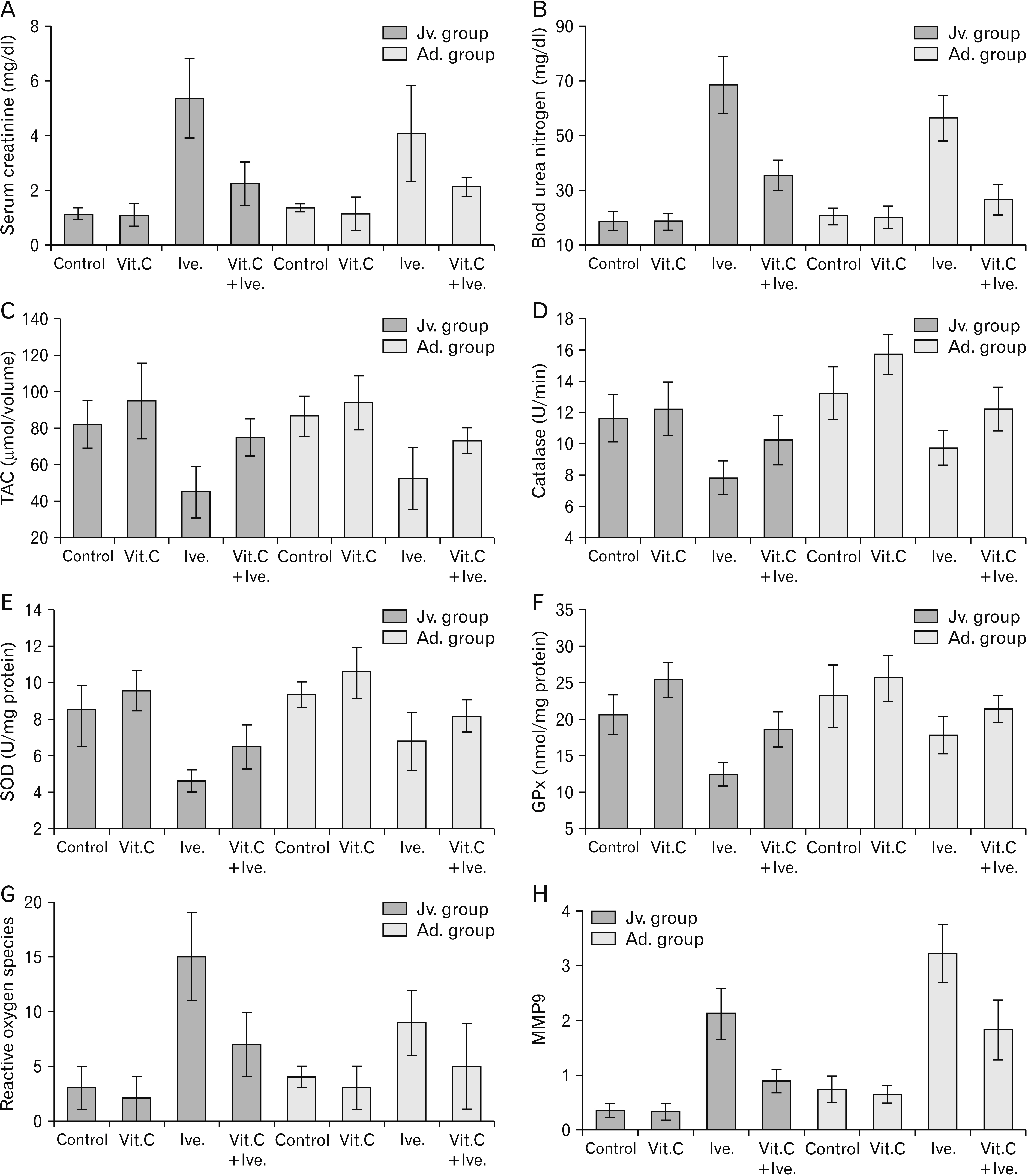
Fig. 2 Biochemical parameters analysis in different study groups showing that rats treated with ivermectin (Ive subgroup) have a highly significant increase in the kidney function marker; Serum creatinine concentrations (A), Blood urea nitrogen (B). Measuring the activities of the TAC (C), antioxidant enzymes catalase assay (CAT) (D), superoxide dismutase assay (SOD) (E), glutathione peroxidase assay (GPx) (F), and reactive oxygen species (G) showing that administration of vitamin C (Vit.C) alone leads to increase in antioxidant enzymes activities, on the other hand, rats treated with ivermectin (Ive subgroups) display a highly significant decrease in the activities of the antioxidant enzymes, comparing juvenile (Jv) and adult (Ad) groups showing that Jv rats have more decline in oxidants activity in treated group and less protected effect of Vit.C. Matrix metalloproteinase 9 (MMP9) is statistically significantly higher in Ive treated subgroups (H). Values are presented as mean±SD. TAC, total antioxidant capacity.

Fig. 3 H&E (×400) stained sections of renal tissues in different experimental groups: control and vitamin C (Vit.C) subgroups (A–D) showing; normal shaped glomeruli (g), normal renal space (S) lined by parietal layer of Bowman’s capsules (B) and renal tubules (dt, distal tubule; pt, proximal tubule) lined by normal cells with vesicular nuclei (short arrow). Ivermectin treated sub groups (Ive subgroup) (E–H) showing; shrunken glomeruli (G), widened urinary space (*), dilated tubules (dt & pt) lined by vacuolated cells with pyknotic nuclei (V), some desquamated cells in the lumen (arrow head), inflammatory cell infiltration (curved arrows) and extravasated blood (H). Vit.C+Ive subgroups (I, J) showing; normal shaped glomeruli (g) and tubules (dt, distal tubule; pt, proximal tubule), some shrunken glomeruli (G), relatively wide renal space (*) and some extravasated blood (H). In (I), few tubules exhibit pyknotic nuclei with cytoplasmic vacuolation (V) and epithelial cell exfoliation (arrow head). Morphometrically, in the Ive subgroups, cortical surface area (L) and glomerular area (K) showing significant decrease with significant increase in urinary space cross sectional (M), these changes are more obvious in juvenile (Jv) group (N, O) than adult (Ad) group.

Fig. 4 PAS stained (×400) sections of renal tissues in different experimental groups (A–D) (control & Vit.C) showing; well-formed tubular basement membranes (arrow heads), Basement membranes of Bowman’s capsules appear as thin regular PAS+Ive (thick arrows) and well developed brush borders of proximal convoluted tubule (arrows). Ive treated sub groups, (E, F) showing; strong positive PAS reaction of thickened basement membrane of both Bowman’s capsule (thick arrows) and renal tubules (arrow heads) and discontinuity PAS reaction at the brush borders of proximal tubular cells (arrows). Vit.C+Ive subgroups (G, H) showing; PAS positive reaction at the thin Bowman’s capsule (thick arrows) and tubular basement membrane (arrow heads) and continued brush border of proximal tubules (arrows). Vit.C, vitamin C; Ive, ivermectin; Jv, juvenile; Ad, adult.

Fig. 5 Bax immune-stained (×400) sections of renal tissues in different experimental groups showing; positive proapoptotic protein in the cytoplasm of renal tubules (arrow heads) is normally expressed in control and Vit.C subgroups (A–D), while Ive treated subgroups (E, F) display patchy expression. Vit.C+Ive subgroups (G, H) showing improvement in the Bax proapoptotic protein expression. Morphometrical analysis of percentages of Bax positivity showing high significant increase in Ive subgroups and significant increase in Vit.C+Ive subgroups (I, J). Vit.C, vitamin C; Ive, ivermectin; Jv, juvenile; Ad, adult.

Fig. 6 Electron micrographs of kidney tubules of control subgroup; juvenile (Jv) (A: proximal tubule, ×12,000; C: distal tubule, ×6,000) and adult (Ad) (B: proximal tubule, ×12,000; D: distal tubule, ×12,000) showing the following features: N, normal nucleus; n, nucleolus; m, mitochondria; arrow head, nuclear envelop; arrow, basal lamina; curved arrow, apical microvilli; Lu, lumen; L, lysosomes; RER, rough endoplasmic reticulum and the electron micrographs of glomerulus in both Jv (E: ×30,000) and Ad (F: ×30,000) of the control subgroup showing; P, podocyte nucleus; BC, blood capillary; arrow, basal lamina; arrow head, foot process of podocytes; zigzag arrow, septum between foot process. Electron micrographs of proximal tubule of Vit.C subgroup of Jv (G: ×6,000), and Ad (H: ×6,000) showing; N, normal nucleus; n, nucleolus; m, mitochondria; L, lysosomes; MV, microvilli; arrow, basement membrane. Vit.C, vitamin C; Ive, ivermectin.

Fig. 7 Electron micrographs of kidney tubules of ivermectin (Ive) treated sub groups; juvenile (Jv) (A: proximal tubule, ×5,000; C: distal tubule, ×6,000) and adult (Ad) (B: proximal tubule, ×6,000; D: distal tubule, ×12,000) showing the following features: N, normal nucleus; aN, apoptotic nucleus; m, ballooned mitochondrial; R, cytoplasmic rarefaction; C, intraluminal casts; L, secondery lysosomes; RER, degenerated rough endoplasmic reticulum; HG, luminal hemorrhage; arrowhead, irregular nuclear envelop; arrow, thick basement membrane. The region of glomerulus in both Jv (E: ×30,000) and Ad (F: ×30,000) of the same subgroup with the following features: P, podosyt; arrow, thick basement membrane; arrowhead, fused foot process of podocytes; zigzag arrow, fused septum between foot process; E, endothelial cells. Vit.C+Ive subgroups of Jv (G: ×6,000; I: ×12,000), and Ad (H: ×6,000; J: ×6,000) rats showing N, normal nucleus; n, nucleolus; aN, apoptotic nucleus m, mitochondria; MV, microvilli; BL, blood capillary; L, lysosomes; arrow, basement membrane; arrow head, regular nuclear envelop in (H) and relatively normal foot process in (I). Moreover, glomerulus in (I) showing P, intact podocyte and E, endothelial cells.
Cited by 1 articles
-
The ameliorating role of sofosbuvir and daclatasvir on thioacetamide-induced kidney injury in adult albino rats
Ahmed H. Moustafa, Heba F. Pasha, Manar A. Abas, Adel M. Aboregela
Anat Cell Biol. 2023;56(1):109-121. doi: 10.5115/acb.22.200.
Reference
-
References
1. Jiang F, Deng L, Zhang L, Cai Y, Cheung CW, Xia Z. 2020; Review of the clinical characteristics of coronavirus disease 2019 (COVID-19). J Gen Intern Med. 35:1545–9. DOI: 10.1007/s11606-020-05762-w. PMID: 32133578. PMCID: PMC7088708.
Article2. Lu H, Stratton CW, Tang YW. 2020; Outbreak of pneumonia of unknown etiology in Wuhan, China: the mystery and the miracle. J Med Virol. 92:401–2. DOI: 10.1002/jmv.25678. PMID: 31950516. PMCID: PMC7166628.
Article3. Sohrabi C, Alsafi Z, O'Neill N, Khan M, Kerwan A, Al-Jabir A, Iosifidis C, Agha R. 2020; World Health Organization declares global emergency: a review of the 2019 novel coronavirus (COVID-19). Int J Surg. 76:71–6. DOI: 10.1016/j.ijsu.2020.02.034. PMID: 32112977. PMCID: PMC7105032.
Article4. Reviglio VE, Osaba M, Reviglio V, Chiaradia P, Kuo IC, O'Brien TP. 2020; COVID-19 and Ophthalmology: a new chapter in an old story. Med Hypothesis Discov Innov Ophthalmol. 9:71–3. PMID: 32490013. PMCID: PMC7134243.5. Caly L, Druce JD, Catton MG, Jans DA, Wagstaff KM. 2020; The FDA-approved drug ivermectin inhibits the replication of SARS-CoV-2 in vitro. Antiviral Res. 178:104787. DOI: 10.1016/j.antiviral.2020.104787. PMID: 32251768. PMCID: PMC7129059.6. Heidary F, Gharebaghi R. 2020; Ivermectin: a systematic review from antiviral effects to COVID-19 complementary regimen. J Antibiot (Tokyo). 73:593–602. DOI: 10.1038/s41429-020-0336-z. PMID: 32533071. PMCID: PMC7290143.
Article7. Omura S, Crump A. 2014; Ivermectin: panacea for resource-poor communities? Trends Parasitol. 30:445–55. DOI: 10.1016/j.pt.2014.07.005. PMID: 25130507.8. Laing R, Gillan V, Devaney E. 2017; Ivermectin - old drug, new tricks? Trends Parasitol. 33:463–72. DOI: 10.1016/j.pt.2017.02.004. PMID: 28285851. PMCID: PMC5446326.
Article9. Yang SNY, Atkinson SC, Wang C, Lee A, Bogoyevitch MA, Borg NA, Jans DA. 2020; The broad spectrum antiviral ivermectin targets the host nuclear transport importin α/β1 heterodimer. Antiviral Res. 177:104760. DOI: 10.1016/j.antiviral.2020.104760. PMID: 32135219.
Article10. Shoumann WM, Hegazy AA, Nafae RM, Ragab MI, Samra SR, Ibrahim DA, AL-Mahrouky TH, Sileem AE. 2021; Use of ivermectin as a potential chemoprophylaxis for COVID-19 in Egypt: a randomized clinical trial. J Clin Diagn Res. 15:OC27–32. DOI: 10.7860/JCDR/2021/46795.14529.11. Atakisi E, Atakisi O, Topcu B, Uzun M. 2009; Effects of therapeutic dose of ivermectin on plasma nitric oxide and total antioxidant capacity in rabbits. Eur Rev Med Pharmacol Sci. 13:425–9. PMID: 20085123.12. Saha D. 2014; Role of free radicals, oxidative stress and xenobiotics in carcinogenesis by environmental pollutants. Av Biomed. 3:84–92.13. Nwani CD, Odo GE, Nwadinigwe AO, Onyeke CC, Atama CI, Ngwu G, Oluah SN, Ukonze JA, Ezeibe BC. 2016; Short-term effects of Albendazole on the oxidative stress markers and hematological parameters in tissues of African Catfish Clarias gariepinus. J Aquat Anim Health. 28:222–8. DOI: 10.1080/08997659.2016.1194908. PMID: 27754768.14. Budge PJ, Herbert C, Andersen BJ, Weil GJ. 2018; Adverse events following single dose treatment of lymphatic filariasis: observations from a review of the literature. PLoS Negl Trop Dis. 12:e0006454. DOI: 10.1371/journal.pntd.0006454. PMID: 29768412. PMCID: PMC5973625.
Article15. Burnham GM. 1993; Adverse reactions to ivermectin treatment for onchocerciasis. Results of a placebo-controlled, double-blind trial in Malawi. Trans R Soc Trop Med Hyg. 87:313–7. DOI: 10.1016/0035-9203(93)90144-F. PMID: 8236405.
Article16. Lukiana T, Mandina M, Situakibanza NH, Mbula MM, Lepira BF, Odio WT, Kamgno J, Boussinesq M. 2006; A possible case of spontaneous Loa loa encephalopathy associated with a glomerulopathy. Filaria J. 5:6. DOI: 10.1186/1475-2883-5-6. PMID: 16686951. PMCID: PMC1471781.
Article17. Kaur U, Chakrabarti SS, Gambhir IS. 2017; Delirium induced by albendazole-ivermectin combination: report of the first case in an older patient. Geriatr Gerontol Int. 17:2618–20. DOI: 10.1111/ggi.13158. PMID: 29265756.
Article18. Veit O, Beck B, Steuerwald M, Hatz C. 2006; First case of ivermectin-induced severe hepatitis. Trans R Soc Trop Med Hyg. 100:795–7. DOI: 10.1016/j.trstmh.2006.02.003. PMID: 16682062.
Article19. Choksi TT, Madison G, Dar T, Asif M, Fleming K, Clarke L, Danilewitz M, Hennawy R. 2016; Multiorgan dysfunction syndrome from strongyloides stercoralis hyperinfection in a patient with human T-cell lymphotropic virus-1 coinfection after initiation of ivermectin treatment. Am J Trop Med Hyg. 95:864–7. DOI: 10.4269/ajtmh.16-0259. PMID: 27527631. PMCID: PMC5062789.
Article20. Almeida MG, Röpke CD, Davino SC, Nobre FAF, Santos E, Barros SBM. 1998; P1C84 - Modulation of hexachlorobenzene induced liver oxidative stress by α-Tocopherol. Toxicol Lett. 95(Suppl 1):57. DOI: 10.1016/S0378-4274(98)80227-0.21. Abubakar MG, Taylor A, Ferns GA. 2004; Regional accumulation of aluminium in the rat brain is affected by dietary vitamin E. J Trace Elem Med Biol. 18:53–9. DOI: 10.1016/j.jtemb.2004.02.001. PMID: 15487764.
Article22. Holford P, Carr AC, Jovic TH, Ali SR, Whitaker IS, Marik PE, Smith AD. 2020; Vitamin C-an adjunctive therapy for respiratory infection, sepsis and COVID-19. Nutrients. 12:3760. DOI: 10.3390/nu12123760. PMID: 33297491. PMCID: PMC7762433.
Article23. El-Demerdash FM, Yousef MI, Zoheir MA. 2005; Stannous chloride induces alterations in enzyme activities, lipid peroxidation and histopathology in male rabbit: antioxidant role of vitamin C. Food Chem Toxicol. 43:1743–52. DOI: 10.1016/j.fct.2005.05.017. PMID: 16051410.
Article24. Kojo S. 2004; Vitamin C: basic metabolism and its function as an index of oxidative stress. Curr Med Chem. 11:1041–64. DOI: 10.2174/0929867043455567. PMID: 15078165.
Article25. Yavuz T, Delibas N, Yildirim B, Altuntas I, Candir O, Cora A, Karaman N, Ibrisim E, Kutsal A. 2004; Vascular wall damage in rats induced by methidathion and ameliorating effect of vitamins E and C. Arch Toxicol. 78:655–9. DOI: 10.1007/s00204-004-0593-9. PMID: 15368089.
Article26. Aversa R, Victoria R, Petrescu V, Apicella A, Petrescu F. 2016; We are addicted to vitamins C and E-A review. Am J Eng Appl Sci. 9:1003–18. DOI: 10.3844/ajeassp.2016.1003.1018.
Article27. Zuhair Z, AL-amri H. 2011; The role of vitamin C in alteration of enzymes responsible of energy metabolism induced by administration of tamoxifen to mouse. Adv Biol Chem. 1:15–23. DOI: 10.4236/abc.2011.12003.
Article28. Arise RO, Malomo SO, Oyewole OI. 2012; Histological changes in selected tissues of ivermectin and/or albendazole treated rats. Int J Toxicol Appl Pharmacol. 2:1–5.29. Khan RA, Khan MR, Sahreen S. 2012; Brain antioxidant markers, cognitive performance and acetylcholinesterase activity of rats: efficiency of Sonchus asper. Behav Brain Funct. 8:21. DOI: 10.1186/1744-9081-8-21. PMID: 22591917. PMCID: PMC3527136.
Article30. Ebrahimi Z, Khazaei MR, Ghanbari E, Khazaei M. 2019; Renal tissue damages and its antioxidant status improved by crab shell extract in streptozotocin-induced diabetic rat. Adv Biomed Res. 8:41. DOI: 10.4103/abr.abr_65_19. PMID: 31360682. PMCID: PMC6621417.
Article31. Maehly AC, Chance B. 1954; The assay of catalases and peroxidases. Methods Biochem Anal. 1:357–424. DOI: 10.1002/9780470110171.ch14. PMID: 13193536.
Article32. Kakkar P, Das B, Viswanathan PN. 1984; A modified spectrophotometric assay of superoxide dismutase. Indian J Biochem Biophys. 21:130–2. PMID: 6490072.33. Mohandas J, Marshall JJ, Duggin GG, Horvath JS, Tiller DJ. 1984; Differential distribution of glutathione and glutathione-related enzymes in rabbit kidney. Possible implications in analgesic nephropathy. Biochem Pharmacol. 33:1801–7. DOI: 10.1016/0006-2952(84)90353-8. PMID: 6145422.34. Bengatta S, Arnould C, Letavernier E, Monge M, de Préneuf HM, Werb Z, Ronco P, Lelongt B. 2009; MMP9 and SCF protect from apoptosis in acute kidney injury. J Am Soc Nephrol. 20:787–97. DOI: 10.1681/ASN.2008050515. PMID: 19329763. PMCID: PMC2663840.
Article35. Schmittgen TD, Livak KJ. 2008; Analyzing real-time PCR data by the comparative C(T) method. Nat Protoc. 3:1101–8. DOI: 10.1038/nprot.2008.73. PMID: 18546601.
Article36. Cakici O, Akat E. 2013; Effects of oral exposure to diazinon on mice liver and kidney tissues: biometric analyses of histopathologic changes. Anal Quant Cytopathol Histpathol. 35:7–16. PMID: 23469619.37. Cui JH, Qiao Q, Guo Y, Zhang YQ, Cheng H, He FR, Zhang J. 2012; Increased apoptosis and expression of FasL, Bax and caspase-3 in human lupus nephritis class II and IV. J Nephrol. 25:255–61. DOI: 10.5301/JN.2011.8451. PMID: 21725926.
Article38. Glauert AM, Lewis PR. Glauert AM, Lewis PR, editors. 2014. Other embedding media. Biological Specimen Preparation for Transmission Electron Microscopy. Princeton University Press;Princeton: p. 279–92.39. Crump A, Ōmura S. 2011; Ivermectin, 'wonder drug' from Japan: the human use perspective. Proc Jpn Acad Ser B Phys Biol Sci. 87:13–28. DOI: 10.2183/pjab.87.13. PMID: 21321478. PMCID: PMC3043740.
Article40. González Canga A, Sahagún Prieto AM, Diez Liébana MJ, Fernández Martínez N, Sierra Vega M, García Vieitez JJ. 2008; The pharmacokinetics and interactions of ivermectin in humans--a mini-review. AAPS J. 10:42–6. DOI: 10.1208/s12248-007-9000-9. PMID: 18446504. PMCID: PMC2751445.
Article41. Al-Jassim KB, Jawad AAH, Al-Masoudi EA, Majeed SK. 2016; Histopathological and biochemical effects of ivermectin on kidney functions, lung and the ameliorative effects of vitamin C in rabbits (Lupus cuniculus). Basrah J Vet Res. 15:110–24.42. Arise RO, Malomo SO. 2009; Effects of ivermectin and albendazole on some liver and kidney function indices in rats. Afr J Biochem Res. 3:190–7.43. Carr AC, Maggini S. 2017; Vitamin C and immune function. Nutrients. 9:1211. DOI: 10.3390/nu9111211. PMID: 29099763. PMCID: PMC5707683.
Article44. Aumailley L, Warren A, Garand C, Dubois MJ, Paquet ER, Le Couteur DG, Marette A, Cogger VC, Lebel M. 2016; Vitamin C modulates the metabolic and cytokine profiles, alleviates hepatic endoplasmic reticulum stress, and increases the life span of Gulo-/- mice. Aging (Albany NY). 8:458–83. DOI: 10.18632/aging.100902. PMID: 26922388. PMCID: PMC4833140.
Article45. LeBert DC, Squirrell JM, Rindy J, Broadbridge E, Lui Y, Zakrzewska A, Eliceiri KW, Meijer AH, Huttenlocher A. 2015; Matrix metalloproteinase 9 modulates collagen matrices and wound repair. Development. 142:2136–46. DOI: 10.1242/dev.121160. PMID: 26015541. PMCID: PMC4483770.
Article46. Catania JM, Chen G, Parrish AR. 2007; Role of matrix metalloproteinases in renal pathophysiologies. Am J Physiol Renal Physiol. 292:F905–11. DOI: 10.1152/ajprenal.00421.2006. PMID: 17190907.
Article47. Khan H, Singh RD, Tiwari R, Gangopadhyay S, Roy SK, Singh D, ivastava V Sr. 2017; Mercury exposure induces cytoskeleton disruption and loss of renal function through epigenetic modulation of MMP9 expression. Toxicology. 386:28–39. DOI: 10.1016/j.tox.2017.05.006. PMID: 28526320.
Article48. Tsur A, Bening Abu-Shach U, Broday L. 2015; ULP-2 SUMO protease regulates E-cadherin recruitment to Adherens junctions. Dev Cell. 35:63–77. DOI: 10.1016/j.devcel.2015.08.019. PMID: 26412237.
Article49. Al Drees A, Salah Khalil M, Soliman M. 2017; Histological and immunohistochemical basis of the effect of aminoguanidine on renal changes associated with hemorrhagic shock in a rat model. Acta Histochem Cytochem. 50:11–9. DOI: 10.1267/ahc.16025. PMID: 28386146. PMCID: PMC5374099.
Article50. Tait SW, Green DR. 2010; Mitochondria and cell death: outer membrane permeabilization and beyond. Nat Rev Mol Cell Biol. 11:621–32. DOI: 10.1038/nrm2952. PMID: 20683470.
Article51. Whelan RS, Konstantinidis K, Wei AC, Chen Y, Reyna DE, Jha S, Yang Y, Calvert JW, Lindsten T, Thompson CB, Crow MT, Gavathiotis E, Dorn GW 2nd, O'Rourke B, Kitsis RN. 2012; Bax regulates primary necrosis through mitochondrial dynamics. Proc Natl Acad Sci U S A. 109:6566–71. DOI: 10.1073/pnas.1201608109. PMID: 22493254. PMCID: PMC3340068.
Article52. Karch J, Kwong JQ, Burr AR, Sargent MA, Elrod JW, Peixoto PM, Martinez-Caballero S, Osinska H, Cheng EH, Robbins J, Kinnally KW, Molkentin JD. 2013; Bax and Bak function as the outer membrane component of the mitochondrial permeability pore in regulating necrotic cell death in mice. Elife. 2:e00772. DOI: 10.7554/eLife.00772. PMID: 23991283. PMCID: PMC3755340.
Article
- Full Text Links
-
- Actions
-
Cited
- CITED
-
- Close
- Share
-
- Similar articles
-
- Nephroprotective effect of astaxanthin against trivalent inorganic arsenic-induced renal injury in wistar rats
- Protective Effect of Urinary Alkalinization on Gentamicin Nephrotoxicity in Rats
- Diallyl disulfide attenuates acetaminophen-induced renal injury in rats
- Protective effects of green tea polyphenol against cisplatin-induced nephrotoxicity in rats
- Protective effect of resveratrol on arsenic trioxide-induced nephrotoxicity in rats
