Anat Cell Biol.
2021 Dec;54(4):436-440. 10.5115/acb.21.101.
Radiologic evaluation of congenital anomalies of anterior and posterior arch of atlas in Omani subjects
- Affiliations
-
- 1Radiology Residency Program, Oman Medical Specialty Board, Muscat, Oman
- 2College of Medicine and Health Sciences, Sultan Qaboos University, Muscat, Oman
- 3Department of Human and Clinical Anatomy, College of Medicine and Health Sciences, Sultan Qaboos University, Muscat, Oman
- 4Department of Radiology and Molecular Imaging, College of Medicine and Health Sciences, Sultan Qaboos University, Muscat, Oman
- 5Department of Family Medicine & Public Health, College of Medicine and Health Sciences, Sultan Qaboos University, Muscat, Oman
- KMID: 2523574
- DOI: http://doi.org/10.5115/acb.21.101
Abstract
- The atlas (C1) is known to present congenital anomalies in its anterior and posterior arches. The reported incidence of C1 anomalies is varied among the ethnic groups. We sought to determine the prevalence and various existing variations of C1 arch congenital anomalies in Omani subjects. This study was carried out by reviewing the cervical spine computed tomography scans of all the patients who had been referred to the Radiology Department, Sultan Qaboos University Hospital.Descriptive statistics and chi-square test were employed to analyse the data. A total of 663 subjects aged ≥18 years were included in the present study. Overall prevalence of C1 arch anomalies was 4.37% with 4.07% of isolated posterior arch anomalies, 0.3% of combined anterior and posterior arch anomalies. Among isolated posterior arch anomalies, type A and type B posterior arch defects were found in 3.77% and 0.3% of cases, respectively. Atlanto-occipital assimilation was noted in one case of total study subjects. The prevalence rate of C1 arch anomalies is relatively high in Omani subjects. The baseline data of C1 arch anomalies reported in the present study has a great impact on clinical practice, due to the fact that studying and evaluating the types of congenital anomalies helps in their accurate diagnosis and early intervention.
Keyword
Figure
-
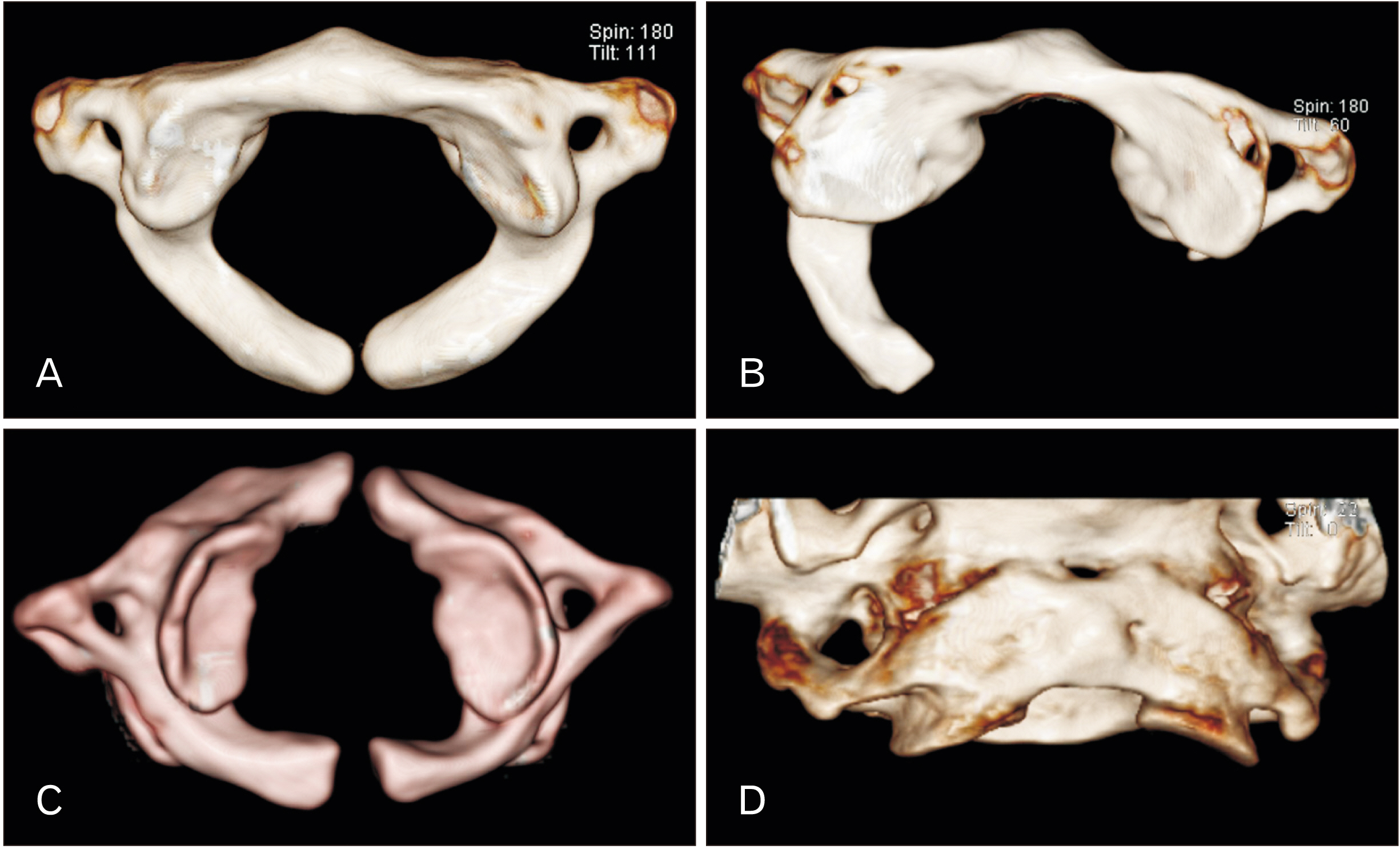
Fig. 1 Three dimensional reconstructed computed tomography scan showing the type A (A), type B (B) posterior arch defects, and combined anterior and posterior arch defect (C). The posterior view (B) of atlanto-occipital assimilation (D) is also seen.
Reference
-
References
1. Currarino G, Rollins N, Diehl JT. 1994; Congenital defects of the posterior arch of the atlas: a report of seven cases including an affected mother and son. AJNR Am J Neuroradiol. 15:249–54. PMID: 8192068. PMCID: PMC8334620.2. Dorne HL, Lander PH. 1986; CT recognition of anomalies of the posterior arch of the atlas vertebra: differentiation from fracture. AJNR Am J Neuroradiol. 7:176–7. PMID: 3082136. PMCID: PMC8334795.3. Karavelioglu E, Kacar E, Karavelioglu A, Gonul Y, Guven M. 2014; Congenital defect of the anterior arch of the atlas: a case report and review of the literature. Neurol India. 62:303–4. DOI: 10.4103/0028-3886.136974. PMID: 25033853.
Article4. Kazanci B, Kahveci R, Ekici MA, Guclu B. 2013; Isolated fracture of anterior arch of atlas in a child: case report and review of the literature. Injury. 44:1956–8. DOI: 10.1016/j.injury.2013.08.004. PMID: 24041431.
Article5. Petre BM, Karp JE, Riley LH 3rd. 2012; Athletic cervical spine injury in the setting of fusion failure of the anterior and posterior atlas. Orthopedics. 35:e1449–52. DOI: 10.3928/01477447-20120822-39. PMID: 22955419.
Article6. Park Y, Kim SM, Lee YT, Yoo JH, Oh HC, Ha JW, Sung SY, Yoon HK, Chang JH, Jung JY. 2014; Congenital anomaly of the atlas misdiagnosed as posterior arch fracture of the atlas and atlantoaxial subluxation. Clin Orthop Surg. 6:96–100. DOI: 10.4055/cios.2014.6.1.96. PMID: 24605195. PMCID: PMC3942609.
Article7. Klimo P Jr, Blumenthal DT, Couldwell WT. 2003; Congenital partial aplasia of the posterior arch of the atlas causing myelopathy: case report and review of the literature. Spine (Phila Pa 1976). 28:E224–8. DOI: 10.1097/01.BRS.0000065492.85852.A9. PMID: 12811285.
Article8. Pasku D, Katonis P, Karantanas A, Hadjipavlou A. 2007; Congenital posterior atlas defect associated with anterior rachischisis and early cervical degenerative disc disease: a case study and review of the literature. Acta Orthop Belg. 73:282–5. PMID: 17515248.9. Geipel P. 1955; Zur Kenntnis der Spaltbildung des Atlas und Epistropheus. IV. Teil [Studies on the fissure formation of the atlas and epistropheus. IV]. Zentralbl Allg Pathol. 94:19–84. German.10. Senoglu M, Safavi-Abbasi S, Theodore N, Bambakidis NC, Crawford NR, Sonntag VK. 2007; The frequency and clinical significance of congenital defects of the posterior and anterior arch of the atlas. J Neurosurg Spine. 7:399–402. DOI: 10.3171/SPI-07/10/399. PMID: 17933313.
Article11. Kwon JK, Kim MS, Lee GJ. 2009; The incidence and clinical implications of congenital defects of atlantal arch. J Korean Neurosurg Soc. 46:522–7. DOI: 10.3340/jkns.2009.46.6.522. PMID: 20062566. PMCID: PMC2803266.
Article12. Guenkel S, Schlaepfer S, Gordic S, Wanner GA, Simmen HP, Werner CM. 2013; Incidence and variants of posterior arch defects of the atlas vertebra. Radiol Res Pract. 2013:957280. DOI: 10.1155/2013/957280. PMID: 24109510. PMCID: PMC3784273.
Article13. Hyun G, Allam E, Sander P, Hasiak C, Zhou Y. 2018; The prevalence of congenital C1 arch anomalies. Eur Spine J. 27:1266–71. DOI: 10.1007/s00586-017-5283-4. PMID: 28849400.
Article14. Martellacci S, Ben Salem D, Méjean N, Sautreaux JL, Krausé D. 2008; A case of foramen magnum syndrome caused by atlanto-occipital assimilation with intracanal fibrosis. Surg Radiol Anat. 30:149–52. DOI: 10.1007/s00276-007-0288-z. PMID: 18259680.
Article15. Junewick JJ, Chin MS, Meesa IR, Ghori S, Boynton SJ, Luttenton CR. 2011; Ossification patterns of the atlas vertebra. AJR Am J Roentgenol. 197:1229–34. DOI: 10.2214/AJR.10.5403. PMID: 22021519.
Article16. Sharma A, Gaikwad SB, Deol PS, Mishra NK, Kale SS. 2000; Partial aplasia of the posterior arch of the atlas with an isolated posterior arch remnant: findings in three cases. AJNR Am J Neuroradiol. 21:1167–71. PMID: 10871035. PMCID: PMC7973897.17. Wang K, Li X, Lou H, Luo B. 2010; Recurrent attacks of headache and neck pain caused by congenital aplasia of the posterior arch of atlas in an adult. BMJ Case Rep. 2010:bcr0520103053. DOI: 10.1136/bcr.05.2010.3053. PMID: 22791780. PMCID: PMC3029836.
Article
- Full Text Links
-
- Actions
-
Cited
- CITED
-
- Close
- Share
-
- Similar articles
-
- Combined Congenital Anterior and Posterior Midline Cleft of the Atlas Associated with Asymptomatic Lateral Atlantoaxial Subluxation
- Cerebellar Ectopia Associated with Unilateral Agenesis of Posterior Arch of Atlas
- Congenital Anomaly of the Atlas Misdiagnosed as Posterior Arch Fracture of the Atlas and Atlantoaxial Subluxation
- Congenital Cleft of Anterior Arch and Partial Aplasia of the Posterior Arch of the C1
- The Incidence and Clinical Implications of Congenital Defects of Atlantal Arch
