Miniscrew insertion sites of infrazygomatic crest and mandibular buccal shelf in different vertical craniofacial patterns: A cone-beam computed tomography study
- Affiliations
-
- 1Department of Orthodontics, Guarulhos University, São Paulo, Brazil
- 2Department of Orthodontics, Faculty of Medicine and Dentistry, University of Alberta, Edmonton, AB, Canada
- 3Department of Orthodontics, School of Dentistry, Universidade Norte do Paraná, Paraná, Brazil
- 4Private Practice, Belo Horizonte, Brazil
- 5Department of Orthodontics, Ingá Dental School, Maringá, Brazil
- 6Private Practice, Rio de Janeiro, Brazil
- 7Department of Social and Pediatric Dentistry, Institute of Science and Technology, São Paulo State University, São José dos Campos, Brazil
- KMID: 2522501
- DOI: http://doi.org/10.4041/kjod.2021.51.6.387
Abstract
Objective
To identify optimal areas for the insertion of extra-alveolar miniscrews into the infrazygomatic crest (IZC) and mandibular buccal shelf (MBS), using cone beam computed tomography (CBCT) imaging in patients with different craniofacial patterns.
Methods
CBCT reconstructions of untreated individuals were used to evaluate the IZC and MBS areas. The participants were divided into three groups, based on the craniofacial pattern, namely, brachyfacial (n = 15; mean age, 23.3 years), mesofacial (n = 15; mean age, 19.24 years), and dolichofacial (n = 15; mean age, 17.79 years). In the IZC, the evaluated areas were at 11, 13, and 15 mm above the buccal cusp tips of the right and left first molars. In the MBS, the evaluated areas were at the projections of the first molars’ distal roots and second molars’ mesial and distal roots, at a 4- and 8-mm distance from the cementoenamel junction. Intergroup comparisons were performed with analysis of variance and the Tukey test.
Results
There was no statistically significant difference in the IZC bone thickness among the groups. For MBS bone availability, some comparisons revealed no difference; meanwhile, other comparisons revealed increased MBS bone thickness in the brachyfacial (first molars distal roots) and dolichofacial (second molars mesial and distal roots) patterns.
Conclusions
There was no significant difference in the IZC bone thickness among the groups. The facial skeletal pattern may affect the availability of ideal bone thickness for the insertion of extra-alveolar miniscrews in the MBS region; however, this variability is unlikely to be clinically meaningful.
Keyword
Figure
-
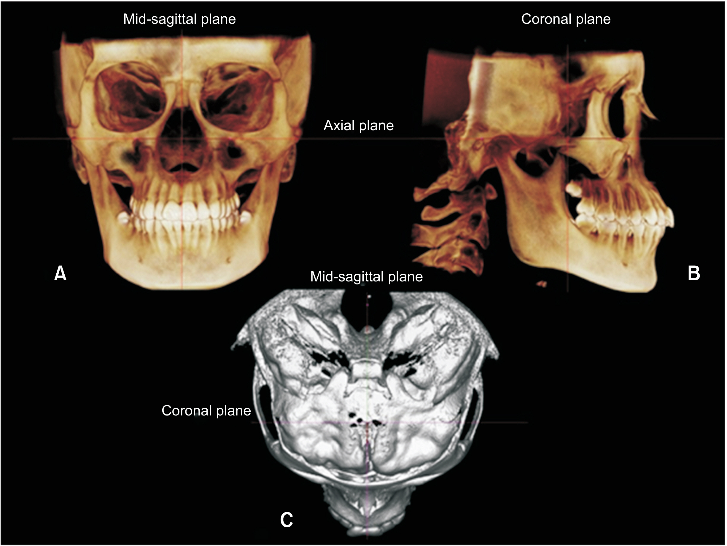
Figure 1 Head standardization. A, Frontal view. B, Sagittal view. C, Axial view.
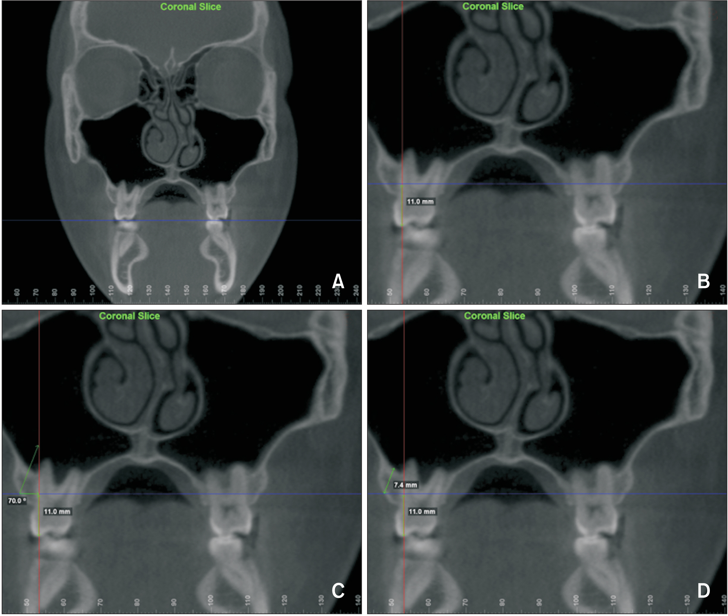
Figure 2 A, Coronal slice consisting of the apex of the distobuccal root of the permanent first maxillary molar. B-D, Measurement of infrazygomatic crest thickness, performed at 11 mm above the buccal tip of the right first maxillary molar at an angle of 70 degrees to the occlusal plane.

Figure 3 Bucco-lingual thicknesses of total bone measured on two horizontal reference lines apically located at 4 mm and 8 mm from the cementoenamel junction.

Figure 4 Color based diagram showing infrazygomatic crest (IZC) areas of adequate cortical bone thickness.
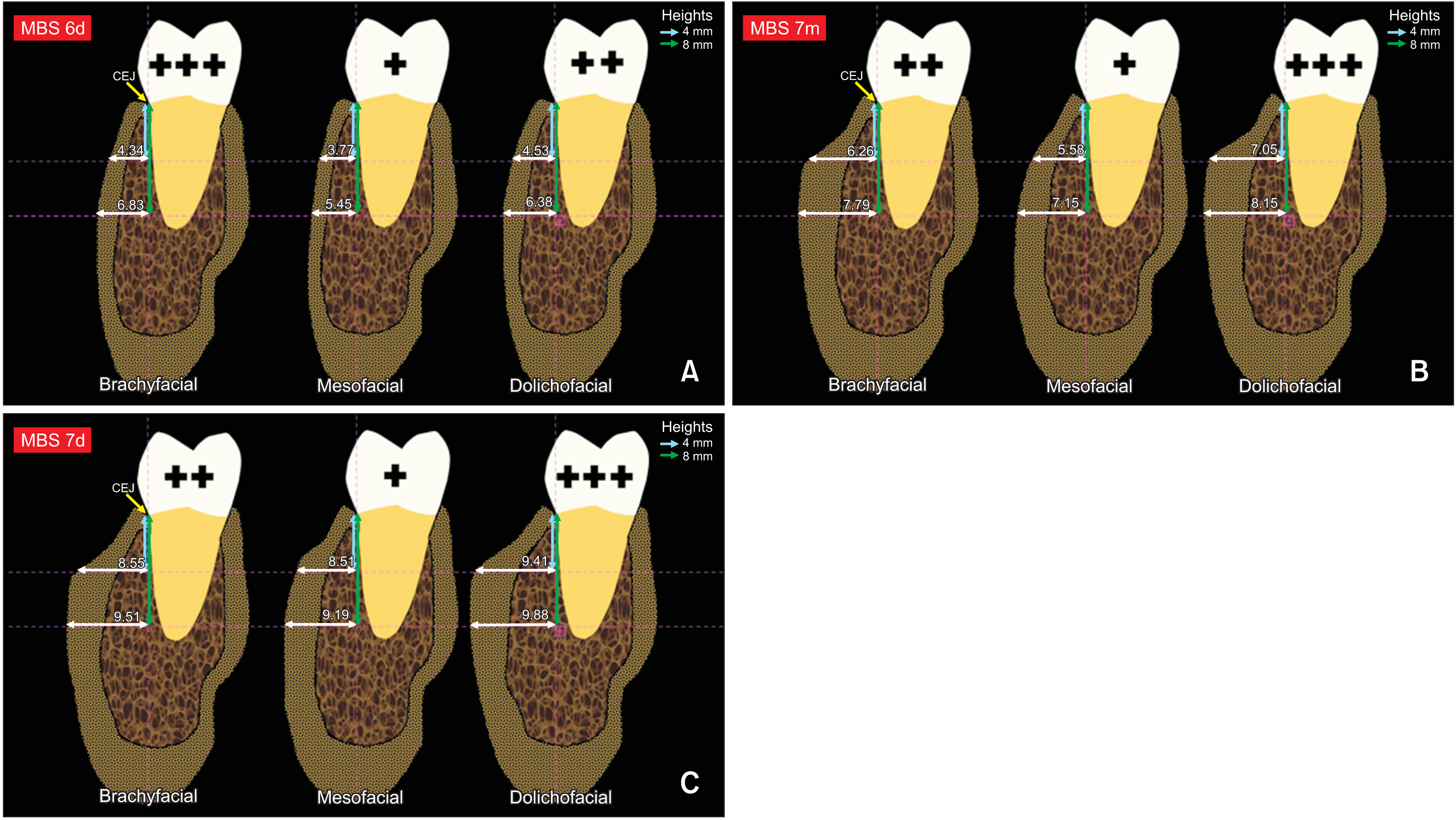
Figure 5 A, Bone thickness for different craniofacial patterns at MBS 6d. B, Bone thickness for different craniofacial patterns at MBS 7m. C, Bone thickness for different craniofacial patterns at MBS 7d. MBS, mandibular buccal shelf; 6, first molar; 7, second molar; m, mesial; d, distal; (+), (++), (+++), amount of cortical bone available in mandibular buccal shelf.
Reference
-
1. Baumgaertel S. 2014; Temporary skeletal anchorage devices: the case for miniscrews. Am J Orthod Dentofacial Orthop. 145:558–64. DOI: 10.1016/j.ajodo.2014.03.009. PMID: 24785918.
Article2. Baumgaertel S. 2011; Cortical bone thickness and bone depth of the posterior palatal alveolar process for mini-implant insertion in adults. Am J Orthod Dentofacial Orthop. 140:806–11. DOI: 10.1016/j.ajodo.2011.05.020. PMID: 22133945.
Article3. Baumgaertel S, Hans MG. 2009; Buccal cortical bone thickness for mini-implant placement. Am J Orthod Dentofacial Orthop. 136:230–5. DOI: 10.1016/j.ajodo.2007.10.045. PMID: 19651353.
Article4. Gracco A, Lombardo L, Cozzani M, Siciliani G. 2008; Quantitative cone-beam computed tomography evaluation of palatal bone thickness for orthodontic miniscrew placement. Am J Orthod Dentofacial Orthop. 134:361–9. DOI: 10.1016/j.ajodo.2007.01.027. PMID: 18774082.
Article5. Baumgaertel S, Hans MG. 2009; Assessment of infrazygomatic bone depth for mini-screw insertion. Clin Oral Implants Res. 20:638–42. DOI: 10.1111/j.1600-0501.2008.01691.x. PMID: 19281501.
Article6. Chang C, Liu SS, Roberts WE. 2015; Primary failure rate for 1680 extra-alveolar mandibular buccal shelf mini-screws placed in movable mucosa or attached gingiva. Angle Orthod. 85:905–10. DOI: 10.2319/092714.695.1. PMID: 25603272.
Article7. Nucera R, Lo Giudice A, Bellocchio AM, Spinuzza P, Caprioglio A, Perillo L, et al. 2017; Bone and cortical bone thickness of mandibular buccal shelf for mini-screw insertion in adults. Angle Orthod. 87:745–51. DOI: 10.2319/011117-34.1. PMID: 28598220. PMCID: PMC8357207.
Article8. Liou EJ, Chen PH, Wang YC, Lin JC. 2007; A computed tomographic image study on the thickness of the infrazygomatic crest of the maxilla and its clinical implications for miniscrew insertion. Am J Orthod Dentofacial Orthop. 131:352–6. DOI: 10.1016/j.ajodo.2005.04.044. PMID: 17346590.
Article9. Chang C, Huang C, Roberts WE. 2016; 3D cortical bone anatomy of the mandibular buccal shelf: a CBCT study to define sites for extra-alveolar bone screws to treat Class III malocclusion. Int J Orthod Implantol. 41:74–82.10. Elshebiny T, Palomo JM, Baumgaertel S. 2018; Anatomic assessment of the mandibular buccal shelf for miniscrew insertion in white patients. Am J Orthod Dentofacial Orthop. 153:505–11. DOI: 10.1016/j.ajodo.2017.08.014. PMID: 29602342.
Article11. Tsunori M, Mashita M, Kasai K. 1998; Relationship between facial types and tooth and bone characteristics of the mandible obtained by CT scanning. Angle Orthod. 68:557–62. DOI: 10.1043/0003-3219(1998)068<0557:RBFTAT>2.3.CO;2. PMID: 9851354.12. Ozdemir F, Tozlu M, Germec-Cakan D. 2013; Cortical bone thickness of the alveolar process measured with cone-beam computed tomography in patients with different facial types. Am J Orthod Dentofacial Orthop. 143:190–6. DOI: 10.1016/j.ajodo.2012.09.013. PMID: 23374925.
Article13. Sadek MM, Sabet NE, Hassan IT. 2015; Alveolar bone mapping in subjects with different vertical facial dimensions. Eur J Orthod. 37:194–201. DOI: 10.1093/ejo/cju034. PMID: 25114124.
Article14. Gandhi V, Upadhyay M, Tadinada A, Yadav S. 2021; Variability associated with mandibular buccal shelf area width and height in subjects with different growth pattern, sex, and growth status. Am J Orthod Dentofacial Orthop. 159:59–70. DOI: 10.1016/j.ajodo.2019.11.020. PMID: 33221093.
Article15. Murugesan A, Sivakumar A. 2020; Comparison of bone thickness in infrazygomatic crest area at various miniscrew insertion angles in Dravidian population - a cone beam computed tomography study. Int Orthod. 18:105–14. DOI: 10.1016/j.ortho.2019.12.001. PMID: 31926867.16. Ricketts RM. 1961; Cephalometric analysis and synthesis. Angle Orthod. 31:141–56.17. Neves LS, Pinzan A, Janson G, Canuto CE, de Freitas MR, Cançado RH. 2005; Comparative study of the maturation of permanent teeth in subjects with vertical and horizontal growth patterns. Am J Orthod Dentofacial Orthop. 128:619–23. DOI: 10.1016/j.ajodo.2004.05.026. PMID: 16286209.
Article18. Li JL, Kau C, Wang M. 2014; Changes of occlusal plane inclination after orthodontic treatment in different dentoskeletal frames. Prog Orthod. 15:41. DOI: 10.1186/s40510-014-0041-1. PMID: 25033937. PMCID: PMC4884032.
Article19. Houston WJ. 1983; The analysis of errors in orthodontic measurements. Am J Orthod. 83:382–90. DOI: 10.1016/0002-9416(83)90322-6. PMID: 6573846.
Article20. Dahlberg G. 1940. Statistical methods for medical and biological students. Allen & Unwin;London:21. Liu H, Wu X, Yang L, Ding Y. 2017; Safe zones for miniscrews in maxillary dentition distalization assessed with cone-beam computed tomography. Am J Orthod Dentofacial Orthop. 151:500–6. DOI: 10.1016/j.ajodo.2016.07.021. PMID: 28257734.
Article22. de Freitas LM, de Freitas KM, Pinzan A, Janson G, de Freitas MR. 2010; A comparison of skeletal, dentoalveolar and soft tissue characteristics in white and black Brazilian subjects. J Appl Oral Sci. 18:135–42. DOI: 10.1590/S1678-77572010000200007. PMID: 20485924. PMCID: PMC5349749.23. Castilla EE, Luquetti DV. 2009; Brazil: public health genomics. Public Health Genomics. 12:53–8. DOI: 10.1159/000153424. PMID: 19023184.
Article24. Cunha A, Nelson-Filho P, Marañón-Vásquez GA, Ramos AGC, Dantas B, Sebastiani AM, et al. 2019; Genetic variants in ACTN3 and MYO1H are associated with sagittal and vertical craniofacial skeletal patterns. Arch Oral Biol. 97:85–90. DOI: 10.1016/j.archoralbio.2018.09.018. PMID: 30366217.
Article25. Janson G, Quaglio CL, Pinzan A, Franco EJ, de Freitas MR. 2011; Craniofacial characteristics of Caucasian and Afro-Caucasian Brazilian subjects with normal occlusion. J Appl Oral Sci. 19:118–24. DOI: 10.1590/S1678-77572011000200007. PMID: 21552712. PMCID: PMC4243749.
Article26. Storniolo-Souza JM, Seminario MP, Pinzan-Vercelino CRM, Pinzan A, Janson G. 2021; McNamara analysis cephalometric parameters in White-Brazilians, Japanese and Japanese-Brazilians with normal occlusion. Dental Press J Orthod. 26:e2119133. DOI: 10.1590/2177-6709.26.1.e2119133.oar. PMID: 33759961. PMCID: PMC8018756.
Article27. Vargas EOA, Lopes de Lima R, Nojima LI. 2020; Mandibular buccal shelf and infrazygomatic crest thicknesses in patients with different vertical facial heights. Am J Orthod Dentofacial Orthop. 158:349–56. DOI: 10.1016/j.ajodo.2019.08.016. PMID: 32862936.28. Masumoto T, Hayashi I, Kawamura A, Tanaka K, Kasai K. 2001; Relationships among facial type, buccolingual molar inclination, and cortical bone thickness of the mandible. Eur J Orthod. 23:15–23. DOI: 10.1093/ejo/23.1.15. PMID: 11296507.
Article
- Full Text Links
-
- Actions
-
Cited
- CITED
-
- Close
- Share
-
- Similar articles
-
- Evaluation of mandibular buccal shelf characteristics in the Colombian population: A cone-beam computed tomography study
- Assessment of lower incisor alveolar bone width using cone-beam computed tomography images in skeletal Class III adults of different vertical patterns
- Radiomorphometric analysis of edentulous posterior mandibular ridges in the first molar region: a cone-beam computed tomography study
- Quantitative evaluation of alveolar cortical bone density in adults with different vertical facial types using cone-beam computed tomography
- Cone beam computed tomography findings of ectopic mandibular third molar in the mandibular condyle: report of a case
