Quantitative evaluation of alveolar cortical bone density in adults with different vertical facial types using cone-beam computed tomography
- Affiliations
-
- 1Department of Orthodontics, Faculty of Dentistry, Yeditepe University, Istanbul, Turkey. dgermec@gmail.com
- KMID: 2273269
- DOI: http://doi.org/10.4041/kjod.2014.44.1.36
Abstract
OBJECTIVE
The purpose of this study was to quantitatively evaluate the cortical bone densities of the maxillary and mandibular alveolar processes in adults with different vertical facial types using cone-beam computed tomography (CBCT) images.
METHODS
CBCT images (n = 142) of adult patients (20-45 years) were classified into hypodivergent, normodivergent, and hyperdivergent groups on the basis of linear and angular S-N/Go-Me measurements. The cortical bone densities (in Hounsfield units) at maxillary and mandibular interdental sites from the distal aspect of the canine to the mesial aspect of the second molar were measured on the images.
RESULTS
On the maxillary buccal side, female subjects in the hyperdivergent group showed significantly decreased bone density, while in the posterior region, male subjects in the hyperdivergent group displayed significantly decreased bone density when compared with corresponding subjects in the other groups (p<0.001). Furthermore, the subjects in the hyperdivergent group had significantly lower bone densities on the mandibular buccal side than hypodivergent subjects. The maxillary palatal bone density did not differ significantly among groups, but female subjects showed significantly denser palatal cortical bone. No significant difference in bone density was found between the palatal and buccal sides in the maxillary premolar region. Overall, the palatal cortical bone was denser anteriorly and buccal cortical bone was denser posteriorly.
CONCLUSION
Adults with the hyperdivergent facial type tend to have less-dense buccal cortical bone in the maxillary and mandibular alveolar processes. Clinicians should be aware of the variability of cortical bone densities at mini-implant placement sites.
MeSH Terms
Figure
-
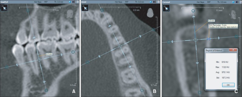
Figure 1 Cortical bone density measurement. A, The vertical reference line bisects the interdental area and is parallel to the long axes of the roots in the sagittal slice. B, The vertical reference line bisects the interdental area in the axial slice. C, The cortical bone density is measured in Hounsfield units (HU) between points (one at the outer surface and the other at the border of the cortical and cancellous bone) on a line perpendicular to the bone surface 4 mm apical to the alveolar crest in the coronal slice.
Cited by 2 articles
-
Quantitative evaluation of palatal bone thickness in patients with normal and open vertical skeletal configurations using cone-beam computed tomography
Piyoros Suteerapongpun, Tanapan Wattanachai, Apirum Janhom, Polbhat Tripuwabhrut, Dhirawat Jotikasthira
Imaging Sci Dent. 2018;48(1):51-57. doi: 10.5624/isd.2018.48.1.51.Differences in the mandibular premolar positions in Angle Class I subjects with different vertical facial types: A cone-beam computed tomography study
Jun Duan, Feng Deng, Wan-Shan Li, Xue-Lei Li, Lei-Lei Zheng, Gui-Yuan Li, Yan-Jie Bai
Korean J Orthod. 2015;45(4):180-189. doi: 10.4041/kjod.2015.45.4.180.
Reference
-
1. Park HM, Kim BH, Yang IH, Baek SH. Preliminary three-dimensional analysis of tooth movement and arch dimension change of the maxillary dentition in Class II division 1 malocclusion treated with first premolar extraction: conventional anchorage vs. mini-implant anchorage. Korean J Orthod. 2012; 42:280–290. PMID: 23323242.
Article2. Schätzle M, Männchen R, Zwahlen M, Lang NP. Survival and failure rates of orthodontic temporary anchorage devices: a systematic review. Clin Oral Implants Res. 2009; 20:1351–1359. PMID: 19793320.
Article3. Miyawaki S, Koyama I, Inoue M, Mishima K, Sugahara T, Takano-Yamamoto T. Factors associated with the stability of titanium screws placed in the posterior region for orthodontic anchorage. Am J Orthod Dentofacial Orthop. 2003; 124:373–378. PMID: 14560266.
Article4. Motoyoshi M, Yoshida T, Ono A, Shimizu N. Effect of cortical bone thickness and implant placement torque on stability of orthodontic mini-implants. Int J Oral Maxillofac Implants. 2007; 22:779–784. PMID: 17974113.5. Tozlu M, Nalbantgil D, Öztoprak MO, Özdemir F. Mini-implantların devrilmesini önlemede yeni bir yaklaşm / A new approach to prevent migration of mini-implants. Turkish J Orthod. 2011; 24:170–180.6. Ozdemir F, Tozlu M, Germec-Cakan D. Cortical bone thickness of the alveolar process measured with cone-beam computed tomography in patients with different facial types. Am J Orthod Dentofacial Orthop. 2013; 143:190–196. PMID: 23374925.
Article7. Cha JY, Kil JK, Yoon TM, Hwang CJ. Miniscrew stability evaluated with computerized tomography scanning. Am J Orthod Dentofacial Orthop. 2010; 137:73–79. PMID: 20122434.
Article8. Iijima M, Takano M, Yasuda Y, Muguruma T, Nakagaki S, Sakakura Y, et al. Effect of the quantity and quality of cortical bone on the failure force of a miniscrew implant. Eur J Orthod. 2013; 35:583–589. PMID: 23041933.
Article9. Lundström A, McWilliam JS. A comparison of vertical and horizontal cephalometric variables with regard to heritability. Eur J Orthod. 1987; 9:104–108. PMID: 3472887.10. Moon CH, Park HK, Nam JS, Im JS, Baek SH. Relationship between vertical skeletal pattern and success rate of orthodontic mini-implants. Am J Orthod Dentofacial Orthop. 2010; 138:51–57. PMID: 20620833.
Article11. Duckmanton NA, Austin BW, Lechner SK, Klineberg IJ. Imaging for predictable maxillary implants. Int J Prosthodont. 1994; 7:77–80. PMID: 8179788.12. Aranyarachkul P, Caruso J, Gantes B, Schulz E, Riggs M, Dus I, et al. Bone density assessments of dental implant sites: 2. Quantitative cone-beam computerized tomography. Int J Oral Maxillofac Implants. 2005; 20:416–424. PMID: 15973953.13. de Oliveira RC, Leles CR, Normanha LM, Lindh C, Ribeiro-Rotta RF. Assessments of trabecular bone density at implant sites on CT images. Oral Surg Oral Med Oral Pathol Oral Radiol Endod. 2008; 105:231–238. PMID: 18230392.
Article14. Nomura Y, Watanabe H, Shirotsu K, Honda E, Sumi Y, Kurabayshi T. Stability of voxel values from cone-beam computed tomography for dental use in evaluating bone mineral content. Clin Oral Implants Res. 2013; 24:543–548. PMID: 22320314.
Article15. Lagravère MO, Fang Y, Carey J, Toogood RW, Packota GV, Major PW. Density conversion factor determined using a cone-beam computed tomography unit NewTom QR-DVT 9000. Dentomaxillofac Radiol. 2006; 35:407–409. PMID: 17082330.
Article16. Mah P, Reeves TE, McDavid WD. Deriving Hounsfield units using grey levels in cone beam computed tomography. Dentomaxillofac Radiol. 2010; 39:323–335. PMID: 20729181.
Article17. Nomura Y, Watanabe H, Honda E, Kurabayashi T. Reliability of voxel values from cone-beam computed tomography for dental use in evaluating bone mineral density. Clin Oral Implants Res. 2010; 21:558–562. PMID: 20443807.
Article18. Marquezan M, Lau TC, Mattos CT, Cunha AC, Nojima LI, Sant'Anna EF, et al. Bone mineral density. Angle Orthod. 2012; 82:62–66. PMID: 21774580.
Article19. Cassetta M, Stefanelli LV, Pacifici A, Pacifici L, Barbato E. How accurate Is CBCT in measuring bone density? A comparative CBCT-CT in vitro study. Clin Implant Dent Relat Res. 2013; 1. 07. [Epub ahead of print].
Article20. Nackaerts O, Maes F, Yan H, Couto Souza P, Pauwels R, Jacobs R. Analysis of intensity variability in multislice and cone beam computed tomography. Clin Oral Implants Res. 2011; 22:873–879. PMID: 21244502.
Article21. Silva IM, Freitas DQ, Ambrosano GM, Bóscolo FN, Almeida SM. Bone density: comparative evaluation of Hounsfield units in multislice and cone-beam computed tomography. Braz Oral Res. 2012; 26:550–556. PMID: 23184166.
Article22. Bryant JA, Drage NA, Richmond S. Study of the scan uniformity from an i-CAT cone beam computed tomography dental imaging system. Dentomaxillofac Radiol. 2008; 37:365–374. PMID: 18812597.
Article23. Pauwels R, Nackaerts O, Bellaiche N, Stamatakis H, Tsiklakis K, Walker A, et al. SEDENTEXCT Project Consortium. Variability of dental cone beam CT grey values for density estimations. Br J Radiol. 2013; 86:20120135. PMID: 23255537.
Article24. Moon SH, Park SH, Lim WH, Chun YS. Palatal bone density in adult subjects: implications for mini-implant placement. Angle Orthod. 2010; 80:137–144. PMID: 19852653.
Article25. Park HS, Lee YJ, Jeong SH, Kwon TG. Density of the alveolar and basal bones of the maxilla and the mandible. Am J Orthod Dentofacial Orthop. 2008; 133:30–37. PMID: 18174068.
Article26. Han S, Bayome M, Lee J, Lee YJ, Song HH, Kook YA. Evaluation of palatal bone density in adults and adolescents for application of skeletal anchorage devices. Angle Orthod. 2012; 82:625–631. PMID: 22077190.
Article27. Choi JH, Park CH, Yi SW, Lim HJ, Hwang HS. Bone density measurement in interdental areas with simulated placement of orthodontic miniscrew implants. Am J Orthod Dentofacial Orthop. 2009; 136:766. PMID: 19962594.
Article28. Mavropoulos A, Kiliaridis S, Bresin A, Ammann P. Effect of different masticatory functional and mechanical demands on the structural adaptation of the mandibular alveolar bone in young growing rats. Bone. 2004; 35:191–197. PMID: 15207756.
Article29. Cheng SJ, Tseng IY, Lee JJ, Kok SH. A prospective study of the risk factors associated with failure of mini-implants used for orthodontic anchorage. Int J Oral Maxillofac Implants. 2004; 19:100–106. PMID: 14982362.
- Full Text Links
-
- Actions
-
Cited
- CITED
-
- Close
- Share
-
- Similar articles
-
- The bone density of mandible as the aging process in Koreans
- Assessment of the relationship between the maxillary molars and adjacent structures using cone beam computed tomography
- Evaluation of imaging reformation with cone beam computed tomography for the assessment of bone density and shape in mandible
- Analysis of the root position of the maxillary incisors in the alveolar bone using cone-beam computed tomography
- Evaluation of alveolar bone density by intraoral periapical radiography
