Apoptin gene delivery by a PAMAM dendrimer modified with a nuclear localization signal peptide as a gene carrier for brain cancer therapy
- Affiliations
-
- 1Department of Physiology, College of Medicine, Cardiovascular and Metabolic Disease Center, Smart Marine Therapeutics Center, Inje University, Busan 47392, Korea.
- 2Division of Applied Medicine, Research Institute for Korea Medicine, School of Korean Medicine, Pusan National University, Busan 50612, Korea.
- 3Department of Biochemistry, College of Natural Sciences, Chungnam National University, Daejeon 34134, Korea.
- KMID: 2519409
- DOI: http://doi.org/10.4196/kjpp.2021.25.5.467
Abstract
- In this study, we aimed to synthesize PAMAMG3 derivatives (PAMAMG3-KRRR and PAMAMG3-HKRRR), using KRRR peptides as a nuclear localization signal and introduced histidine residues into the KRRR-grafted PAMAMG3 for delivering a therapeutic, carcinoma cell-selective apoptosis gene, apoptin into human primary glioma (GBL-14) cells and human dermal fibroblasts. We examined their cytotoxicity and gene expression using luciferase activity and enhanced green fluorescent protein PAMAMG3 derivatives in both cell lines. We treated cells with PAMAMG3 derivative/apoptin complexes and investigated their intracellular distribution using confocal microscopy. The PAMAMG3-KRRR and PAMAMG3-HKRRR dendrimers were found to escape from endolysosomes into the cytosol. The JC-1 assay, glutathione levels, and Annexin V staining results showed that apoptin triggered cell death in GBL-14 cells. Overall, these findings indicated that the PAMAMG3-HKRRR/apoptin complex is a potential candidate for an effective nonviral gene delivery system for brain tumor therapy in vitro.
Keyword
Figure
-

Fig. 1 Synthesis of nonviral gene delivery systems. (A) 1H NMR spectra of PAMAMG3, PAMAMG3-KRRR, and PAMAMG3-HKRRR. (B) The synthesis scheme of PAMAM derivates conjugated with nuclear localization signal (NLS) peptides. (C) Scheme 1. Diagram of the endocytic pathway and the formation of PAMAMG3-HKRRR/pJDK-apoptin complex and apoptin gene delivery into the nucleus. Apoptin promotes apoptotic cell death of GBL-14 cells.

Fig. 2 PicoGreen assays of PAMAMG3 derivative/apoptin complexes. Complexes were prepared using PAMAMG3, PAMAMG3-KRRR, and PAMAMG3-HKRRR with pJDK or pJDK-apoptin at different weight ratios. The fluorescence intensity was normalized to 100% with pJDK and pJDK-apoptin DNA. Values are expressed as the mean ± SD of three independent experiments (n = 3).

Fig. 3 Cytotoxicity assay of the PAMAMG3 derivatives. (A, B) GBL-14 cells and (C, D) human dermal fibroblasts (HDFs) were exposed to various concentrations of PAMAMG3, PAMAMG3-KRRR, and PAMAMG3-HKRRR. Cytotoxicity was assessed by WST-1 assay after 24 h (A, C) and 48 h (B, D). Values are expressed as the mean ± SD of three independent experiments (n = 3). (E, F) GBL-14 cells and (G, H) HDFs were incubated under similar condition as in WST-1 assay. Cytotoxicity was assessed using LDH assay after 24 h (E, G) and 48 h (F, H). Values are expressed as the mean ± SD of three independent experiments (n = 3).

Fig. 4 In vitro transfection efficiency and cell viability of the PAMAMG3 derivatives. (A) GBL-14 cells and (C) human dermal fibroblasts (HDFs) were exposed to PAMAMG3, PAMAMG3-KRRR, and PAMAMG3-HKRRR complexes with pJDK/Luci at different weight ratios for 24 h. (B, D) Cytotoxicity of PAMAMG3 derivative/Luci complexes. Viability of the complexes was assessed using the WST-1 assay. Values are expressed as the mean ± SD of three independent experiments (n = 3).
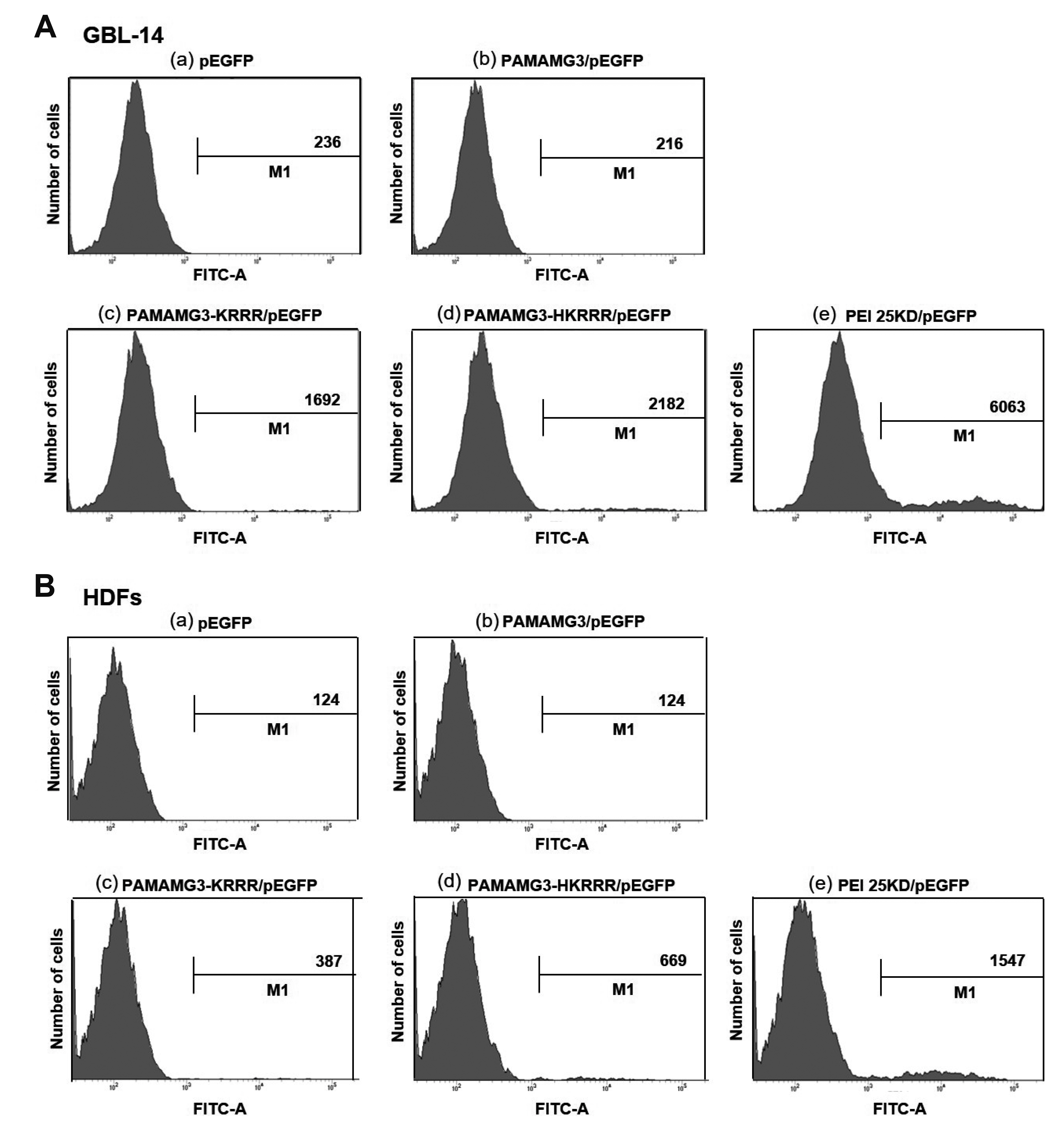
Fig. 5 GFP expression by the PAMAMG3 derivatives. (A) GBL-14 cells and (B) human dermal fibroblasts (HDFs) were exposed to pEGFP, PAMAMG3/pEGFP, PAMAMG3-KRRR/pEGFP, and PAMAMG3-HKRRR/pEGFP for 24 h. The cells were harvested, and the cell fluorescence intensity of GFP-positive cells was quantitatively analyzed by flow cytometry. The x-axis shows fluorescence intensity and y-axis shows number of cells.

Fig. 6 Uptake and intracellular localization of the PAMAMG3 derivatives. (A) GBL-14 cells and (B) human dermal fibroblasts (HDFs) were treated with PAMAMG3, PAMAMG3-KRRR, and PAMAMG3-HKRRR labeled with Alexa Fluor 546 and pJDK or pJDK-apoptin at the ratio of 4:1 for 24 h. After 24 h post-exposure, the images were obtained by confocal microscopy. Red fluorescence indicated DNA-labeled with Alexa Fluor 546 dye and blue fluorescence indicated nuclei stained with DAPI. (C, D) Each cell was exposed to Alexa Fluor 488-labeled PAMAMG3, PAMAMG3-KRRR, and PAMAMG3-HKRRR with pJDK or pJDK-apoptin at the ratio of 4:1 for 24 h. After 24 h post-exposed, the cells were harvested. The live cells were stained with LysoTracker Red for lysosomes and counterstained with DAPI for nuclei.

Fig. 7 Mitochondrial membrane depolarization and ROS levels induced by PAMAMG3 derivative/pJDK-apoptin. (A) GBL-14 cells and (B) human dermal fibroblasts (HDFs) were exposed to PAMAMG3, PAMAMG3-KRRR, and PAMAMG3-HKRRR with pJDK or pJDK-apoptin for 24 h. The cells were analyzed for mitochondrial membrane potential (MMP) using flow cytometry after JC-1 staining. The number in the bottom right quadrant indicates the percentage of MMP loss. (C, D) Each cell was exposed to similar conditions as those in panel (A). After 24 h post-exposure, intracellular ROS levels were assessed using GSH assay as descried in methods. Values are indicted as the mean ± SD of three independent experiments (n = 3). Statistical analyses were conducted using the unpaired two-tailed Students t-test. Asterisks indicate statistically significant values (**p < 0.01, *p < 0.1, and not significant [n.s.] [p > 0.05]).

Fig. 8 Cytotoxicity of the PAMAMG3 derivative/apoptin complexes. (A, B) GBL-14 cells and (C, D) human dermal fibroblasts (HDFs) were incubated with PAMAMG3, PAMAMG3-KRRR, and PAMAMG3-HKRRR with pJDK or pJDK-apoptin for 24 h. After 24 h post-exposure, cytotoxicity of each polymer and polyplex was determined using the WST-1 assay. Values are expressed as mean ± SD of three independent experiments (n = 3). Statistical analyses were conducted using the unpaired two-tailed Students t-test. Asterisks indicate statistically significant values (**p < 0.01, *p < 0.1, and not significant [n.s.] [p > 0.05]).
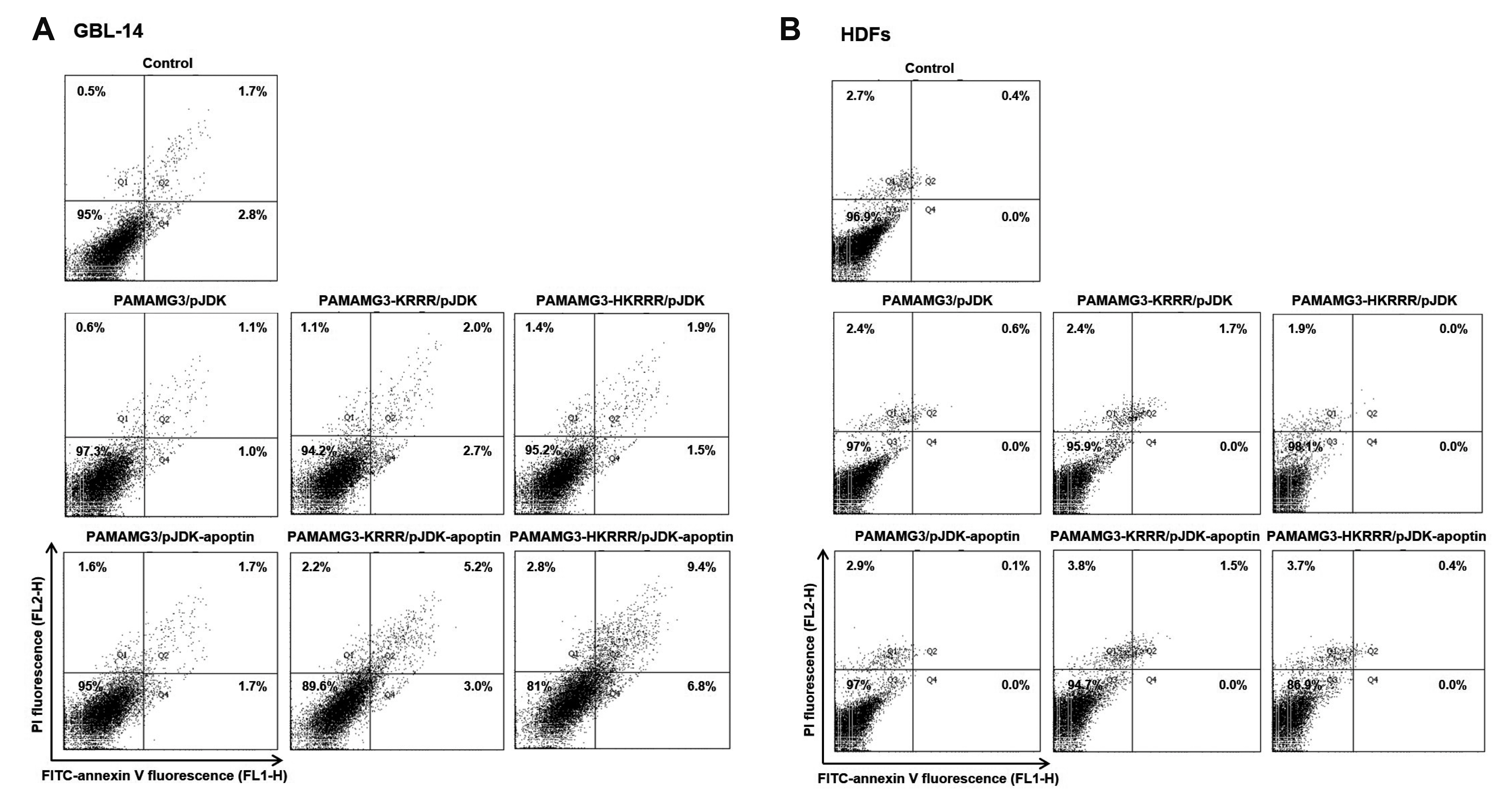
Fig. 9 Apoptosis induced the PAMAMG3 derivative/pJDK-apoptin. (A, B) Annexin V staining of PAMAMG3 derivative/pJDK-apoptin using flow cytometry. GBL-14 cells and human dermal fibroblasts (HDFs) were incubated with PAMAMG3, PAMAMG3-KRRR, and PAMAMG3-HKRRR with pJDK and pJDK-apoptin. After 24 h post-exposure, apoptosis levels for each polyplex were assessed by flow cytometry. Representative histograms showing four quadrants (Q1: percentage of necrosis, Q2: percentage of late apoptosis, Q3: percentage of live cells, and Q4: percentage of early apoptosis).
Reference
-
1. Zeng W, Tang Z, Li Y, Yin G, Liu Z, Gao J, Chen Y, Chen F. 2020; Patient-derived xenografts of different grade gliomas retain the heterogeneous histological and genetic features of human gliomas. Cancer Cell Int. 20:1. DOI: 10.1186/s12935-019-1086-5. PMID: 31908598. PMCID: PMC6941273.
Article2. Assi H, Candolfi M, Baker G, Mineharu Y, Lowenstein PR, Castro MG. 2012; Gene therapy for brain tumors: basic developments and clinical implementation. Neurosci Lett. 527:71–77. DOI: 10.1016/j.neulet.2012.08.003. PMID: 22906921. PMCID: PMC3462660.
Article3. Reardon DA, Wen PY. 2006; Therapeutic advances in the treatment of glioblastoma: rationale and potential role of targeted agents. Oncologist. 11:152–164. DOI: 10.1634/theoncologist.11-2-152. PMID: 16476836.
Article4. Taylor OG, Brzozowski JS, Skelding KA. 2019; Glioblastoma multiforme: an overview of emerging therapeutic targets. Front Oncol. 9:963. DOI: 10.3389/fonc.2019.00963. PMID: 31616641. PMCID: PMC6775189.
Article5. Pourgholi F, Hajivalili M, Farhad JN, Kafil HS, Yousefi M. 2016; Nanoparticles: novel vehicles in treatment of glioblastoma. Biomed Pharmacother. 77:98–107. DOI: 10.1016/j.biopha.2015.12.014. PMID: 26796272.
Article6. Lin G, Zhang H, Huang L. 2015; Smart polymeric nanoparticles for cancer gene delivery. Mol Pharm. 12:314–321. DOI: 10.1021/mp500656v. PMID: 25531409. PMCID: PMC4319689.
Article7. Balakrishnan B, David E. 2019; Biopolymers augment viral vectors based gene delivery. J Biosci. 44:84. DOI: 10.1007/s12038-019-9905-3. PMID: 31502562.
Article8. Dunbar CE, High KA, Joung JK, Kohn DB, Ozawa K, Sadelain M. 2018; Gene therapy comes of age. Science. 359:eaan4672. DOI: 10.1126/science.aan4672. PMID: 29326244.
Article9. Ramamoorth M, Narvekar A. 2015; Non viral vectors in gene therapy-an overview. J Clin Diagn Res. 9:GE01–GE06. DOI: 10.7860/JCDR/2015/10443.5394. PMID: 25738007. PMCID: PMC4347098.10. Choi YS, Lee MY, David AE, Park YS. 2014; Nanoparticles for gene delivery: therapeutic and toxic effects. Mol Cell Toxicol. 10:1–8. DOI: 10.1007/s13273-014-0001-3.
Article11. Wang LH, Wu T, Wu DC, You YZ. 2016; Bioreducible gene delivery vector capable of self-scavenging the intracellular-generated ROS exhibiting high gene transfection. ACS Appl Mater Interfaces. 8:19238–19244. DOI: 10.1021/acsami.6b04327. PMID: 27420138.
Article12. Nitta SK, Numata K. 2013; Biopolymer-based nanoparticles for drug/gene delivery and tissue engineering. Int J Mol Sci. 14:1629–1654. DOI: 10.3390/ijms14011629. PMID: 23344060. PMCID: PMC3565338.
Article13. Hu J, Zhu M, Liu K, Fan H, Zhao W, Mao Y, Zhang Y. 2016; A biodegradable polyethylenimine-based vector modified by trifunctional peptide R18 for enhancing gene transfection efficiency in vivo. PLoS One. 11:e0166673. DOI: 10.1371/journal.pone.0166673. PMID: 27935984. PMCID: PMC5147860.
Article14. Abbasi E, Aval SF, Akbarzadeh A, Milani M, Nasrabadi HT, Joo SW, Hanifehpour Y, Nejati-Koshki K, Pashaei-Asl R. 2014; Dendrimers: synthesis, applications, and properties. Nanoscale Res Lett. 9:247. DOI: 10.1186/1556-276X-9-247. PMID: 24994950. PMCID: PMC4074873.
Article15. Taghavi PAN, Mutlu P, Khodadust R, Gunduz U. 2013; Poly amidoamine PAMAM nanoparticles: synthesis and biomedical applications. Hacet J Biol Chem. 41:289–299.16. Dutta T, Jain NK, McMillan NA, Parekh HS. 2010; Dendrimer nanocarriers as versatile vectors in gene delivery. Nanomedicine. 6:25–34. DOI: 10.1016/j.nano.2009.05.005. PMID: 19450708.17. Kolhatkar RB, Kitchens KM, Swaan PW, Ghandehari H. 2007; Surface acetylation of polyamidoamine (PAMAM) dendrimers decreases cytotoxicity while maintaining membrane permeability. Bioconjug Chem. 18:2054–2060. DOI: 10.1021/bc0603889. PMID: 17960872.
Article18. Li J, Han Y, Lu Y, Song B, Zhao M, Hu H, Chen D. 2018; A novel disulfide bond-mediated cleavable RGD-modified PAMAM nanocomplex containing nuclear localization signal HMGB1 for enhancing gene transfection efficiency. Int J Nanomedicine. 13:7135–7153. DOI: 10.2147/IJN.S182445. PMID: 30464464. PMCID: PMC6228086.
Article19. Li J, Liang H, Liu J, Wang Z. 2018; Poly (amidoamine) (PAMAM) dendrimer mediated delivery of drug and pDNA/siRNA for cancer therapy. Int J Pharm. 546:215–225. DOI: 10.1016/j.ijpharm.2018.05.045. PMID: 29787895.
Article20. Lee J, Jung J, Kim YJ, Lee E, Choi JS. 2014; Gene delivery of PAMAM dendrimer conjugated with the nuclear localization signal peptide originated from fibroblast growth factor 3. Int J Pharm. 459:10–18. DOI: 10.1016/j.ijpharm.2013.11.027. PMID: 24275448.
Article21. Lee J, Lee S, Kwon YE, Kim YJ, Choi JS. 2019; Gene delivery by PAMAM dendrimer conjugated with the nuclear localization signal peptide derived from influenza B virus nucleoprotein. Macromol Res. 27:360–368. DOI: 10.1007/s13233-019-7057-9.
Article22. Rollano Peñaloza OM, Lewandowska M, Stetefeld J, Ossysek K, Madej M, Bereta J, Sobczak M, Shojaei S, Ghavami S, Łos MJ. 2014; Apoptins: selective anticancer agents. Trends Mol Med. 20:519–528. DOI: 10.1016/j.molmed.2014.07.003. PMID: 25164066.
Article23. Noteborn MH, van der Eb AJ. 1998; Apoptin-induced apoptosis: potential for antitumor therapy. Drug Resist Updat. 1:99–103. DOI: 10.1016/S1368-7646(98)80024-1. PMID: 16904395.
Article24. Maddika S, Booy EP, Johar D, Gibson SB, Ghavami S, Los M. 2005; Cancer-specific toxicity of apoptin is independent of death receptors but involves the loss of mitochondrial membrane potential and the release of mitochondrial cell-death mediators by a Nur77-dependent pathway. J Cell Sci. 118(Pt 19):4485–4493. DOI: 10.1242/jcs.02580. PMID: 16179607.
Article25. Hou Z, Mao J, Lu Y, Li L. 2018; rApoptin induces apoptosis in human breast cancer cells via phosphorylation of Nur77 and Akt. Biochem Biophys Res Commun. 498:221–227. DOI: 10.1016/j.bbrc.2018.02.204. PMID: 29501489.
Article26. An S, Nam K, Choi S, Bai CZ, Lee Y, Park JS. 2013; Nonviral gene therapy in vivo with PAM-RG4/apoptin as a potential brain tumor therapeutic. Int J Nanomedicine. 8:821–834. DOI: 10.2147/IJN.S39072. PMID: 23589689. PMCID: PMC3622651.27. Bae Y, Green ES, Kim GY, Song SJ, Mun JY, Lee S, Park JI, Park JS, Ko KS, Han J, Choi JS. 2016; Dipeptide-functionalized polyamidoamine dendrimer-mediated apoptin gene delivery facilitates apoptosis of human primary glioma cells. Int J Pharm. 515:186–200. DOI: 10.1016/j.ijpharm.2016.09.083. PMID: 27732896.
Article28. Bae Y, Jung MK, Song SJ, Green ES, Lee S, Park HS, Jeong SH, Han J, Mun JY, Ko KS, Choi JS. 2017; Functional nanosome for enhanced mitochondria-targeted gene delivery and expression. Mitochondrion. 37:27–40. DOI: 10.1016/j.mito.2017.06.005. PMID: 28669809.
Article29. Holder AL, Goth-Goldstein R, Lucas D, Koshland CP. 2012; Particle-induced artifacts in the MTT and LDH viability assays. Chem Res Toxicol. 25:1885–1892. DOI: 10.1021/tx3001708. PMID: 22799765. PMCID: PMC3446248.
Article30. Liu BR, Lo SY, Liu CC, Chyan CL, Huang YW, Aronstam RS, Lee HJ. 2013; Endocytic trafficking of nanoparticles delivered by cell-penetrating peptides comprised of nona-arginine and a penetration accelerating sequence. PLoS One. 8:e67100. DOI: 10.1371/journal.pone.0067100. PMID: 23840594. PMCID: PMC3694042.
Article31. Elefantova K, Lakatos B, Kubickova J, Sulova Z, Breier A. 2018; Detection of the mitochondrial membrane potential by the cationic dye JC-1 in L1210 cells with massive overexpression of the plasma membrane ABCB1 drug transporter. Int J Mol Sci. 19:1985. DOI: 10.3390/ijms19071985. PMID: 29986516. PMCID: PMC6073605.
Article32. Dubey A, Goswami M, Yadav K, Chaudhary D. 2015; Oxidative stress and nano-toxicity induced by TiO2 and ZnO on WAG cell line. PLoS One. 10:e0127493. DOI: 10.1371/journal.pone.0127493. PMID: 26011447. PMCID: PMC4444277.
Article33. Uram Ł, Szuster M, Gargasz K, Filipowicz A, Wałajtys-Rode E, Wołowiec S. 2013; In vitro cytotoxicity of the ternary PAMAM G3-pyridoxal-biotin bioconjugate. Int J Nanomedicine. 8:4707–4720. DOI: 10.2147/IJN.S53254. PMID: 24376351. PMCID: PMC3864882.34. Martin ME, Rice KG. 2007; Peptide-guided gene delivery. AAPS J. 9:E18–E29. DOI: 10.1208/aapsj0901003. PMID: 17408236. PMCID: PMC2751301.
Article35. Al-Dosari MS, Gao X. 2009; Nonviral gene delivery: principle, limitations, and recent progress. AAPS J. 11:671–681. DOI: 10.1208/s12248-009-9143-y. PMID: 19834816. PMCID: PMC2782077.
Article
- Full Text Links
-
- Actions
-
Cited
- CITED
-
- Close
- Share
-
- Similar articles
-
- Monitoring Gene Therapy by Radionuclide Approaches
- Modulation of Electroosmotic Flow through Skin: Effect of Poly(Amidoamine) Dendrimers
- The molecular mechanism for nuclear transport and its application
- A immunohistochemical study of localization of calcitonin gene related peptide in the rats cochlear nucleus and superior olivary complex
- Molecular Imaging of Biological Gene Delivery Vehicles for Targeted Cancer Therapy: Beyond Viral Vectors
