Luteolin inhibits H2O2 -induced cellular senescence via modulation of SIRT1 and p53
- Affiliations
-
- 1Department of Biochemistry, Wonkwang University School of Medicine, Iksan 54538, Korea
- KMID: 2516869
- DOI: http://doi.org/10.4196/kjpp.2021.25.4.297
Abstract
- Luteolin, a sort of flavonoid, has been reported to be involved in neuroprotective function via suppression of neuroinflammation. In this study, we investigated the protective effect of luteolin against oxidative stress-induced cellular senescence and its molecular mechanism using hydrogen peroxide (H2O2)-induced cellular senescence model in House Ear Institute-Organ of Corti 1 cells (HEI-OC1). Our results showed that luteolin attenuated senescent phenotypes including alterations of morphology, cell proliferation, senescence-associated β-galactosidase expression, DNA damage, as well as related molecules expression such as p53 and p21 in the oxidant challenged model. Interestingly, we found that luteolin induces expression of sirtuin 1 in dose- and time-dependent manners and it has protective role against H2O2 -induced cellular senescence by upregulation of sirtuin 1 (SIRT1). In contrast, the inhibitory effect of luteolin on cellular senescence under oxidative stress was abolished by silencing of SIRT1. This study indicates that luteolin effectively protects against oxidative stress-induced cellular senescence through p53 and SIRT1. These results suggest that luteolin possesses therapeutic potentials against age-related hearing loss that are induced by oxidative stress.
Figure
-

Fig. 1 Luteolin reduces hydrogen peroxide (H2O2)-induced cellular senescence in House Ear Institute-Organ of Corti 1 (HEI-OC1) cells. (A–C) Cells were pretreated with luteolin for 12 h, then incubated in 30 μM H2O2 for 3 days. Cell viability was determined by MTT assay (A). The percentage of SA-β-gal positive cells out of total cells was counted and the average data was obtained from three independent experiments (B). The senescent phenotype of HEI-OC1 cells was detected by the SA-β-gal assay (×100) (C). (D) Cell growth was evaluated by MTT assay at various time points indicated in the figure after addition of H2O2. Data represents means values of triple experiments. *p < 0.05 vs. control, #p < 0.05 vs. H2O2. Piperine was used as a positive control.

Fig. 2 Protective effect of luteolin against hydrogen peroxide (H2O2)-induced cellular senescence is dependent on p53. (A) Cells were pretreated with 2 μM luteolin for 12 h, then incubated in 30 μM H2O2 for 3 days. (B) Cells were transfected with various concentrations siRNAs of control or p53, then incubated with 30 μM H2O2 for 3 days. The protein levels were detected by Western blotting and quantified by densitometry based on immunoblot images (A, B). (C) Representative images of SA-β-gal staining of HEI-OC1 (×100). (D) The percentage of senescent cells were calculated in (D). Data represents means values of triple experiments. *p < 0.05 vs. control; #p < 0.05 vs. H2O2-treated cells.

Fig. 3 Effects of luteolin on sirtuin 1 (SIRT1) expression in House Ear Institute-Organ of Corti 1 cells (HEI-OC1). (A, C) HEI-OC1 cells were treated with luteolin (0–4 μM, 12 h). (B, D) Cells were treated with luteolin (2 μM, 0–12 h). The proteins level (A, B) and SIRT1 activity (C, D) were performed with Western blotting and SIRT1 fluorometric kit, respectively. The proteins were quantified by densitometry based on immunoblot images. Data represents means values of triple experiments. Significance vs. control: *p < 0.05.
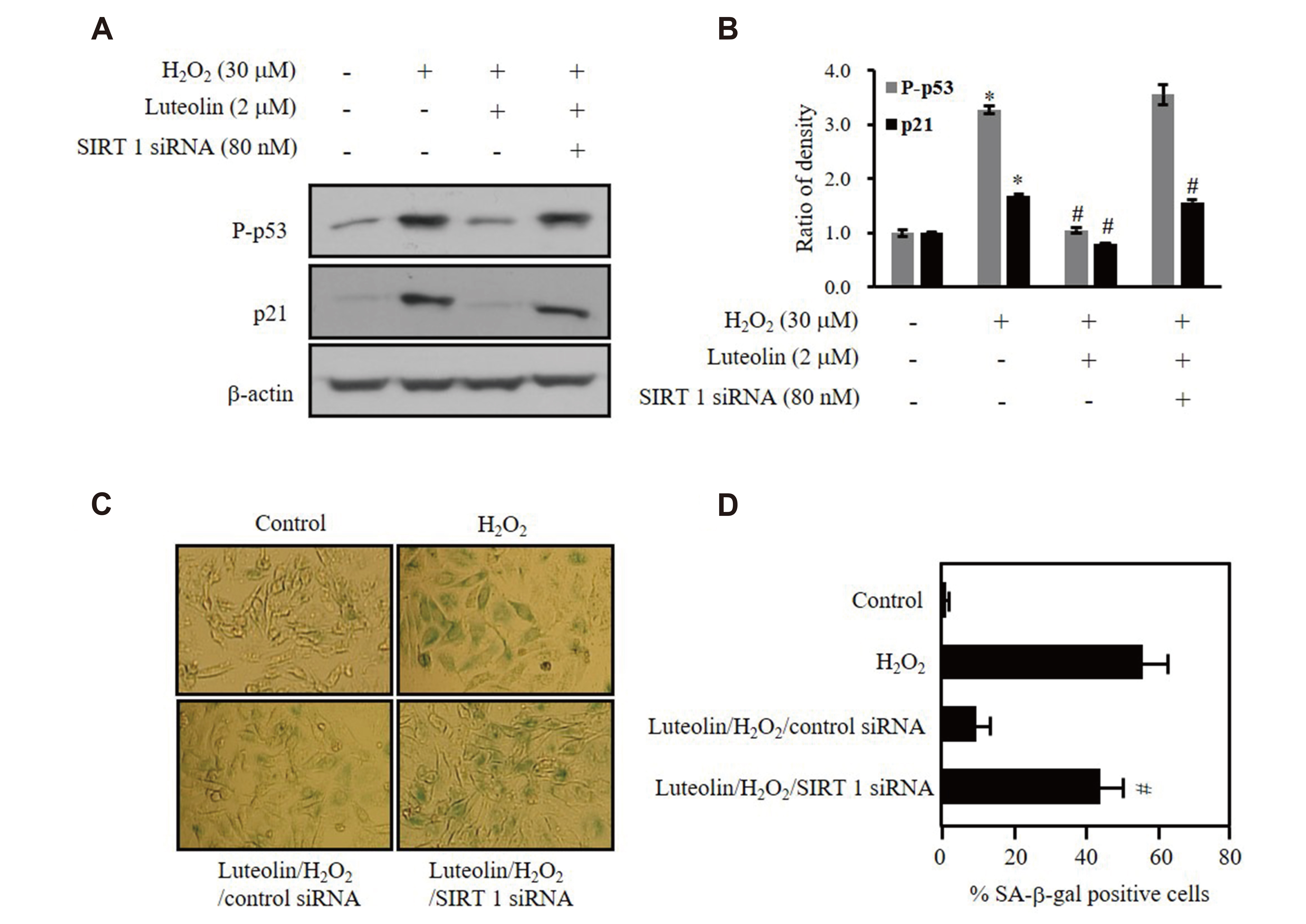
Fig. 4 Luteolin protects against hydrogen peroxide (H2O2)-induced p53 phosphorylation via sirtuin 1 (SIRT1) protein expression. House Ear Institute-Organ of Corti 1 (HEI-OC1) cells were transfected with 80 nM siRNAs of control or SIRT1, then incubated in 2 μM luteolin for 12 h, followed by 30 μM H2O2 treatment for additional 3 days. (A, B) The protein levels were detected by Western blotting and quantified by densitometry based on immunoblot images. (C) Representative images of SA-β-gal staining of HEI-OC1 (×100). (D) The percentage of senescent cells were calculated in (D). Data represents means values of triple experiments. *p < 0.05 vs. control; #p < 0.05 vs. H2O2-treated cells.

Fig. 5 Luteolin prevents DNA from hydrogen peroxide (H2O2)-induced damage. (A, B) House Ear Institute-Organ of Corti 1 (HEI-OC1) cells were transfected with 80 nM siRNAs of sirtuin 1 (SIRT1) or p53, then incubated in 2 μM luteolin for 12 h, followed by 30 μM H2O2 treatment for additional 3 days. Comet assay was performed as described in Methods. Data show the results of one of three independent experiments. *p < 0.05 vs. H2O2, #p < 0.05 vs. luteolin/H2O2/control siRNA.
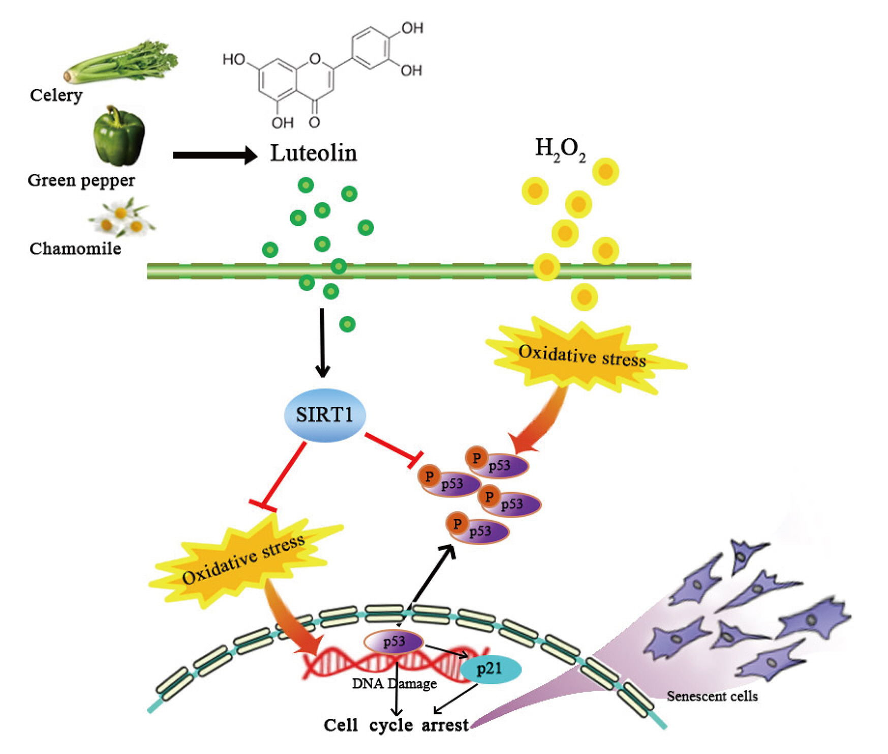
Fig. 6 A schematic illustration summarizing the protective effect of Luteolin against hydrogen peroxide (H2O2)-induced premature senescence in House Ear Institute-Organ of Corti 1 (HEI-OC1) cells.
Reference
-
1. Qian Y, Chen X. 2010; Tumor suppression by p53: making cells senescent. Histol Histopathol. 25:515–526. DOI: 10.14670/HH-25.515. PMID: 20183804. PMCID: PMC2841029.2. Chen QM, Bartholomew JC, Campisi J, Acosta M, Reagan JD, Ames BN. 1998; Molecular analysis of H2O2-induced senescent-like growth arrest in normal human fibroblasts: p53 and Rb control G1 arrest but not cell replication. Biochem J. 332(Pt 1):43–50. DOI: 10.1042/bj3320043. PMID: 9576849. PMCID: PMC1219449.3. Frippiat C, Dewelle J, Remacle J, Toussaint O. 2002; Signal transduction in H2O2-induced senescence-like phenotype in human diploid fibroblasts. Free Radic Biol Med. 33:1334–1346. DOI: 10.1016/S0891-5849(02)01044-4. PMID: 12419465.4. MacNee W. 2005; Pulmonary and systemic oxidant/antioxidant imbalance in chronic obstructive pulmonary disease. Proc Am Thorac Soc. 2:50–60. DOI: 10.1513/pats.200411-056SF. PMID: 16113469.
Article5. Rahman I, Biswas SK, Kode A. 2006; Oxidant and antioxidant balance in the airways and airway diseases. Eur J Pharmacol. 533:222–239. DOI: 10.1016/j.ejphar.2005.12.087. PMID: 16500642.
Article6. Han J, Miyamae Y, Shigemori H, Isoda H. 2010; Neuroprotective effect of 3,5-di-O-caffeoylquinic acid on SH-SY5Y cells and senescence-accelerated-prone mice 8 through the up-regulation of phosphoglycerate kinase-1. Neuroscience. 169:1039–1045. DOI: 10.1016/j.neuroscience.2010.05.049. PMID: 20570715.
Article7. Luo J, Nikolaev AY, Imai S, Chen D, Su F, Shiloh A, Guarente L, Gu W. 2001; Negative control of p53 by Sir2alpha promotes cell survival under stress. Cell. 107:137–148. DOI: 10.1016/S0092-8674(01)00524-4. PMID: 11672522.8. Ota H, Akishita M, Eto M, Iijima K, Kaneki M, Ouchi Y. 2007; SIRT1 modulates premature senescence-like phenotype in human endothelial cells. J Mol Cell Cardiol. 43:571–579. DOI: 10.1016/j.yjmcc.2007.08.008. PMID: 17916362.
Article9. Yi J, Luo J. 2010; SIRT1 and p53, effect on cancer, senescence and beyond. Biochim Biophys Acta. 1804:1684–1689. DOI: 10.1016/j.bbapap.2010.05.002. PMID: 20471503. PMCID: PMC2989880.
Article10. Ota H, Tokunaga E, Chang K, Hikasa M, Iijima K, Eto M, Kozaki K, Akishita M, Ouchi Y, Kaneki M. 2006; SIRT1 inhibitor, Sirtinol, induces senescence-like growth arrest with attenuated Ras-MAPK signaling in human cancer cells. Oncogene. 25:176–185. DOI: 10.1038/sj.onc.1209049. PMID: 16170353.
Article11. Ju W, Wang X, Shi H, Chen W, Belinsky SA, Lin Y. 2007; A critical role of luteolin-induced reactive oxygen species in blockage of tumor necrosis factor-activated nuclear factor-kappaB pathway and sensitization of apoptosis in lung cancer cells. Mol Pharmacol. 71:1381–1388. DOI: 10.1124/mol.106.032185. PMID: 17296806.12. Shi RX, Ong CN, Shen HM. 2004; Luteolin sensitizes tumor necrosis factor-alpha-induced apoptosis in human tumor cells. Oncogene. 23:7712–7721. DOI: 10.1038/sj.onc.1208046. PMID: 15334063.13. Xagorari A, Papapetropoulos A, Mauromatis A, Economou M, Fotsis T, Roussos C. 2001; Luteolin inhibits an endotoxin-stimulated phosphorylation cascade and proinflammatory cytokine production in macrophages. J Pharmacol Exp Ther. 296:181–187. PMID: 11123379.14. Lin Y, Shi R, Wang X, Shen HM. 2008; Luteolin, a flavonoid with potential for cancer prevention and therapy. Curr Cancer Drug Targets. 8:634–646. DOI: 10.2174/156800908786241050. PMID: 18991571. PMCID: PMC2615542.
Article15. Kang KP, Park SK, Kim DH, Sung MJ, Jung YJ, Lee AS, Lee JE, Ramkumar KM, Lee S, Park MH, Roh SG, Kim W. 2011; Luteolin ameliorates cisplatin-induced acute kidney injury in mice by regulation of p53-dependent renal tubular apoptosis. Nephrol Dial Transplant. 26:814–822. DOI: 10.1093/ndt/gfq528. PMID: 20817674.
Article16. Jang S, Dilger RN, Johnson RW. 2010; Luteolin inhibits microglia and alters hippocampal-dependent spatial working memory in aged mice. J Nutr. 140:1892–1898. DOI: 10.3945/jn.110.123273. PMID: 20685893. PMCID: PMC2937579.
Article17. Xu B, Li XX, He GR, Hu JJ, Mu X, Tian S, Du GH. 2010; Luteolin promotes long-term potentiation and improves cognitive functions in chronic cerebral hypoperfused rats. Eur J Pharmacol. 627:99–105. DOI: 10.1016/j.ejphar.2009.10.038. PMID: 19857483.
Article18. Kalinec GM, Webster P, Lim DJ, Kalinec F. 2003; A cochlear cell line as an in vitro system for drug ototoxicity screening. Audiol Neurootol. 8:177–189. DOI: 10.1159/000071059. PMID: 12811000.19. Yu HB, Li DY, Zhang HF, Xue HZ, Pan CE, Zhao SH, Wang L. 2010; Resveratrol inhibits invasion and metastasis of hepatocellular carcinoma cells. J Anim Vet Adv. 9:3117–3124. DOI: 10.3923/javaa.2010.3117.3124.
Article20. Lee DS, Li B, Keo S, Kim KS, Jeong GS, Oh H, Kim YC. 2013; Inhibitory effect of 9-hydroxy-6,7-dimethoxydalbergiquinol from Dalbergia odorifera on the NF-κB-related neuroinflammatory response in lipopolysaccharide-stimulated mouse BV2 microglial cells is mediated by heme oxygenase-1. Int Immunopharmacol. 17:828–835. DOI: 10.1016/j.intimp.2013.08.024. PMID: 24055019.
Article21. Dimri GP, Lee X, Basile G, Acosta M, Scott G, Roskelley C, Medrano EE, Linskens M, Rubelj I, Pereira-Smith O, Peacocke M, Campisi J. 1995; A biomarker that identifies senescent human cells in culture and in aging skin in vivo. Proc Natl Acad Sci U S A. 92:9363–9367. DOI: 10.1073/pnas.92.20.9363. PMID: 7568133. PMCID: PMC40985.
Article22. Cheng XS, Li MS, Du J, Jiang QY, Wang L, Yan SY, Yu DM, Deng JB. 2011; Neuronal apoptosis in the developing cerebellum. Anat Histol Embryol. 40:21–27. DOI: 10.1111/j.1439-0264.2010.01033.x. PMID: 21231956.
Article23. Olive PL, Banáth JP. 2006; The comet assay: a method to measure DNA damage in individual cells. Nat Protoc. 1:23–29. DOI: 10.1038/nprot.2006.5. PMID: 17406208.
Article24. Muller M. 2009; Cellular senescence: molecular mechanisms, in vivo significance, and redox considerations. Antioxid Redox Signal. 11:59–98. DOI: 10.1089/ars.2008.2104. PMID: 18976161.25. Andrade LN, Nathanson JL, Yeo GW, Menck CF, Muotri AR. 2012; Evidence for premature aging due to oxidative stress in iPSCs from Cockayne syndrome. Hum Mol Genet. 21:3825–3834. DOI: 10.1093/hmg/dds211. PMID: 22661500. PMCID: PMC3412382.
Article26. Tilstra JS, Robinson AR, Wang J, Gregg SQ, Clauson CL, Reay DP, Nasto LA, St Croix CM, Usas A, Vo N, Huard J, Clemens PR, Stolz DB, Guttridge DC, Watkins SC, Garinis GA, Wang Y, Niedernhofer LJ, Robbins PD. 2012; NF-κB inhibition delays DNA damage-induced senescence and aging in mice. J Clin Invest. 122:2601–2612. DOI: 10.1172/JCI45785. PMID: 22706308. PMCID: PMC3386805.
Article27. Wölfle U, Esser PR, Simon-Haarhaus B, Martin SF, Lademann J, Schempp CM. 2011; UVB-induced DNA damage, generation of reactive oxygen species, and inflammation are effectively attenuated by the flavonoid luteolin in vitro and in vivo. Free Radic Biol Med. 50:1081–1093. DOI: 10.1016/j.freeradbiomed.2011.01.027. PMID: 21281711.28. Rodier F, Campisi J, Bhaumik D. 2007; Two faces of p53: aging and tumor suppression. Nucleic Acids Res. 35:7475–7484. DOI: 10.1093/nar/gkm744. PMID: 17942417. PMCID: PMC2190721.
Article29. Mao GX, Wang Y, Qiu Q, Deng HB, Yuan LG, Li RG, Song DQ, Li YY, Li DD, Wang Z. 2010; Salidroside protects human fibroblast cells from premature senescence induced by H2O2 partly through modulating oxidative status. Mech Ageing Dev. 131:723–731. DOI: 10.1016/j.mad.2010.10.003. PMID: 21035481.30. Duan J, Duan J, Zhang Z, Tong T. 2005; Irreversible cellular senescence induced by prolonged exposure to H2O2 involves DNA-damage-and-repair genes and telomere shortening. Int J Biochem Cell Biol. 37:1407–1420. DOI: 10.1016/j.biocel.2005.01.010. PMID: 15833273.31. Rajendran R, Garva R, Krstic-Demonacos M, Demonacos C. 2011; Sirtuins: molecular traffic lights in the crossroad of oxidative stress, chromatin remodeling, and transcription. J Biomed Biotechnol. 2011:368276. DOI: 10.1155/2011/368276. PMID: 21912480. PMCID: PMC3168296.
Article32. Chua KF, Mostoslavsky R, Lombard DB, Pang WW, Saito S, Franco S, Kaushal D, Cheng HL, Fischer MR, Stokes N, Murphy MM, Appella E, Alt FW. 2005; Mammalian SIRT1 limits replicative life span in response to chronic genotoxic stress. Cell Metab. 2:67–76. DOI: 10.1016/j.cmet.2005.06.007. PMID: 16054100.
Article33. Lim CS, Potts M, Helm RF. 2006; Nicotinamide extends the replicative life span of primary human cells. Mech Ageing Dev. 127:511–514. DOI: 10.1016/j.mad.2006.02.001. PMID: 16545428.
Article34. Chung S, Yao H, Caito S, Hwang JW, Arunachalam G, Rahman I. 2010; Regulation of SIRT1 in cellular functions: role of polyphenols. Arch Biochem Biophys. 501:79–90. DOI: 10.1016/j.abb.2010.05.003. PMID: 20450879. PMCID: PMC2930135.
Article35. Kaeberlein M, McDonagh T, Heltweg B, Hixon J, Westman EA, Caldwell SD, Napper A, Curtis R, DiStefano PS, Fields S, Bedalov A, Kennedy BK. 2005; Substrate-specific activation of sirtuins by resveratrol. J Biol Chem. 280:17038–17045. DOI: 10.1074/jbc.M500655200. PMID: 15684413.
Article36. de Boer VC, de Goffau MC, Arts IC, Hollman PC, Keijer J. 2006; SIRT1 stimulation by polyphenols is affected by their stability and metabolism. Mech Ageing Dev. 127:618–627. DOI: 10.1016/j.mad.2006.02.007. PMID: 16603228.
Article37. Davis JM, Murphy EA, Carmichael MD, Davis B. 2009; Quercetin increases brain and muscle mitochondrial biogenesis and exercise tolerance. Am J Physiol Regul Integr Comp Physiol. 296:R1071–R1077. DOI: 10.1152/ajpregu.90925.2008. PMID: 19211721.
Article
- Full Text Links
-
- Actions
-
Cited
- CITED
-
- Close
- Share
-
- Similar articles
-
- A Novel Role of Hyaluronic Acid and Proteoglycan Link Protein 1 (HAPLN1) in Delaying Vascular Endothelial Cell Senescence
- Tenovin-1 Induces Senescence and Decreases Wound-Healing Activity in Cultured Rat Primary Astrocytes
- Nervonic Acid Inhibits Replicative Senescence of Human Wharton’s Jelly-Derived Mesenchymal Stem Cells
- SIRT1 Inhibits p53 but not NF-kappaB Transcriptional Activity during Differentiation of Mouse Embryonic Stem Cells into Embryoid Bodies
- Enhanced Viral Replication by Cellular Replicative Senescence
