Feasibility of Improving the Accuracy of Dose Calculation Using Hybrid Computed Tomography Images: A Phantom Study
- Affiliations
-
- 1Department of Radiation Oncology and Research Institute for Convergence of Biomedical Science and Technology, Pusan National University Yangsan Hospital, Yangsan, Korea
- 2Department of Radiation Oncology, Pusan National University School of Medicine, Yangsan, Korea
- 3Department of Radiation Oncology, Pusan National University Hospital, Busan, Korea
- KMID: 2514822
- DOI: http://doi.org/10.14316/pmp.2021.32.1.18
Abstract
- Purpose
Kilovoltage computed tomography (kV-CT) is essential for radiation treatment planning. However, kV-CT images are significantly distorted by artifacts when a metallic prosthesis is present in the patient's body. Thus, the accuracies of target delineation and treatment dose calculation are inevitably lowered. We evaluated the accuracy of the calculated doses using an image restoration method with hybrid CT, which was introduced in our previous study.
Methods
A cylindrical phantom containing four metals, namely, silver, copper, tin, and tungsten, was scanned using kV-CT and megavoltage CT to produce hybrid CT images. We created six verification plans for three head and neck patients on kV-CT and hybrid CT images of the phantom and calculated their doses. The actual doses were measured with film patches during beam delivery using tomotherapy. We used the gamma evaluation method to compare dose distribution between kV-CT and hybrid CT with three gamma criteria, namely, 3%/3 mm, 2%/2 mm, and 1%/1 mm.
Results
The gamma pass rates decreased as the gamma criteria were strengthened, and the pass rate of hybrid CT was higher than that of kV-CT in all cases. When the 1%/1 mm criterion was used, the difference in gamma pass rates between them was up to 13%p.
Conclusions
According to our findings, we expect that the use of hybrid CT can be a suitable approach to avoid the effect of severe metal artifacts on the accuracy of dose calculation and contouring.
Figure
-
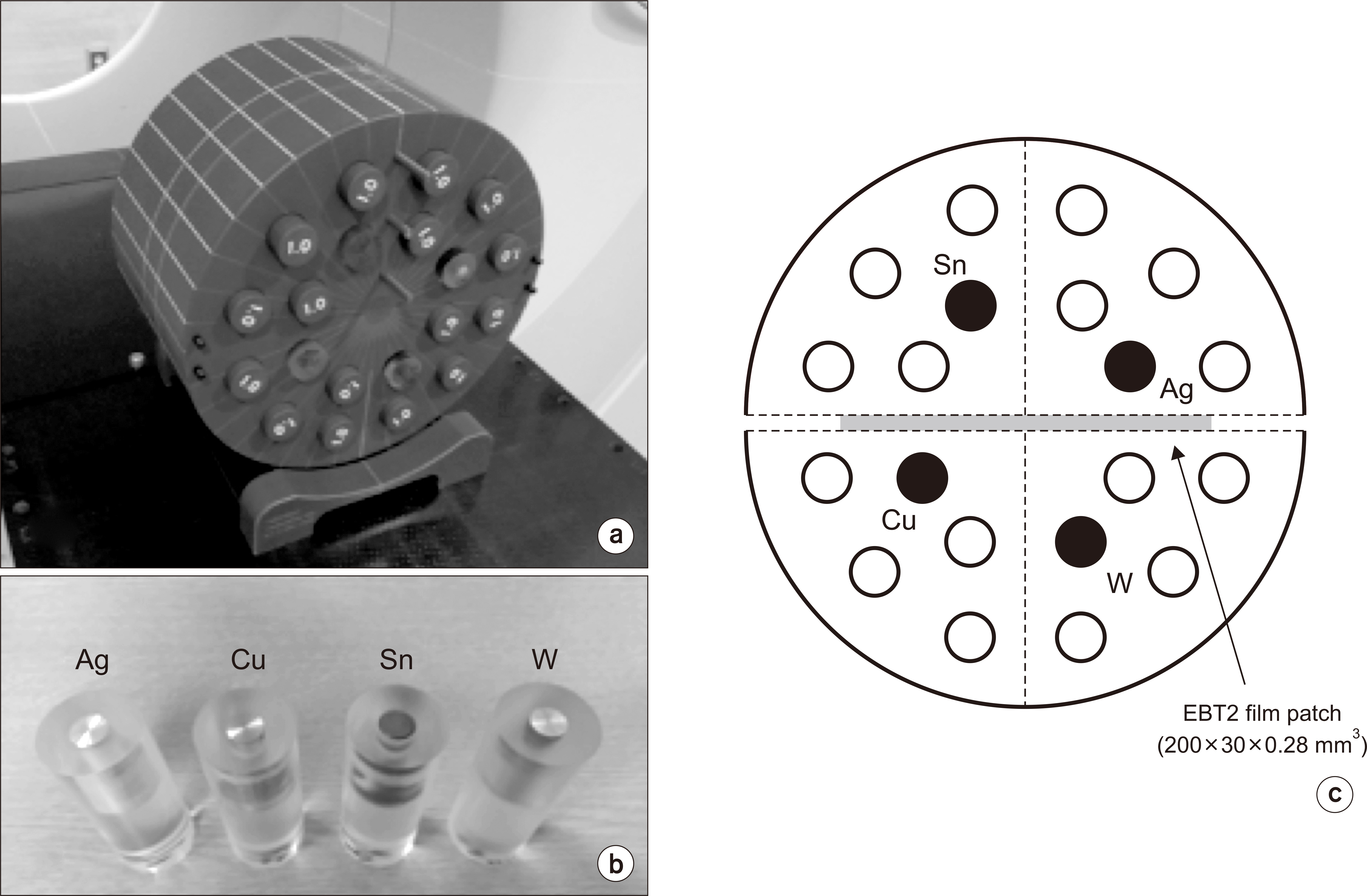
Fig. 1 Experimental setup of (a) the cylindrical phantom containing (b) four metal inserts. (c) Transverse view of the phantom. Ag, silver; Cu, Copper; Sn, tin; W, tungsten.

Fig. 2 Density distributions of the phantom (a) scanned using kilovoltage computed tomography (kV-CT) and (b) generated using hybrid CT, expressed in g/cm3. (c, d) They are also plotted on three-dimensional axes. Ag, silver; Cu, Copper; Sn, tin; W, tungsten.
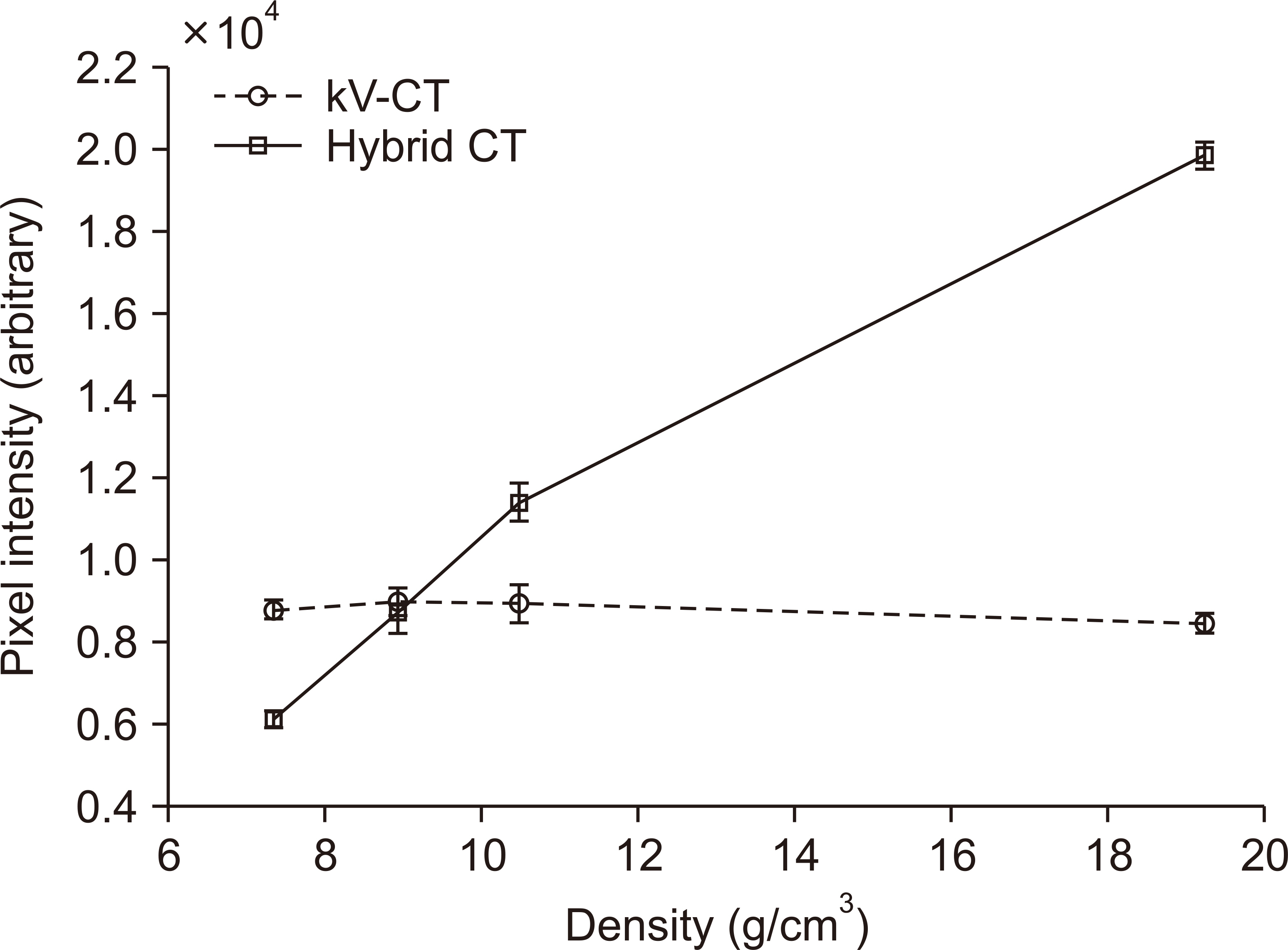
Fig. 3 Density-intensity curves in the reconstructed images of kilovoltage computed tomography (kV-CT) and hybrid CT.
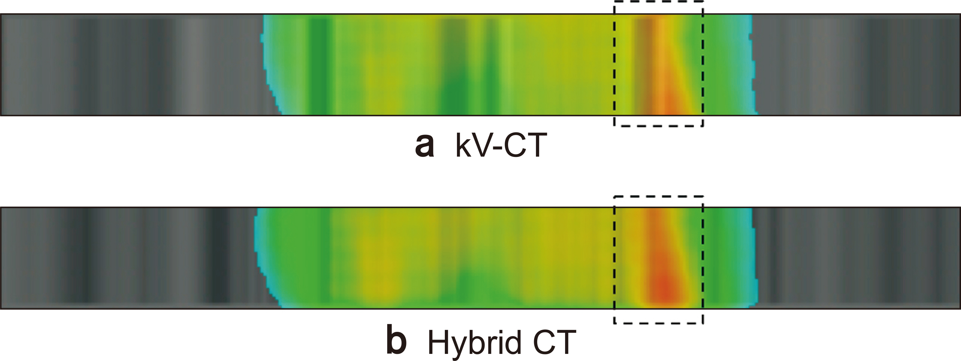
Fig. 4 Calculated dose distributions on the phantom obtained using (a) kilovoltage computed tomography (kV-CT) and (b) hybrid CT in the coronal view in patient 1.
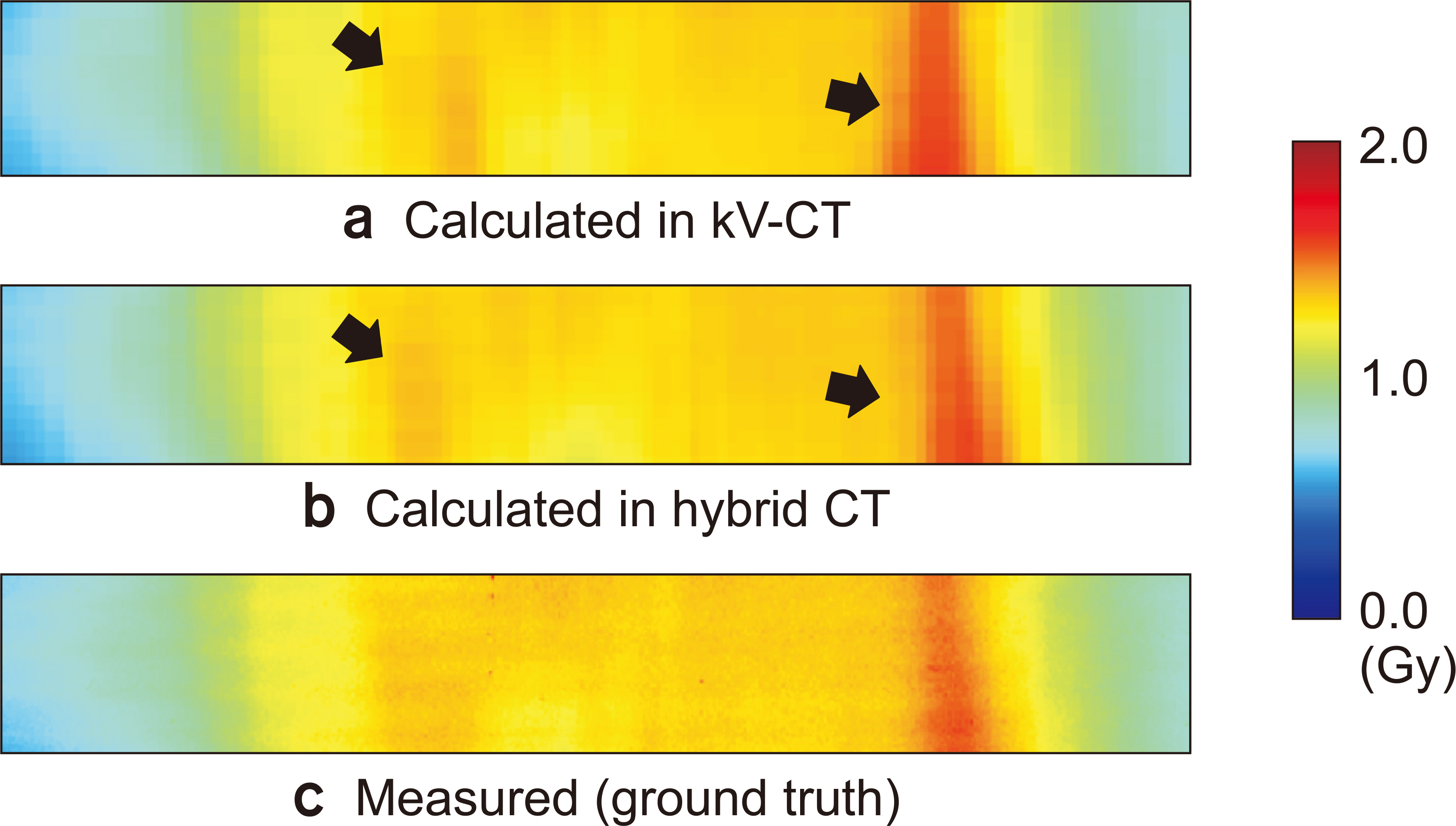
Fig. 5 Dose distributions calculated using (a) kilovoltage computed tomography (kV-CT) and (b) hybrid CT, and (c) measured using film in the coronal view in patient 1. The size of the film is 200×30 mm2.
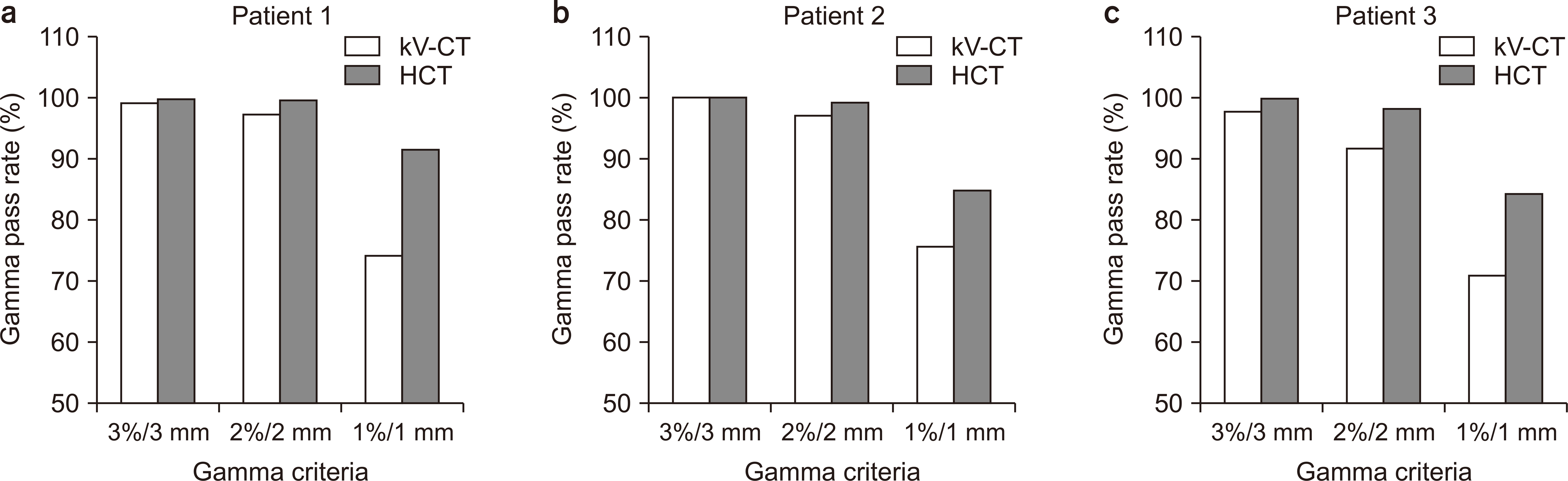
Fig. 6 Comparison of gamma pass rates at different gamma criteria in the three patients. kV-CT, kilovoltage computed tomography; HCT, hybrid CT.
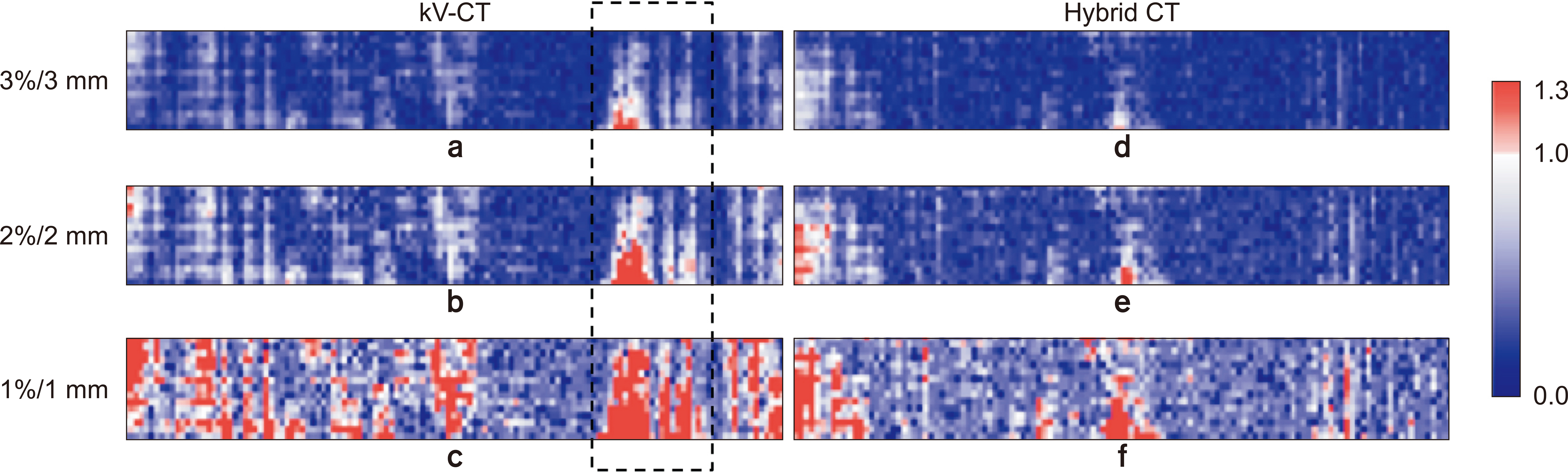
Fig. 7 Gamma evaluation results of (a–c) kilovoltage computed tomography (kV-CT) and (d–f) hybrid CT with the three gamma criteria in patient 1.
Reference
-
References
1. Glover GH, Pelc NJ. 1981; An algorithm for the reduction of metal clip artifacts in CT reconstructions. Med Phys. 8:799–807. DOI: 10.1118/1.595032. PMID: 7322078.
Article2. Yazdi M, Gingras L, Beaulieu L. 2005; An adaptive approach to metal artifact reduction in helical computed tomography for radiation therapy treatment planning: experimental and clinical studies. Int J Radiat Oncol Biol Phys. 62:1224–1231. DOI: 10.1016/j.ijrobp.2005.02.052. PMID: 15927413.3. Zhao S, Robertson DD, Wang G, Whiting B, Bae KT. 2000; X-ray CT metal artifact reduction using wavelets: an application for imaging total hip prostheses. IEEE Trans Med Imaging. 19:1238–1247. DOI: 10.1109/42.897816. PMID: 11212372.
Article4. Kalender WA, Hebel R, Ebersberger J. 1987; Reduction of CT artifacts caused by metallic implants. Radiology. 164:576–577. DOI: 10.1148/radiology.164.2.3602406. PMID: 3602406.
Article5. Mahnken AH, Raupach R, Wildberger JE, Jung B, Heussen N, Flohr TG, et al. 2003; A new algorithm for metal artifact reduction in computed tomography: in vitro and in vivo evaluation after total hip replacement. Invest Radiol. 38:769–775. DOI: 10.1097/01.rli.0000086495.96457.54. PMID: 14627894.6. Yu H, Zeng K, Bharkhada DK, Wang G, Madsen MT, Saba O, et al. 2007; A segmentation-based method for metal artifact reduction. Acad Radiol. 14:495–504. DOI: 10.1016/j.acra.2006.12.015. PMID: 17368220. PMCID: PMC1995751.
Article7. Zhang Y, Zhang L, Zhu XR, Lee AK, Chambers M, Dong L. 2007; Reducing metal artifacts in cone-beam CT images by preprocessing projection data. Int J Radiat Oncol Biol Phys. 67:924–932. DOI: 10.1016/j.ijrobp.2006.09.045. PMID: 17161556.
Article8. Boas FE, Fleischmann D. 2011; Evaluation of two iterative techniques for reducing metal artifacts in computed tomography. Radiology. 259:894–902. DOI: 10.1148/radiol.11101782. PMID: 21357521.
Article9. Mehranian A, Ay MR, Rahmim A, Zaidi H. 2013; X-ray CT metal artifact reduction using wavelet domain L0 sparse regularization. IEEE Trans Med Imaging. 32:1707–1722. DOI: 10.1109/TMI.2013.2265136. PMID: 23744669.10. Zhang X, Wang J, Xing L. 2011; Metal artifact reduction in x-ray computed tomography (CT) by constrained optimization. Med Phys. 38:701–711. DOI: 10.1118/1.3533711. PMID: 21452707. PMCID: PMC3033877.
Article11. Stayman JW, Otake Y, Prince JL, Khanna AJ, Siewerdsen JH. 2012; Model-based tomographic reconstruction of objects containing known components. IEEE Trans Med Imaging. 31:1837–1848. DOI: 10.1109/TMI.2012.2199763. PMID: 22614574. PMCID: PMC4503263.
Article12. Ruchala KJ, Olivera GH, Schloesser EA, Mackie TR. 1999; Megavoltage CT on a tomotherapy system. Phys Med Biol. 44:2597–2621. DOI: 10.1088/0031-9155/44/10/316. PMID: 10533931.
Article13. Meeks SL, Harmon JF Jr, Langen KM, Willoughby TR, Wagner TH, Kupelian PA. 2005; Performance characterization of megavoltage computed tomography imaging on a helical tomotherapy unit. Med Phys. 32:2673–2681. DOI: 10.1118/1.1990289. PMID: 16193798.
Article14. Jeon H, Park D, Youn H, Nam J, Lee J, Kim W, et al. 2015; Generation of hybrid sinograms for the recovery of kV-CT images with metal artifacts for helical tomotherapy. Med Phys. 42:4654–4667. DOI: 10.1118/1.4926552. PMID: 26233193.
Article15. Ahnesjö A, Aspradakis MM. 1999; Dose calculations for external photon beams in radiotherapy. Phys Med Biol. 44:R99–R155. DOI: 10.1088/0031-9155/44/11/201. PMID: 10588277.
Article16. Ahnesjö A. 1989; Collapsed cone convolution of radiant energy for photon dose calculation in heterogeneous media. Med Phys. 16:577–592. DOI: 10.1118/1.596360. PMID: 2770632.
Article17. Lu W, Olivera GH, Chen ML, Reckwerdt PJ, Mackie TR. 2005; Accurate convolution/superposition for multi-resolution dose calculation using cumulative tabulated kernels. Phys Med Biol. 50:655–680. DOI: 10.1088/0031-9155/50/4/007. PMID: 15773626.
Article18. Low DA, Harms WB, Mutic S, Purdy JA. 1998; A technique for the quantitative evaluation of dose distributions. Med Phys. 25:656–661. DOI: 10.1118/1.598248. PMID: 9608475.
Article19. Heilemann G, Poppe B, Laub W. 2013; On the sensitivity of common gamma-index evaluation methods to MLC misalignments in Rapidarc quality assurance. Med Phys. 40:031702. DOI: 10.1118/1.4789580. PMID: 23464297.
Article20. Fredh A, Scherman JB, Fog LS. Munck af Rosenschöld P. 2013; Patient QA systems for rotational radiation therapy: a comparative experimental study with intentional errors. Med Phys. 40:031716. DOI: 10.1118/1.4788645. PMID: 23464311.
Article
- Full Text Links
-
- Actions
-
Cited
- CITED
-
- Close
- Share
-
- Similar articles
-
- Improvement of the Dose Calculation Accuracy Using MVCBCT Image Processing
- Study of Scatter Influence of kV-Conebeam CT Based Calculation for Pelvic Radiotherapy
- Dosimetric Result of Magnetic Resonance/ Computed Tomography Compatible Hybrid Phantom for Magnetic Resonance Guided Radiotherapy
- Dosimetric Effects of Low Dose 4D CT Using a Commercial Iterative Reconstruction on Dose Calculation in Radiation Treatment Planning: A Phantom Study
- Intervention Planning Using a Laser Navigation System for CT-Guided Interventions: A Phantom and Patient Study
