Fetal cervical zygapophysial joint with special reference to the associated synovial tissue: a histological study using near-term human fetuses
- Affiliations
-
- 1Department of Histology and Embryology, Tokyo Dental College, Tokyo, Japan
- 2Department of Anatomy, Division of Basic Medical Science, Tokai University School of Medicine, Kanagawa, Japan
- 3Department of Anatomy, Wuxi School of Medicine, Jiangnan University, Wuxi, China
- 4Department of Anatomy, Tokyo Dental College, Tokyo, Japan
- 5Division of Internal Medicine, Jikou-kai Clinic of Home Visits, Sapporo, Japan
- 6Department of Anatomy and Embryology, School of Medicine, Complutense University, Madrid, Spain
- KMID: 2514586
- DOI: http://doi.org/10.5115/acb.20.265
Abstract
- Human fetal cervical vertebrae are characterized by the large zygapophysial joint (ZJ) extending posteriorly. During our recent studies on regional differences in the shape, extent, and surrounding tissue of the fetal ZJ, we incidentally found a cervical-specific structure of synovial tissues. This study aimed to provide a detailed evaluation of the synovial structure using sagittal and horizontal sections of 20 near-term fetuses. The cervical ZJ consistently had a large cavity with multiple recesses at the margins and, especially at the anterior end, the recess interdigitated with or were located close to tree-like tributaries of the veins of the external vertebral plexus. In contrast to the flat and thin synovial cell lining of the recess, the venous tributary had cuboidal endothelial cells. No or few elastic fibers were identified around the ZJ. The venous-synovial complex seems to be a transient morphology at and around birth, and it may play a role in the stabilization of the growing cervical ZJ against frequent spontaneous dislocation reported radiologically in infants. The venous-synovial complex in the cervical region should be lost and replaced by elastic fibers in childhood or adolescence. However, the delayed development of the ligament flavum is also likely to occur in the lumbar ZJ in spite of no evidence of a transient venous-synovial structure. The cuboidal venous endothelium may simply represent the high proliferation rate for the growing complex.
Keyword
Figure
-

Fig. 1 Sagittal sections of the cervical vertebrae from a fetus with a crown-rump length of 280 mm. Hematoxylin and eosin staining. Joints between the second and seventh vertebrae are included; numbers 2–7 indicate the vertebral level. The joints between the articular processes, laminae, and bases of the spinous processes are shown in (A–C), respectively. (D, E) exhibit higher-magnification views of the square in (A, C), respectively. The joint cavity creates a thin recess at the anterior and posterior ends (arrows in A). Arrowheads in (D) indicate daughter recesses of the cavity between the fourth and fifth vertebrae. (A–C) and (D, E) were prepared at the same magnification, respectively. Scale bars in (A, D)=1 mm. DRG, dorsal root ganglion.

Fig. 2 Sagittal sections of the cervical vertebrae from a fetus with a crown-rump length of 302 mm. Hematoxylin and eosin staining. Joints between the laminae from the first to the seventh vertebrae (A); numbers 1–7 indicate the vertebral level. (B–F) display higher-magnification views of the squares in (A). (B–D) display the anterior end of the joints in levels between the second and third, third and fourth, and fifth and sixth vertebrae, respectively. (B) shows venous tributaries (arrows). (D) shows a cross-section of a vein in the synovial tissue. € shows a synovial fold at the posterior end of the joint between the third and fourth vertebrae. (F) exhibits slit-like or recess-like venous tributaries in the anterior synovial tissue (arrows). Arrowheads in (C, F) indicate a thin daughter recess with a rough surface. Scale bars=1 mm in (A, F); 0.1 mm in (B–E). DRG, dorsal root ganglion; VA, vertebral artery.

Fig. 3 Sagittal sections of the cervical vertebrae from a fetus with a crown-rump length of 285 mm. Elastica-Masson staining. Joints between the laminae from the second to the sixth vertebrae and a single joint between the articular processes of the sixth and seventh vertebrae (A); numbers 2–7 indicate the vertebral level. (B, D, and G) display higher-magnification views of the squares in (A). (C, E, F, and I) are higher-magnification views of the squares at the anterior end of the joints in (B, C, D, and G), respectively. (B) contains a thin recess of the joint cavity. (C) shows no elastic fibers. (D) exhibits recesses of the joint cavity and a vein at the anterior end of the joint between the fourth and fifth vertebrae. In this area, neither the periosteum (E) nor the vein (F) contains or accompanies elastic fibers. (G) shows recesses of the joint cavity between the fifth and sixth vertebrae; the sixth vertebrae has a few elastic fibers (arrows in I). (H) (same magnification as I) shows the internal elastic lamina of an artery outside of (A). Scale bars=1 mm in (A, D); 0.1 mm in (B, C, E–G, and I). DRG, dorsal root ganglion.

Fig. 4 Horizontal sections of the mid-cervical vertebrae from a fetus with a crown-rump length of 310 mm. Hematoxylin and eosin staining (A–D) and elastica-Masson staining (E–H). A shows the topographical anatomy of a joint between the fourth and fifth vertebrae (4, 5); at the joint (right), the lower vertebral element is located anterior to the upper adjacent vertebra. (B) is a higher-magnification view of a joint on the left side of (A). (B, C) exhibit planes 0.2 and 0.4 mm, respectively, below (A). A recess of the joint cavity originates in (B) (arrowhead) and extends infero-anteriorly in (B, C). (E) shows a higher-magnification view of the square in (C). (F, G) are higher-magnification views of squares in (A). There are no evident elastic fibers around the recesses of the joint cavity (E, F). (G) displays a venous plexus in synovial tissue on the medial side of the joint. (H) exhibits highly wavy elastic fibers in a wall of the vertebral artery (VA). The thick bundle just below the VA corresponds to the internal lamina, whereas the other thin bundles are embedded in the future smooth muscle layer. Scale bars=2 mm in (A); 1 mm in (B–D); 0.1 mm in (E–H). DRG, dorsal root ganglion; VA, vertebral artery.
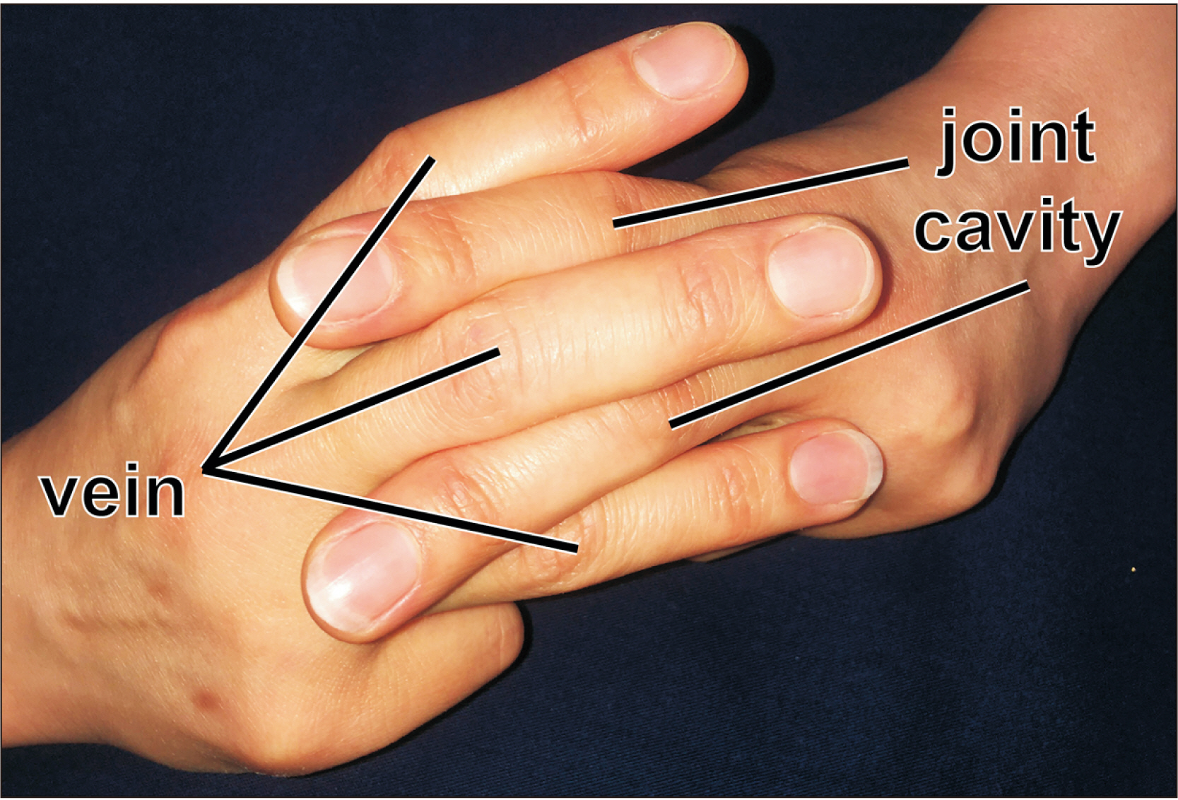
Fig. 5 Hand-finger model showing the topographical relationship between the venous tributaries and joint cavity of the cervical zygapophysial joints. Venous tributaries (vein) intercalate with recesses of the joint cavity, especially at the anterior margin of the latter.
Reference
-
References
1. Bogduk N. 2016; Functional anatomy of the spine. Handb Clin Neurol. 136:675–88. DOI: 10.1016/B978-0-444-53486-6.00032-6. PMID: 27430435.
Article2. Gray H, Williams PL. 1995. Gray's anatomy. 38th ed. Livingstone;New York: p. 516–22. p. 1431–6.3. Cattell HS, Filtzer DL. 1965; Pseudosubluxation and other normal variations in the cervical spine in children. A study of one hundred and sixty children. J Bone Joint Surg Am. 47:1295–309. DOI: 10.2106/00004623-196547070-00001. PMID: 5837630.4. Castellana C, Kósa F. 1999; Morphology of the cervical vertebrae in the fetal-neonatal human skeleton. J Anat. 194(Pt 1):147–52. DOI: 10.1046/j.1469-7580.1999.19410147.x. PMID: 10227677. PMCID: PMC1467903.
Article5. Müller F, O'Rahilly R. 2003; Segmentation in staged human embryos: the occipitocervical region revisited. J Anat. 203:297–315. DOI: 10.1046/j.1469-7580.2003.00219.x. PMID: 14529047. PMCID: PMC1571167.6. O'Rahilly R, Muller F, Meyer DB. 1980; The human vertebral column at the end of the embryonic period proper. 1. The column as a whole. J Anat. 131(Pt 3):565–75. PMID: 7216919. PMCID: PMC1233253.7. San Román P, Palma JC, Oteo MD, Nevado E. 2002; Skeletal maturation determined by cervical vertebrae development. Eur J Orthod. 24:303–11. DOI: 10.1093/ejo/24.3.303. PMID: 12143094.8. Bagnall KM, Harris PF, Jones PR. 1982; A radiographic study of the growth in width of the human fetal vertebral column. Anat Rec. 204:265–70. DOI: 10.1002/ar.1092040311. PMID: 7158830.
Article9. Cheng PJ, Huang SY, Shaw SW, Chueh HY, Soong YK. 2010; Evaluation of fetal spine biometry between 11 and 14 weeks of gestation. Ultrasound Med Biol. 36:1060–5. DOI: 10.1016/j.ultrasmedbio.2010.04.007. PMID: 20620693.
Article10. Glenn OA, Barkovich AJ. 2006; Magnetic resonance imaging of the fetal brain and spine: an increasingly important tool in prenatal diagnosis, part 1. AJNR Am J Neuroradiol. 27:1604–11. PMID: 16971596.11. Kitamura K, Cho KH, Yamamoto M, Ishii M, Murakami G, Rodríguez-Vázquez JF, Abe SI. 2019; Suboccipital myodural bridges revisited: application to cervicogenic headaches. Clin Anat. 32:914–28. DOI: 10.1002/ca.23411. PMID: 31116454.
Article12. Sakanaka K, Yamamoto M, Hirouchi H, Kim JH, Murakami G, Rodríguez Vázquez JF, Abe SI. 2019; A temporary disc-like structure at the median atlanto-axial joint in human fetuses. Anat Cell Biol. 52:436–42. DOI: 10.5115/acb.19.128. PMID: 31949983. PMCID: PMC6952699.
Article13. Kitamura K, Kim JH, Cho KH, Murakami G, Rodríguez-Vázquez JF, Yamamoto H. 2020; Oct. 9. Regional differences in zygapophysial joint cavities: a histological study of human fetuses. Anat Rec (Hoboken). [Epub]. https://doi.org/10.1002/ar.24532. DOI: 10.1002/ar.24532. PMID: 33034079.
Article14. Cho KH, Jin ZW, Abe H, Shibata S, Murakami G, Rodríguez-Vázquez JF. 2016; Neural-dural transition at the thoracic and lumbar spinal nerve roots: a histological study of human late-stage fetuses. Biomed Res Int. 2016:8163519. DOI: 10.1155/2016/8163519. PMID: 27069926. PMCID: PMC4812201.
Article15. Khorooshi MH, Fischer Hansen B, Keeling J, Nolting D, Kjaer I. 2001; Prenatal localization of the dorsal root ganglion in different segments of the normal human vertebral column. Spine (Phila Pa 1976). 26:1–5. DOI: 10.1097/00007632-200101010-00002. PMID: 11148637.
Article16. O'Rahilly R, Müller F, Meyer DB. 1990; The human vertebral column at the end of the embryonic period proper. 3. The thoracicolumbar region. J Anat. 168:81–93. PMID: 2323997. PMCID: PMC1256892.17. Hayashi T, Kumasaka T, Mitani K, Yao T, Suda K, Seyama K. 2010; Loss of heterozygosity on tuberous sclerosis complex genes in multifocal micronodular pneumocyte hyperplasia. Mod Pathol. 23:1251–60. DOI: 10.1038/modpathol.2010.114. PMID: 20526286.
Article18. Motohashi O, Suzuki M, Shida N, Umezawa K, Ohtoh T, Sakurai Y, Yoshimoto T. 1995; Subarachnoid haemorrhage induced proliferation of leptomeningeal cells and deposition of extracellular matrices in the arachnoid granulations and subarachnoid space. Immunhistochemical study. Acta Neurochir (Wien). 136:88–91. DOI: 10.1007/BF01411441. PMID: 8748833.19. Barbolini G, Tischendorf F, Curri SB. 1971; Histology, histochemistry, and function of the human digital arteriovenous anastomoses (Hoyer-Grosser's organs, Masson's glomera). I. The possible relationship with Pacinian corpuscles. Microvasc Res. 3:142–53. DOI: 10.1016/0026-2862(71)90018-5.20. Jin ZW, Cho KH, Jang HS, Murakami G, Rodríguez-Vázquez JF, Yamamoto M, Abe SI. 2017; Coccygeal body revisited: an immunohistochemical study using donated elderly cadavers. Anat Rec (Hoboken). 300:1826–37. DOI: 10.1002/ar.23615. PMID: 28545163.
Article21. Hurley HJ Jr, Mescon H, Moretti G. 1956; The anatomy and histochemistry of the arteriovenous anastomosis in human digital skin. J Invest Dermatol. 27:133–45. DOI: 10.1038/jid.1956.85. PMID: 13367521.22. Yamashita T, Minaki Y, Ozaktay AC, Cavanaugh JM, King AI. 1996; A morphological study of the fibrous capsule of the human lumbar facet joint. Spine (Phila Pa 1976). 21:538–43. DOI: 10.1097/00007632-199603010-00002. PMID: 8852306.
Article23. Kinoshita H, Umezawa T, Omine Y, Kasahara M, Rodríguez-Vázquez JF, Murakami G, Abe S. 2013; Distribution of elastic fibers in the head and neck: a histological study using late-stage human fetuses. Anat Cell Biol. 46:39–48. DOI: 10.5115/acb.2013.46.1.39. PMID: 23560235. PMCID: PMC3615611.
Article24. Kawamoto A, Honkura Y, Suzuki R, Abe H, Abe S, Murakami G, Katori Y. 2016; Cricothyroid articulation in elderly Japanese with special reference to morphology of the synovial and capsular tissues. J Voice. 30:538–48. DOI: 10.1016/j.jvoice.2015.07.011. PMID: 26687542.
Article25. Rodríguez-Vázquez JF, Yamamoto M, Kim JH, Jin ZW, Katori Y, Murakami G. 2020; The incudopetrosal joint of the human middle ear: a transient morphology in fetuses. J Anat. 237:176–87. DOI: 10.1111/joa.13181. PMID: 32159229. PMCID: PMC7309281.26. Takanashi Y, Shibata S, Katori Y, Murakami G, Abe S, Rodríguez-Vázquez JF, Kawase T. 2013; Fetal development of the elastic-fiber-mediated enthesis in the human middle ear. Ann Anat. 195:441–8. DOI: 10.1016/j.aanat.2013.03.010. PMID: 23706648.
Article27. Kawase T, Shibata S, Katori Y, Ohtsuka A, Murakami G, Fujimiya M. 2012; Elastic fiber-mediated enthesis in the human middle ear. J Anat. 221:331–40. DOI: 10.1111/j.1469-7580.2012.01542.x. PMID: 22803514. PMCID: PMC3458252.
Article28. Clausen JD, Goel VK, Traynelis VC, Scifert J. 1997; Uncinate processes and Luschka joints influence the biomechanics of the cervical spine: quantification using a finite element model of the C5-C6 segment. J Orthop Res. 15:342–7. DOI: 10.1002/jor.1100150305. PMID: 9246079.
Article29. Hur JW, Bae T, Ye S, Kim JH, Lee S, Kim K, Lee SH, Kim JS, Lee JB, Cho TH, Park JY, Hur JK. 2017; Myofibroblast in the ligamentum flavum hypertrophic activity. Eur Spine J. 26:2021–30. DOI: 10.1007/s00586-017-4981-2. PMID: 28180980.
Article30. Sairyo K, Biyani A, Goel V, Leaman D, Booth R Jr, Thomas J, Gehling D, Vishnubhotla L, Long R, Ebraheim N. 2005; Pathomechanism of ligamentum flavum hypertrophy: a multidisciplinary investigation based on clinical, biomechanical, histologic, and biologic assessments. Spine (Phila Pa 1976). 30:2649–56. DOI: 10.1097/01.brs.0000188117.77657.ee. PMID: 16319751.
Article31. Mwaka ES, Yayama T, Uchida K, Kobayashi S, Kokubo Y, Nakajima H, Sato R, Orwotho NT, Baba H. 2009; Calcium pyrophosphate dehydrate crystal deposition in the ligamentum flavum of the cervical spine: histopathological and immunohistochemical findings. Clin Exp Rheumatol. 27:430–8. PMID: 19604435.32. Nakamura T, Okada T, Endo M, Kadomatsu T, Taniwaki T, Sei A, Odagiri H, Masuda T, Fujimoto T, Nakamura T, Oike Y, Mizuta H. 2014; Angiopoietin-like protein 2 induced by mechanical stress accelerates degeneration and hypertrophy of the ligamentum flavum in lumbar spinal canal stenosis. PLoS One. 9:e85542. DOI: 10.1371/journal.pone.0085542. PMID: 24465594. PMCID: PMC3894965.
Article33. Viejo-Fuertes D, Liguoro D, Rivel J, Midy D, Guerin J. 1998; Morphologic and histologic study of the ligamentum flavum in the thoraco-lumbar region. Surg Radiol Anat. 20:171–6. DOI: 10.1007/BF01628891. PMID: 9706675.
Article34. Okuda T, Baba I, Fujimoto Y, Tanaka N, Sumida T, Manabe H, Hayashi Y, Ochi M. 2004; The pathology of ligamentum flavum in degenerative lumbar disease. Spine (Phila Pa 1976). 29:1689–97. DOI: 10.1097/01.BRS.0000132510.25378.8C. PMID: 15284518.
Article35. McDermott LJ. 1943; Development of the human knee joint. Arch Surg. 46:705–19. DOI: 10.1001/archsurg.1943.01220110121021.
Article36. Gray DJ, Gardner E. 1950; Prenatal development of the human knee and superior tibiofibular joints. Am J Anat. 86:235–87. DOI: 10.1002/aja.1000860204. PMID: 15410671.
Article37. Isogai S, Murakami G, Wada T, Ishii S. 2001; Which morphologies of synovial folds result from degeneration and/or aging of the radiohumeral joint: an anatomic study with cadavers and embryos. J Shoulder Elbow Surg. 10:169–81. DOI: 10.1067/mse.2001.112956. PMID: 11307082.
Article
- Full Text Links
-
- Actions
-
Cited
- CITED
-
- Close
- Share
-
- Similar articles
-
- A temporary disc-like structure at the median atlanto-axial joint in human fetuses
- Cervical nerve roots and the dural sheath: a histological study using human fetuses near term
- Synovial Chondromatosis: Report of 4 cases
- A clinical Study of Synovial Chondromatosis
- Giant Extra-Capsular Synovial Chondroma of the knee joint: A Case Report
