A temporary disc-like structure at the median atlanto-axial joint in human fetuses
- Affiliations
-
- 1Department of Anatomy, Tokyo Dental College, Tokyo, Japan. yamamotomasahito@ tdc.ac.jp
- 2Department of Anatomy, Chonbuk National University School of Medicine, Jeonju, Korea.
- 3Division of Internal Medicine, Jikou-kai Clinic for Home Visits, Sapporo, Japan.
- 4Department of Anatomy and Embryology, School of Medicine, Complutense University, Madrid, Spain.
- KMID: 2466697
- DOI: http://doi.org/10.5115/acb.19.128
Abstract
- During observations of mid-term and late-stage fetuses, we found a joint disk-like structure at the anterior component of the median atlanto-axial joint. At mid-term, the disk-like structure was thick (0.1-0.15 mm) relative to the sizes of bones surrounding the joint. However, it did not completely separate the joint cavity, and was absent in the inferior and/or central part of the cavity. This morphology was similar to the so-called fibroadipose meniscoid of the lumbar zygapophysial joint that is usually seen in adults. In mid-term fetuses, there was evidence suggesting that a mesenchymal tissue plate was separated from a roof of the joint cavity. In late-stage fetuses, the thickness (less than 0.15 mm) was usually the same as, or less than that at mid-term, and the disk-like structure was often flexed, folded and fragmented. Therefore, in contrast to the zygapophysial meniscoid as a result of aging, the present disk-like structure was most likely a temporary product during the cavitation process. It seemed to be degenerated in late-stage fetuses and possibly also in newborns. Anomalies at the craniocervical junction such as Chiari malformations might accompany this disk-like structure at the median atlanto-axial joint even in childhood.
Keyword
Figure
-

Fig. 1 Sagittal sections of the median atlanto-axial joint from two mid-term specimens. Panels A–C display a specimen of 82 mm crown-rump length (CRL), while panels D and E show a specimen of 100 mm CRL. Panel A (D) is the most medial side of the figure, whereas panel C (E) is the most lateral side. (A–C) A disk-like structure (arrows) is thick in lateral part of the joint cavity (C) and thin in the medial part (A). Likewise, in panels D and E, the structure appears to be complete at the medial part (D). Arrows in D and E indicate a disk-like structure. All panels were prepared at the same magnification. Scale bar=1 mm (D).

Fig. 2 Horizontal sections of the median atlanto-axialjoint from two mid-term specimens. Panels A–E display a specimen of 96 mm crown-rump length (CRL), while panels F–I show a specimen of 100 mm CRL. Panel A is the most superior side of the figure, whereas panel E is the most inferior side. In the former specimen, a disk-like structure (D, E; arrows) is continuous with mesenchymal tissue at the irregular superior margin of the joint cavity (A, B). In the latter specimen, the roof of the joint cavity is separated from the atlas between panels F and G. In both specimens, the structure was not a complete septum but was considered to be a superior marginal fold (E, I). Arrows in G, H, and I indicate a disk-like structure. All panels were prepared at the same magnification. Scale bar=1 mm (A).
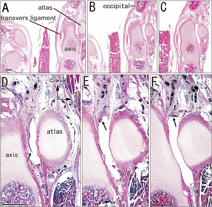
Fig. 3 Sagittal sections of short flexed fold in the median atlanto-axial joint from a late-stage specimen. Panels A–F display a specimen of 270 mm crown-rump length. Panel A is the most lateral side of the figure, Panel C is the most medial side. The right-hand side of the figure corresponds to the anterior side of the head. Panels A–C show lower-magnification views to illustrate the topographical anatomy. Panels D–F show higher-magnification views of the anterior component of the joint. A short fold (arrows) is attached to the superior margin of the joint cavity at the medial or nearly median site (F). Scale bars=1 mm (A, D).

Fig. 4 Sagittal sections of thin but large disk-like structure in the median atlanto-axial joint from a late-stage specimen. Panels A–F display a specimen of 280 mm crown-rump length. Panel A is the most lateral side of the figure, panel C is the most medial side. The right-hand side of the figure corresponds to the anterior side of the head. Panels A–C show lower-magnification views to illustrate the topographical anatomy. Asterisk indicates the posterior atlanto-occipital membrane that has become separated from the vertebrae during histological procedures. Note the relatively low position of the upper tip of the dens relative to the anterior atlantal arch. Panels D–F show higher-magnification views of the anterior component of the median atlanto-axial joint. Arrows in D and E indicate a short membranous structure. A short membranous structure (arrows) is interrupted in the medial or nearly median aspect (F). Scale bars=1 mm (A, D).
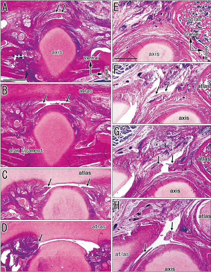
Fig. 5 Horizontal and sagittal sections of two late-stage specimens. The sectional plane of panels A–D was horizontal, while that of panels E–H was sagittal. Panels A–E display a specimen of 270 mm crown-rump length (CRL), while panels F–H show a specimen of 280 mm CRL. In the former specimen, panel A is the most superior side of the figure, whereas panel D is the most inferior side. The upper side of the figure corresponds to the anterior side of the head. In the latter specimen, panel E is the most lateral side of the figure, whereas panel H is the most medial side. A membranous structure (arrows) is a short, thin synovial fold projecting from the roof of the joint cavity in association wi th ti ssue fragments scattered within the cavity (A–D). Arrow in E indicates a membranous structure. The membranous structure (arrows) is interrupted in the medial or nearly median aspect (F–H). All panels were prepared at the same magnification. Scale bars=5 mm (A, E).
Cited by 2 articles
-
Fetal development of the human trapezius and sternocleidomastoid muscles
Kwang Ho Cho, Ichiro Morimoto, Masahito Yamamoto, Shinya Hanada, Gen Murakami, Jose Francisco Rodríguez-Vázquez, Shinichi Abe
Anat Cell Biol. 2020;53(4):405-410. doi: 10.5115/acb.20.202.Fetal cervical zygapophysial joint with special reference to the associated synovial tissue: a histological study using near-term human fetuses
Kei Kitamura, Shogo Hayashi, Zhe Wu Jin, Masahito Yamamoto, Gen Murakami, José Francisco Rodríguez-Vázquez, Hitoshi Yamamoto
Anat Cell Biol. 2021;54(1):65-73. doi: 10.5115/acb.20.265.
Reference
-
1. Cave AJ. On the occipito-atlanto-axial articulations. J Anat. 1934; 68:416–423.2. Cave AJ. The morphology of the mammalian cervical pleurapophysis. J Zool. 1975; 177:377–393.3. Abe H, Ishizawa A, Cho KH, Suzuki R, Fujimiya M, Rodríguez-Vázquez JF, Murakami G. Fetal development of the transverse atlantis and alar ligaments at the craniovertebral junction. Clin Anat. 2012; 25:714–721.4. Engel R, Bogduk N. The menisci of the lumbar zygapophysial joints. J Anat. 1982; 135:795–809.5. Isogai S, Murakami G, Wada T, Ishii S. Which morphologies of synovial folds result from degeneration and/or aging of the radiohumeral joint: an anatomic study with cadavers and embryos. J Shoulder Elbow Surg. 2001; 10:169–181.6. Mérida-Velasco JA, Sánchez-Montesinos I, Espín-Ferra J, Mérida-Velasco JR, Rodríguez-Vázquez JF, Jiménez-Collado J. Development of the human elbow joint. Anat Rec. 2000; 258:166–175.7. Gray DJ, Gardner E. Prenatal development of the human knee and superior tibiofibular joints. Am J Anat. 1950; 86:235–287.8. Azahraa Haddad F, Qaisi I, Joudeh N, Dajani H, Jumah F, Elmashala A, Adeeb N, Chern JJ, Tubbs RS. The newer classifications of the chiari malformations with clarifications: an anatomical review. Clin Anat. 2018; 31:314–322.9. Pang D, Thompson DN. Embryology and bony malformations of the craniovertebral junction. Childs Nerv Syst. 2011; 27:523–564.10. Prescher A. The craniocervical junction in man, the osseous variations, their significance and differential diagnosis. Ann Anat. 1997; 179:1–19.11. Shoja MM, Johal J, Oakes WJ, Tubbs RS. Embryology and pathophysiology of the Chiari I and II malformations: a comprehensive review. Clin Anat. 2018; 31:202–215.12. Shoja MM, Ramdhan R, Jensen CJ, Chern JJ, Oakes WJ, Tubbs RS. Embryology of the craniocervical junction and posterior cranial fossa, part I: development of the upper vertebrae and skull. Clin Anat. 2018; 31:466–487.
- Full Text Links
-
- Actions
-
Cited
- CITED
-
- Close
- Share
-
- Similar articles
-
- A Case of Grisel Syndrome Showing No Underlying Laxity of the Atlanto-axial Joint
- Atlanto-Axial Joint Block: Case reports
- Quadriplegia and Dyspnea Caused by Os Odontoideum in a Down Syndrome Patient: A case report
- Combined Chronic Occipito-atlantal and Atlanto-axial Rotator Fixation with Cerebral Palsy
- Normal range of atlanto-denal interval
