A Case of Lymphocyte-Rich Hepatocellular Carcinoma in a Patient Who Was Treated for Colon Cancer
- Song JW1,2
- Chun HS1,2
- Lee JS1,2,3
- Lee HW1,2,3
- Kim BK1,2,3
- Kim SU1,2,3
- Park JY1,2,3
- Ahn SH1,2,3
- Park YN4
- Han DH5
- Kim DY
 1,2,3
1,2,3
- Affiliations
-
- 1Department of Internal Medicine, Yonsei University College of Medicine, Seoul, Korea
- 2Institute of Gastroenterology, Yonsei University College of Medicine, Seoul, Korea
- 3Yonsei Liver Center, Severance Hospital, Seoul, Korea
- 4Department of Pathology, Yonsei University College of Medicine, Seoul, Korea
- 5Department of Surgery, Yonsei University College of Medicine, Seoul, Korea
- KMID: 2514236
- DOI: http://doi.org/10.17998/jlc.21.1.69
Abstract
- Hepatocellular carcinoma (HCC) primarily originates in the liver with hepatic differentiation. However, HCCs are not homogenous, and approximately 35% of HCC cases are classified as histopathological variants that present distinct pathologic characteristics. In particular, the lymphocyte-rich variant is the rarest subtype accounting for less than 1% of HCCs, which is not well known to date about molecular features and pathophysiology. Herein, we present a case of a patient who was suspected of metastatic liver cancer and confirmed as lymphocyte-rich HCC pathologically. A 78-year-old woman who underwent a right hemicolectomy for colon cancer was referred to our hospital for a newly detected liver mass. We could not make a decision because of insufficient evidence for diagnosis from imaging studies. After resection, we found that it was a lymphocyte-rich HCC. The pathologic features and prognostic trends of this subtype are also discussed.
Figure
-

Figure 1 Initial computed tomography scan findings. A 3.6 cm sized lobulated mass is observed in segment VII, showing hypodensity compared to normal liver tissue in the pre-contrast phase (A) and peripheral rim enhancement in contrast images (B).

Figure 2 Dynamic liver magnetic resonance imaging findings. The mass is enhanced heterogeneously and also shows peripheral rim enhancement in the arterial phase (A). Wash-out in portal phase (B). Hypointensity in hepato-biliary phase (C). In T2-weighted images, it presents moderate hyperintensity (D).

Figure 3 Positron emission tomography-computed tomography findings. Increased FDG uptake in the liver mass is observed.
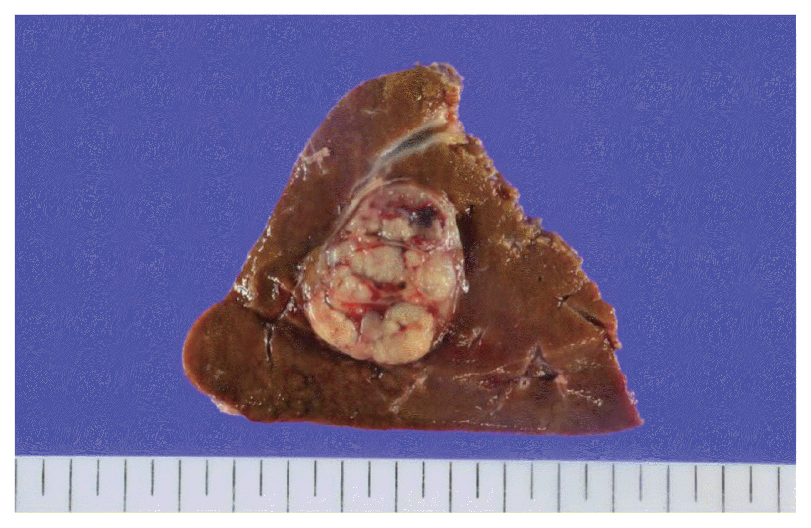
Figure 4 Macroscopic findings of the specimen from surgical resection. It exhibits a 3.5×2.5×2.5 cm sized lobulated mass with a thin capsule encapsulating the mass. It contains a small portion of peliosis and necrotic change. Portal vein and bile duct invasion are not seen.

Figure 5 Findings on microscopy. Hematoxylin and eosin (H&E) staining showing moderately differentiated hepatic tumor cells and large numbers of intra-tumoral infiltration of lymphocytes, which is referred to as a lymphoepithelioma-like pattern (H&E; ×100 [A], ×400 [B]).

Figure 6 Findings on microscopy. Immunohistochemistry showing weakly positive arginase-1 (original magnification, ×200) (A). The cluster of differentiation 3 and 8 (CD3 and CD8) showing diffusely positive T-lymphocyte markers (original magnification, ×100) (B, showing the result of CD8). Programmed death-ligand 1 is positive with 90% of tumor proportion score (original magnification, ×200) (C). Negative findings of Epstein-Barr virus-encoded RNA in-situ hybridization (original magnification, ×200) (D).
Reference
-
1. Bray F, Ferlay J, Soerjomataram I, Siegel RL, Torre LA, Jemal A. Global cancer statistics 2018: GLOBOCAN estimates of incidence and mortality worldwide for 36 cancers in 185 countries. CA Cancer J Clin. 2018; 68:394–424.2. Hong S, Won YJ, Park YR, Jung KW, Kong HJ, Lee ES, et al. Cancer statistics in Korea: incidence, mortality, survival, and prevalence in 2017. Cancer Res Treat. 2020; 52:335–350.3. Shin HY, Kim J, Lee S, Park MS, Park S, Huh S. Cause-of-death statistics in 2018 in the Republic of Korea. Journal of the Korean Medical Association. 2020; 63:286–297.4. Torbenson MS. Morphologic subtypes of hepatocellular carcinoma. Gastroenterol Clin North Am. 2017; 46:365–391.5. Lo RCL. An update on the histological subtypes of hepatocellular carcinoma. Hepatoma Res. 2019; 5:41.6. Vyas M, Zhang X. Hepatocellular carcinoma: role of pathology in the era of precision medicine. Clin Liver Dis. 2020; 24:591–610.7. Kim H, Jang M, Park YN. Histopathological variants of hepatocellular carcinomas: an update according to the 5th edition of the WHO classification of digestive system tumors. J Liver Cancer. 2020; 20:17–24.8. Torbenson MS, Ng IOL, Park YN, Roncalli M, Sakamoto M. Hepatocellular carcinoma. WHO Classification of Tumours Editorial Board. Digestive system tumours WHO classification of tumours series. 5th ed. Lyon: International Agency for Research on Cancer;2019. p. 229–239.9. Karadag Soylu N. Update on hepatocellular carcinoma: a brief review from pathologist standpoint. J Gastrointest Cancer. 2020; 51:1176–1186.10. Calderaro J, Ziol M, Paradis V, Zucman-Rossi J. Molecular and histological correlations in liver cancer. J Hepatol. 2019; 71:616–630.11. Zhang K, Tao C, Tao Z, Wu F, An S, Wu J, et al. Lymphoepithelioma-like carcinoma in liver not associated with Epstein-Barr virus: a report of 3 cases and literature review. Diagn Pathol. 2020; 15:115.12. Wada Y, Nakashima O, Kutami R, Yamamoto O, Kojiro M. Clinicopathological study on hepatocellular carcinoma with lymphocytic infiltration. Hepatology. 1998; 27:407–414.13. Miyasaka C, Ishida M, Ito H, Kaibori M, Uemura Y, Tsuta K. Lymphoepithelioma-like hepatocellular carcinoma: a case report with emphasis on the cytological features. Int J Clin Exp Pathol. 2017; 10:7893–7897.14. Chan AWH, Tong JHM, Pan Y, Chan SL, Wong GLH, Wong VWS, et al. Lymphoepithelioma-like hepatocellular carcinoma: an uncommon variant of hepatocellular carcinoma with favorable outcome. Am J Surg Pathol. 2015; 39:304–312.15. Patel KR, Liu TC, Vaccharajani N, Chapman WC, Brunt EM. Characterization of inflammatory (lymphoepithelioma-like) hepatocellular carcinoma: a study of 8 cases. Arch Pathol Lab Med. 2014; 138:1193–1202.16. Labgaa I, Stueck A, Ward SC. Lymphoepithelioma-like carcinoma in liver. Am J Pathol. 2017; 187:1438–1444.17. Calderaro J, Rousseau B, Amaddeo G, Mercey M, Charpy C, Costentin C, et al. Programmed death ligand 1 expression in hepatocellular carcinoma: relationship With clinical and pathological features. Hepatology. 2016; 64:2038–2046.18. Chew V, Tow C, Teo M, Wong HL, Chan J, Gehring A, et al. Inflammatory tumour microenvironment is associated with superior survival in hepatocellular carcinoma patients. J Hepatol. 2010; 52:370–379.19. Chan AW, Zhang Z, Chong CC, Tin EK, Chow C, Wong N. Genomic landscape of lymphoepithelioma-like hepatocellular carcinoma. J Pathol. 2019; 249:166–172.20. Harding JJ, Nandakumar S, Armenia J, Khalil DN, Albano M, Ly M, et al. Prospective genotyping of hepatocellular carcinoma: clinical implications of next-generation sequencing for matching patients to targeted and immune therapies. Clin Cancer Res. 2019; 25:2116–2126.
- Full Text Links
-
- Actions
-
Cited
- CITED
-
- Close
- Share
-
- Similar articles
-
- Hepatocellular carcinoma with colon metastasis
- Intraductal Lipid-Rich Carcinoma of the Breast with a Component of Glycogen-Rich Carcinoma
- Up-to-date Knowledge on the Pathological Diagnosis of Hepatocellular Carcinoma
- Fibrolamellar hepatocellular carcinoma that was successfully treated with surgical resection: a case report
- A case of double primary cancer of hepatocellular carcinoma and mucoepidermoid carcinoma in the liver
