Pancreatic metastasis from malignant phyllodes tumor of the breast
- Affiliations
-
- 1Department of Radiology, Yeungnam University College of Medicine, Daegu, Korea
- 2Department of Pathology, Yeungnam University College of Medicine, Daegu, Korea
- KMID: 2512072
- DOI: http://doi.org/10.12701/yujm.2020.00759
Abstract
- Pancreatic metastasis from malignant phyllodes tumor (PT) of the breast is rare, and only a few cases have been reported in the literature. Here, we report a case of pancreatic metastasis from malignant PT of the breast in a 48-year-old woman. She had had three episodes of recurrence of malignant PT in her right breast. She presented with epigastric pain for 2 months. Computed tomography and magnetic resonance imaging revealed a 6 cm-sized, well-defined, heterogeneous mass with peripheral enhancement in the body of the pancreas. Endoscopic ultrasonography-guided fine-needle aspiration was performed, and the pathologic report suggested spindle cell mesenchymal neoplasm. Subsequently, surgical excision was performed, and the mass was confirmed as a metastatic malignant PT. The imaging findings are discussed and the literature is briefly reviewed in this report.
Keyword
Figure
-
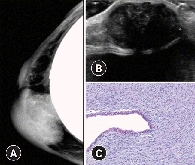
Fig. 1. Radiologic and pathologic findings of recurrent malignant phyllodes tumor of the breast in a 48-year-old woman. (A) Mammography of the right breast reveals an oval, circumscribed, hyperdense mass in the right inner breast, above the implant, with an approximate size of 3.6 cm. (B) Ultrasonography reveals a 3.6×3.0×2.0 cm-sized, oval, microlobulated, hypoechoic mass at the 3 o’clock location of the periareolar area. (C) The recurred breast mass shows stromal overgrowth and hypercellularity. The stromal cells are spindle-shaped and show significant nuclear atypia. Benign epithelial component is present (hematoxylin and eosin stain, ×100).
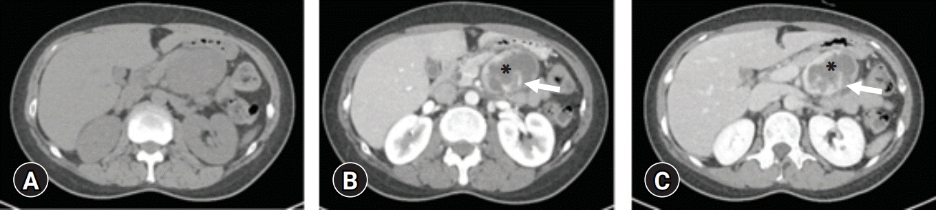
Fig. 2. Computed tomography imaging findings of pancreatic metastasis from malignant phyllodes tumor of the breast. Precontrast (A), arterial (B), and portal (C) phase of contrast-enhanced abdominal computed tomography show a 6 cm-sized, well-defined heterogeneous mass involving the body of the pancreas. The mass reveals central cystic or necrotic portion (asterisk) and internal septa (arrow). The mass shows progressive, peripheral, and septal enhancement.

Fig. 3. Magnetic resonance imaging findings of pancreatic metastasis from malignant phyllodes tumor of the breast. The mass shows low signal intensity on T1-weighted image (A) and high signal intensity and internal septa with low signal intensities (arrows) on T2-weighted images (B, C). (D–F) Dynamic studies after the administration of gadolinium show thick irregular peripheral enhancement and septal enhancement with central poorly enhancing areas.
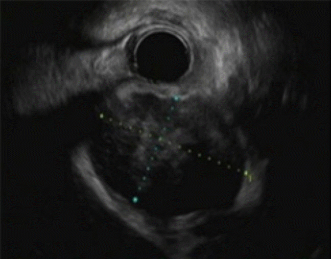
Fig. 4. Endoscopic ultrasonography (EUS) finding of pancreatic metastasis from malignant phyllodes tumor of the breast. EUS revealed a 6-cm mixed and heterogeneous hypoechoic mass in the body of the pancreas.

Fig. 5. (A) Gross photograph of the pancreatic tumor shows a 6.0×5.0-cm, grayish white, myxoid and solid cut surface with hemorrhage. (B) Histologic photographs of the pancreatic tumor show proliferation of spindle-shaped tumor cells in the myxoid stroma. (C) The tumor cells have elongated hyperchromatic nuclei, with fascicular pattern (hematoxylin and eosin stain, ×100 [B] and ×200 [C]).
Reference
-
References
1. Telli ML, Horst KC, Guardino AE, Dirbas FM, Carlson RW. Phyllodes tumors of the breast: natural history, diagnosis, and treatment. J Natl Compr Canc Netw. 2007; 5:324–30.
Article2. Mishra SP, Tiwary SK, Mishra M, Khanna AK. Phyllodes tumor of breast: a review article. ISRN Surg. 2013; 2013:361469.
Article3. Zhou ZR, Wang CC, Yang ZZ, Yu XL, Guo XM. Phyllodes tumors of the breast: diagnosis, treatment and prognostic factors related to recurrence. J Thorac Dis. 2016; 8:3361–8.
Article4. Kessinger A, Foley JF, Lemon HM, Miller DM. Metastatic cystosarcoma phyllodes: a case report and review of the literature. J Surg Oncol. 1972; 4:131–47.
Article5. Amir RA, Rabah RS, Sheikh SS. Malignant phyllodes tumor of the breast with metastasis to the pancreas: a case report and review of literature. Case Rep Oncol Med. 2018; 2018:6491675.
Article6. Ang TL, Ng VW, Fock KM, Teo EK, Chong CK. Diagnosis of a metastatic phyllodes tumor of the pancreas using EUS-FNA. JOP. 2007; 8:35–8.7. Bachert SE, Stewart RL, Samayoa L, Massarweh SA. Malignant phyllodes tumor metastatic to pancreas. Breast J. 2020; 26:1627–8.
Article8. Bednar F, Scheiman JM, McKenna BJ, Simeone DM. Breast cancer metastases to the pancreas. J Gastrointest Surg. 2013; 17:1826–31.
Article9. Serikawa M, Sasaki T, Kobayashi K, Itsuki H, Kamigaki M, Minami T, et al. Malignant phyllodes tumor metastatic to the pancreas: a case report. Nihon Shokakibyo Gakkai Zasshi. 2012; 109:795–803.10. Yukawa M, Watatani M, Isono S, Shiono H, Hasegawa H, Okajima K, et al. Pancreatic metastasis from phyllodes tumor presenting initially as acute retroperitoneal hemorrhage. Int Canc Conf J. 2013; 2:238–42.
Article11. Asoglu O, Karanlik H, Barbaros U, Yanar H, Kapran Y, Kecer M, et al. Malignant phyllode tumor metastatic to the duodenum. World J Gastroenterol. 2006; 12:1649–51.
Article12. Karczmarek-Borowska B, Bukala A, Syrek-Kaplita K, Ksiazek M, Filipowska J, Gradalska-Lampart M. A rare case of breast malignant phyllodes tumor with metastases to the kidney: case report. Medicine (Baltimore). 2015; 94:e1312.13. Khangembam BC, Sharma P, Singla S, Singhal A, Dhull VS, Bal C, et al. Malignant phyllodes tumor of the breast metastasizing to the vulva: (18)F-FDG PET-CT demonstrating rare metastasis from a rare tumor. Nucl Med Mol Imaging. 2012; 46:232–3.
Article14. Low G, Panu A, Millo N, Leen E. Multimodality imaging of neoplastic and nonneoplastic solid lesions of the pancreas. Radiographics. 2011; 31:993–1015.
Article15. Mituś JW, Blecharz P, Walasek T, Reinfuss M, Jakubowicz J, Kulpa J. Treatment of patients with distant metastases from phyllodes tumor of the breast. World J Surg. 2016; 40:323–8.
Article
- Full Text Links
-
- Actions
-
Cited
- CITED
-
- Close
- Share
-
- Similar articles
-
- Patterns of p53 expression in phyllodes tumors of the breast: an immunohistochemical study
- DNA Ploidy Analysis as a Prognostic Indicator in Phyllodes Tumor of the Breast
- Clinical Characteristics and Recurrence Patterns of Malignant Phyllodes Tumors
- Cutaneous Scalp Metastases of Malignant Phyllodes Tumor of the Breast
- Malignant Phyllodes Tumor of the Breast Metastasizing to the Vulva: 18F-FDG PET-CT Demonstrating Rare Metastasis from a Rare Tumor
