J Rhinol.
2020 Nov;27(2):140-144. 10.18787/jr.2020.00330.
Melanotic Oncocytic Metaplasia of the Nasopharynx in the Patient with Suspicious Hemoptysis: Case Report
- Affiliations
-
- 1Department of Otorhinolaryngology-Head and Neck Surgery, Ilsan Paik Hospital, Inje University College of Medicine, Goyang, Korea
- 2Department of Pathology, Ilsan Paik Hospital, Inje University College of Medicine, Goyang, Korea
- KMID: 2508961
- DOI: http://doi.org/10.18787/jr.2020.00330
Abstract
- Melanotic oncocytic metaplasia (MOM) in the nasopharyngeal space is a very rare entity. Only 35 cases have been reported in the English literature, and most patients were East Asian males between 60 and 70 years of age. MOM presents as a brown or black lesion with slight elevation of the mucosa. These lesions are benign and defined as cellular enlargement with eosinophilic granular melanin-pigmented cytoplasm caused by mitochondrial accumulation. However, such presentation can lead physicians to misjudge MOM as a malignant lesion. Recently, we experienced a case of MOM of the nasopharynx. A 58-year-old woman was admitted to the internal medicine department with small-volume hemoptysis and referred to the ENT department for evaluation. She was a regular smoker without any medical history. Sinus endoscopy showed black pigmented lesions on both the torus tubaris and left posterior tonsillar pillar, with low bleeding risk. Excisional biopsy of the lesion was performed, and oncocytic metaplasia was confirmed pathologically. Hemoptysis showed spontaneous remission and no recurrence or other symptoms over 12 months of follow up. Melanotic oncocytic metaplasia in the nasopharynx should be clinically recognized to avoid misdiagnosis as a malignancy like melanoma.
Keyword
Figure
-
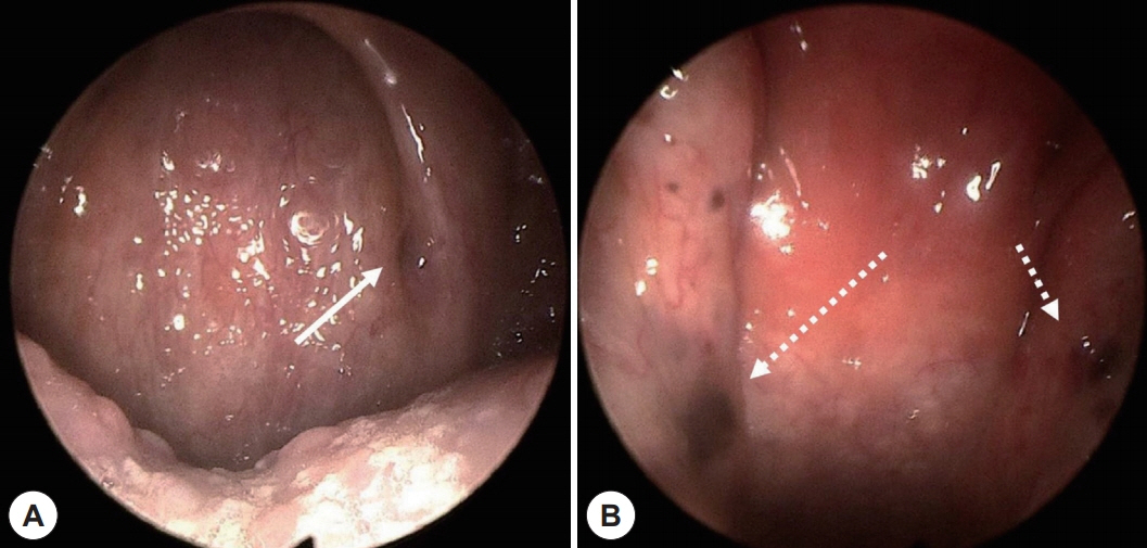
Fig. 1. Endoscopic findings. A: Endoscopic finding of melanotic oncocytic metaplasia on left anterior pharyngeal pillar (white arrow). B: Endoscopic finding of melanotic oncocytic metaplasia on both torus tubaris (white dotted arrows).
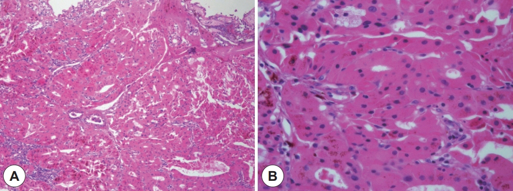
Fig. 2. Pathologic results of the case. A: Microscopic finding (×100) shows deeply homogeneous eosinophilic epitheial cells, noted with lumen formation. B: Scattered melanin pigment granules are found in eosinophilic cytoplasm (×400).

Fig. 3. Endoscopic findings after 12 months. A: Rt torus tubaris. B: Lt torus tubaris.
Reference
-
References
1. Shek TWH, Luk ISC, Nicholls JM, Fok KO. Melanotic oncocytic metaplasia of the nasopharynx. Histopathology. 1995; 26(3):273–5.2. Tajima S, Ohkubo A, Yoshida M, Koda K, Nameki I. Melanotic oncocytic metaplasia of the nasopharynx: a case report with a focus on immunohistochemical analyses and literature review. International Journal of Clinical and Experimental Pathology. 2015; 8(2):2103–10.3. Antonio C, Pieter J, Nina G, Alessandro F. Pathology of the Head and Neck. 2nd ed. Berlin, Heidelberg: Springer;2016.4. Mills S. Histology for Pathologists. Philadelphia: Lipincott Williams & Wilkins;2012.5. Xue WC, Hui YZ. Melanotic oncocytic metaplasia of the nasopharynx. Histopathology. 1999; 35(5):481–2.6. Hirakawa E, Miki H, Ohmori M, Kobayashi S, Haba R, Nagai Y. Melanin pigmented oncocytic metaplasia of the nasopharynx. Virchows Archiv. 1999; 434(5):455–7.7. Takano KI, Sato J, Shirasaki H, Yamazaki N, Hoki K, Himi T. Melanin pigmented oncocytic metaplasia of the nasopharynx. Auris Nasus Larynx. 2004; 31(2):161–3.8. Kurihara K, Nakagawa K. Pigmented variant of benign oncocytic lesion of the pharynx. Pathology International. 1997; 47(5):315–7.9. Sakaki M, Shek TWH, Hirokawa M, Kashima K, Daa T, Gamachi A, et al. Melanotic oncocytic metaplasia of the nasopharynx: a report of seven cases and review of the literature. Virchows Archiv. 2004; 444(4):345–9.10. Liao CT, Kuo TT. Melanotic Oncocytic Metaplasia of the Nasopharynx. International Journal of Surgical Pathology. 2005; 13(3):279.11. Lui PCW, Chan ABW, Chan KF, Choi CH, Tse GMK. Melanocytic and non-melanocytic oncocytic metaplasia of the nasopharynx. Pathology. 2004; 36(5):504–5.12. Li Y, Lu ZH, Lü W, Chen J. Images for diagnosis. Melanotic oncocytic metaplasia of nasopharynx: a case report with review. Chin Med J (Engl). 2010; 123(9):1230–2.13. Kondo T, Mori K, Oka S, Morinaka S. Melanotic oncocytic metaplasia of the nasopharynx as a benign mimicker of malignant melanoma: a case report. Diagnostic Pathology. 2010; 5:5.14. Na JY, Kim YH, Choi YD, Lee JS. Melanotic oncocytic metaplasia of the nasopharynx: a report of three cases and review of the literature. Korean Journal of Pathology. 2012; 46(2):201–4.15. Chang IW, Wang CC, Liu KW, Lan CH, Hung CH. Melanotic oncocytic metaplasia of the nasopharynx. Polish Journal of Pathology. 2014; 65(2):162–5.16. Uehara K, Usami Y, Imai Y, Shimizu M. Melanotic oncocytic metaplasia of the nasopharynx. Pathology International. 2015; 65(3):144–7.17. Li JJX, Ng JKM, Chan ABW. Clinicopathological features of melanotic and non-melanotic oncocytic lesions of the nasopharynx. Pathology. 2019; 51(6):600–4.18. Chen HY, Gule MF, Chang IW. Melanotic oncocytic metaplasia of the nasopharynx: a case report with review of literature. Ear Nose Throat J. 2020; 145561320907427.
