Arch Hand Microsurg.
2020 Dec;25(4):314-319. 10.12790/ahm.20.0059.
Free Flap Reconstruction of a Challenging Defect in the Posterior Cervicothoracic Region Using the Transverse Cervical Artery
- Affiliations
-
- 1Department of Plastic and Reconstructive Surgery, Chonbuk National University Medical School, Jeonju, Korea
- 2Research Institute of Clinical Medicine of Jeonbuk National University-Biomedical Research Institute of Jeonbuk National University Hospital, Jeonju, Korea
- KMID: 2508947
- DOI: http://doi.org/10.12790/ahm.20.0059
Abstract
- Reconstructions of large defects located in the posterior cervicothoracic region still present challenges to plastic surgeons. The local or regional flap is preferred in the posterior cervicothoracic region and many surgeons are reluctant to perform reconstruction using a microvascular free flap because of various reasons including vascular paucity. We report a case of a 60-year-old patient with the chronic wound at posterior cervicothoracic region. An anterolateral thigh free flap was considered the best available reconstructive method due to the size of the defect and the possibility of damaging the dorsal scapular artery. We used the transverse cervical artery and jugular vein as recipient vessels and the better result was shown than that of regional or local flaps. In our report, we presented that the transverse cervical artery which didn’t have commonly used can provide a reliable and advantageous recipient artery for the microvascular free flap reconstruction of posterior cervicothoracic defects.
Figure
-

Fig. 1. (A) Magnetic resonance imaging visualized T5 and T6 cord compression by a recurred tumor mass at the T6 vertebra. (B) C6 to T10 spinal stabilization with orthopedic hardware after surgical decompression.

Fig. 2. The wound was grossly 2.5×2.5 cm in size but had a 20×30 cm-sized rhomboid dead space (line).

Fig. 3. (A) The size of the defect after resection and debridement was 18×12 cm with a horizontal axis. And in the right neck area, the transverse cervical artery (arrowhead) and external jugular vein (arrow) was explored for the microvascular free flap (B).
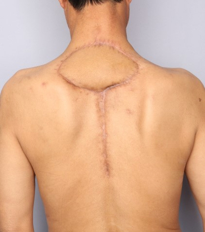
Fig. 4. The wound was followed at 1 month postoperatively and showed a satisfactory result.
Reference
-
1. Liu Y, Yu S, Song B, Yang L, Zhu S, Jin J. Reconstruction of posterior lumbar defects in oncologic patients using two island flaps of the back in series. Ann Plast Surg. 2010; 65:326–9.
Article2. Yoon SK, Song SH, Kang N, Yoon YH, Koo BS, Oh SH. Reconstruction of the head and neck region using lower trapezius musculocutaneous flaps. Arch Plast Surg. 2012; 39:626–30.
Article3. Mathes DW, Thornton JF, Rohrich RJ. Management of posterior trunk defects. Plast Reconstr Surg. 2006; 118:73e–83e.
Article4. Haas F, Weiglein A, Schwarzl F, Scharnagl E. The lower trapezius musculocutaneous flap from pedicled to free flap: anatomical basis and clinical applications based on the dorsal scapular artery. Plast Reconstr Surg. 2004; 113:1580–90.
Article5. Klink BK, Thurman RT, Wittpenn GP, Lauerman WC, Cain JE. Muscle flap closure for salvage of complex back wounds. Spine (Phila Pa 1976). 1994; 19:1467–70.
Article6. Dumanian GA, Ondra SL, Liu J, Schafer MF, Chao JD. Muscle flap salvage of spine wounds with soft tissue defects or infection. Spine (Phila Pa 1976). 2003; 28:1203–11.
Article7. Komagoe S, Watanabe T, Komatsu S, Kimata Y. Pedicled flaps versus free flaps for back reconstruction. Ann Plast Surg. 2018; 81:702–7.
Article8. Luca N, Santana MJ, Festa BM, Collurà F, Righini S. Transverse cervical artery perforator flap: standardized surgical technique and multiple reconstructive opportunity in head and neck surgery. Ann Plast Surg. 2017; 79:577–82.
- Full Text Links
-
- Actions
-
Cited
- CITED
-
- Close
- Share
-
- Similar articles
-
- A propeller superficial transverse cervical artery perforator flap for defect coverage of the submental area: a case report
- Transverse Cervical Artery and Appropriate Veins as Recipient Vessels in Head and Neck Reconstruction
- Reconstruction of a soft tissue defect in the toe using a serratus anterior fascia free flap: a case report
- Chondrocutaneous posterior auricular artery perforator free flap for single-stage reconstruction of the nasal tip: a case report
- Reconstruction of the Hand Using Fabricated Great Toe Pulp and Anterolateral Thigh Chimeric Free Flap
