Endovascular Approach in Patients with Acute Complete Occlusion Due to Middle Cerebral Artery Dissection
- Affiliations
-
- 1Departments of Neurosurgery and Research Institute of Clinical Medicine of Jeonbuk National University-Biomedical Research Institute of Jeonbuk National University Hospital, Jeonju, Korea
- 2Korea Department of Radiology and Research Institute of Clinical Medicine of Jeonbuk National University-Biomedical Research Institute of Jeonbuk National University Hospital, Jeonju, Korea
- KMID: 2508601
- DOI: http://doi.org/10.3340/jkns.2020.0053
Abstract
Objective
: Dissection of the middle cerebral artery (MCA) is less common than dissection of vessels in the vertebrobasilar system or carotid artery. Acute complete occlusion related to MCA dissection is extremely rare. We report an endovascular approach in patients with acute complete occlusion due to MCA dissection.
Methods
: We reviewed retrospectively the endovascular procedure and clinical results for acute-stroke patients who underwent recanalization from October 2014 through December 2018. Initial imaging findings and the endovascular procedure were analyzed for patients with acute complete occlusion due to MCA dissection.
Results
: We undertook first-line aspiration thrombectomy using a Penumbra catheter in 294 patients with acute occlusion of the M1 segment. Of these patients, seven were confirmed to have acute complete occlusion due to MCA dissection. All patients had angiographic findings of an intimal flap at the proximal occlusion site of the MCA. One patient complained of severe headache during microcatheter passage through the occluded lesion and died due to massive bleeding caused by rupture of the false lumen. The remaining patients underwent initial contact aspiration thrombectomy without microcatheter passage. After aspiration thrombectomy, six patients had delayed flow through the MCA. One patient underwent stenting of the MCA because of progressive symptoms.
Conclusion
: An intimal flap at the proximal portion of an occluded MCA can suggest the possibility of MCA dissection. Contrast aspiration thrombectomy without microcatheter passage can reduce the risk of false lumen rupture in cases of MCA dissection.
Keyword
Figure
-

Fig. 1. A 48-year-old woman with complete occlusion of left M1. A : Left internal carotid angiogram showing complete obstruction of the left middle cerebral artery with focal intimal flap (arrow). B : Left internal carotid angiogram using microcather after patient reported severe headache during catheter passage through the obstructive lesion shows extension of the false lumen (arrows) and contrast stagnation (open arrow). C : Noncontrast computed tomography scan after interventional procedure showing massive subarachnoid hemorrhage with contrast extravasation caused by middle cerebral artery rupture.
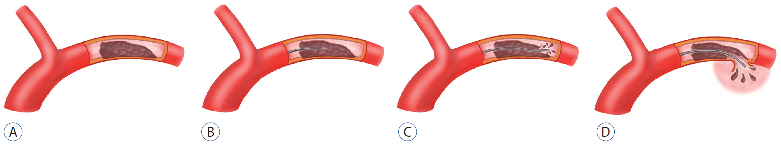
Fig. 2. A : Middle cerebral dissection. B : False lumen location of microcatheter. C : Re-entry of false lumen into the middle cerebral artery. D : Rupture of middle cerebral artery due to microcatheter in the false lumen.

Fig. 3. A 45-year-old man with complete occlusion of left M1. A : Left internal carotid angiogram showing complete obstruction of the left middle cerebral artery with focal intimal flap (arrow). B : Left internal carotid angiogram after initial contrast aspiration thrombectomy using a Penumbra catheter showing a double lumen and intimal flap (arrows). The patient was discharged without neurological complications.
Reference
-
References
1. Ahn SS, Kim BM, Suh SH, Kim DJ, Kim DI, Shin YS, et al. Spontaneous symptomatic intracranial vertebrobasilar dissection: initial and follow-up imaging findings. Radiology. 264:196–202. 2012.
Article2. Asaithambi G, Saravanapavan P, Rastogi V, Khan S, Bidari S, Khanna AY, et al. Isolated middle cerebral artery dissection: a systematic review. Int J Emerg Med. 7:44. 2014.
Article3. Beletsky V, Norris JW. Spontaneous dissection of the carotid and vertebral arteries. N Engl J Med. 345:467. 2001.
Article4. Doijiri R, Yokota C, Suzuki R, Toyoda K, Minematsu K. Intravenous recombinant tissue plasminogen activator thrombolysis in acute ischemic stroke due to middle cerebral artery dissection. J Stroke Cerebrovasc Dis. 21:915.E7–915.E9. 2012.
Article5. Houser OW, Mokri B, Sundt TM Jr, Baker HL Jr, Reese DF. Spontaneous cervical cephalic arterial dissection and its residuum: angiographic spectrum. AJNR Am J Neuroradiol. 5:27–34. 1984.6. Kurino M, Yoshioka S, Ushio Y. Spontaneous dissecting aneurysms of anterior and middle cerebral artery associated with brain infarction: a case report and review of the literature. Surg Neurol. 57:428–436. discussion 436-438. 2002.
Article7. Kwak HS, Hwang SB, Chung GH, Jeong SK. High-resolution magnetic resonance imaging of symptomatic middle cerebral artery dissection. J Stroke Cerebrovasc Dis. 23:550–553. 2014.
Article8. Lee HO, Kwak HS, Chung GH, Hwang SB. Diagnostic usefulness of high resolution cross sectional MRI in symptomatic middle cerabral arterial dissection. J Korean Neurosurg Soc. 49:370–372. 2011.
Article9. Lee JS, Bang OY, Lee PH, Kim BM, Yong SW. Two cases of spontaneous middle cerebral arterial dissection causing ischemic stroke. J Neurol Sci. 250:162–166. 2006.
Article10. Ohkuma H, Suzuki S, Kikkawa T, Shimamura N. Neuroradiologic and clinical features of arterial dissection of the anterior cerebral artery. AJNR Am J Neuroradiol. 24:691–699. 2003.11. Ohkuma H, Suzuki S, Shimamura N, Nakano T. Dissecting aneurysms of the middle cerebral artery: neuroradiological and clinical features. Neuroradiology. 45:143–148. 2003.
Article12. Vertinsky AT, Schwartz NE, Fischbein NJ, Rosenberg J, Albers GW, Zaharchuk G. Comparison of multidetector CT angiography and MR imaging of cervical artery dissection. AJNR Am J Neuroradiol. 29:1753–1760. 2008.
Article
- Full Text Links
-
- Actions
-
Cited
- CITED
-
- Close
- Share
-
- Similar articles
-
- Is the retrograde access for endovascular treatment of a traumatic carotid cavernous fistula associated with dissection of the ipsilateral carotid possible?
- Endovascular recanalization therapy for patients with acute ischemic stroke with hidden aortic dissection: A case series
- Successful Intra-arterial Stent Thrombectomy in Acute Infarction Caused by Spontaneous Middle Cerebral Artery Dissection
- Mechanical thrombectomy for acute ischemic stroke with occlusion of the M2 segment of the middle cerebral artery: A literature review
- Revision Superficial Temporal Artery-Middle Cerebral Artery Bypass Surgery for Recurrent Acute Ischemic Stroke Due to Delayed Occlusion of the Bypass Graft
