Assessment of bone density changes following two-jaw surgery using multidetector computed tomography: A pilot study
- Affiliations
-
- 1Department of Orthodontics, Wonkwang University College of Dentistry, Iksan, Korea
- 2Postgraduate Orthodontic Program, Arizona School of Dentistry and Oral Health, A.T. Still University, Mesa, AZ, USA
- 3Graduate School of Dentistry, Kyung Hee University, Seoul, Korea
- 4Department of Orthodontics, Wonkwang Dental Research Institute, Wonkwang University College of Dentistry, Iksan, Korea
- 5Department of Orthodontics, Seoul National University Gwanak Dental Hospital, Seoul, Korea
- 6Department of Oral and Maxillofacial Surgery, Wonkwang University Daejeon Dental Hospital, Daejeon, Korea
- 7School of Big Data and Financial Statistics, Wonkwang University College of Natural Sciences, Iksan, Korea
- 8Department of Orthodontics, Yonsei University College of Dentistry, Seoul, Korea
- KMID: 2502625
- DOI: http://doi.org/10.4041/kjod.2020.50.3.157
Abstract
Objective
The aim of this retrospective study was to evaluate the pre- and postsurgical bone densities at alveolar and extra-alveolar sites following twojaw orthognathic surgery.
Methods
The sample consisted of 10 patients (mean age, 23.2 years; range, 18.0–27.8 years; 8 males, 2 females) who underwent two-jaw orthognathic surgery. A three-dimensional imaging program (Invivo 5) was used with multidetector computed tomography images taken preand postoperatively (obtained 32.3 ± 6.0 days before surgery and 5.8 ± 2.6 days after surgery, respectively) for the measurement of bone densities at the following sites: (1) alveolar bone in the maxilla and mandible, (2) extra-alveolar sites, such as the top of the head, menton (Me), condyle, and the fourth cervical vertebrae (C4).
Results
When pre- and postsurgical bone densities were compared, an overall tendency of decrease in bone density was noted. Statistically significant reductions were observed in the densities of cancellous bone at several areas of the maxillary alveolar bone; cortical and cancellous bone in most areas of the mandibular alveolar bone; cortical bone in Me; and cancellous bone in C4. There was no statistically significant difference in bone density in relation to the depth of the alveolar bone. In a comparison of the bone densities between groups with and without genioplasty, there was almost no statistically significant difference.
Conclusions
Accelerated tooth movement following orthognathic surgery may be confirmed with reduced bone density. In addition, this study could offer insights into bone metabolism changes following orthognathic surgery, providing direction for further investigations in this field.
Keyword
Figure
-

Figure 1 Frankfurt horizontal (FH) plane orientation of computed tomography images. All images were reoriented to the FH plane using both porions and the right orbitale on the red line. A, Right lateral view. B, Frontal view. C, Left lateral view.
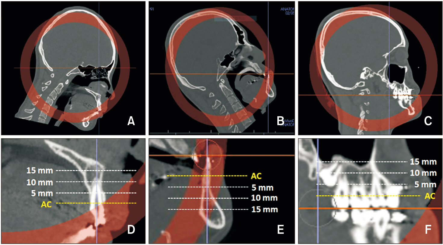
Figure 2 Reorientation and measurement of alveolar bone density in the sagittal views. A, B, Computed tomography images were reoriented to align the axis of the left maxillary and mandibular central incisors perpendicular to the horizontal plane. C, Posterior occlusal plane was aligned so it was parallel to the horizontal plane. D–F, Bone densities were measured at the alveolar crest (AC, yellow line) and at each depth (dotted lines; 5, 10, 15 mm from AC).
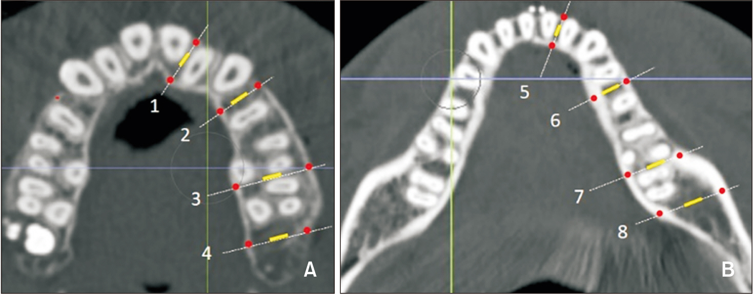
Figure 3 Measurement sites in the alveolar bone. A, B, Measurement sites in the maxilla and mandible. 1, 5: Between central and lateral incisors. 2, 6: Between the first and second premolars or between canines and remaining premolars when premolar extraction was performed. 3, 7: Between the second and first molars. 4: Maxillary tuberosity. 8: Retromolar pad. All measurements were acquired from the left side of the arch. Measurement areas of cortical and cancellous bone are shown as red dots and a yellow line, respectively.
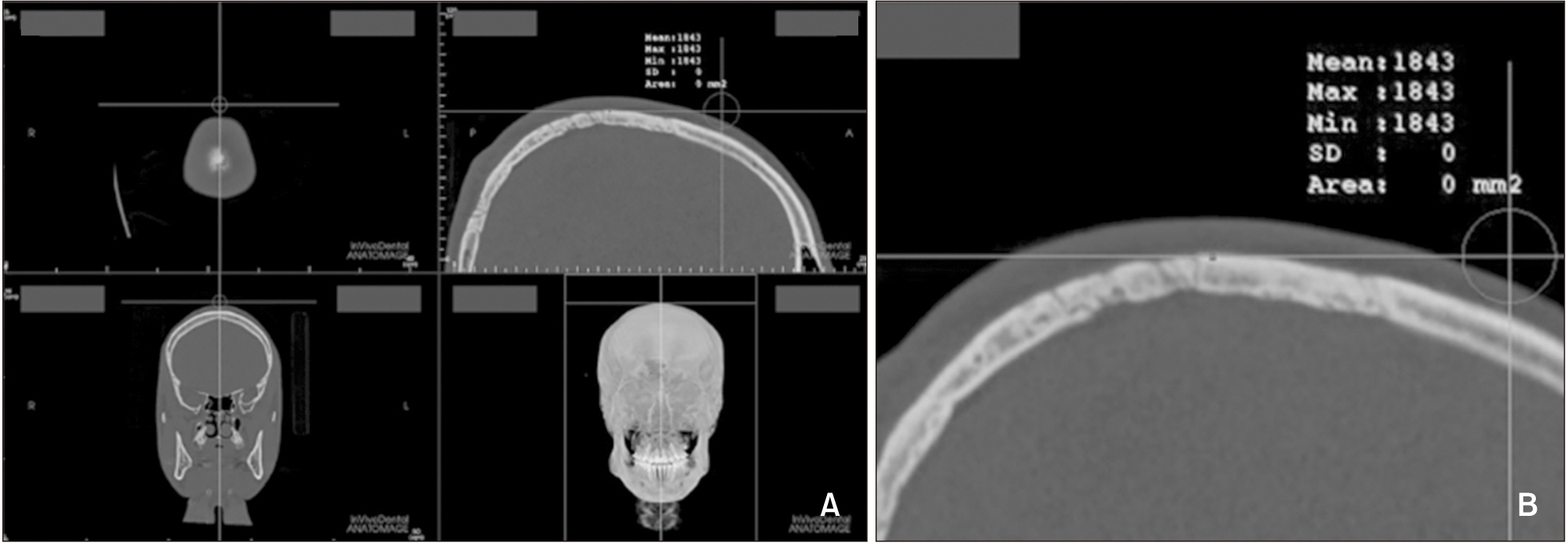
Figure 4 Bone density measurement at the top point of the head. A, The most superior point was selected in the midsagittal view. B, Magnified view of the measurement site.

Figure 5 Bone density measurement at menton (Me). A, Presurgical image, the blue line represents the vertical slice drawn over Me (yellow arrow). B, Presurgical sagittal slice at Me (red dot). C, Postsurgical image. D, Postsurgical sagittal slice at Me (red dot).
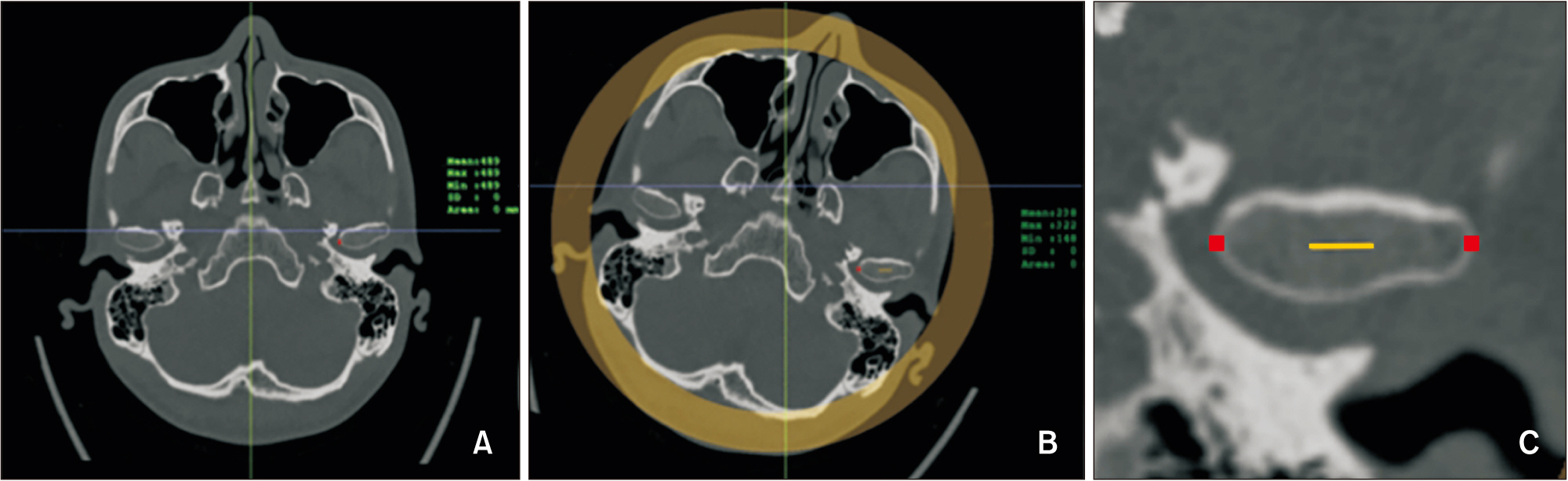
Figure 6 Bone density measurement at the condyle. A, The axial slice with the largest mediolateral diameter of the mandibular condyle was selected. B, The condyle was re-oriented as the most medial and lateral points aligned in a horizontal line. C, Cortical bone densities at the medial and lateral points were measured (red dots), and cancellous bone density was measured in the middle 5 mm upon the line connecting the medial and lateral points.
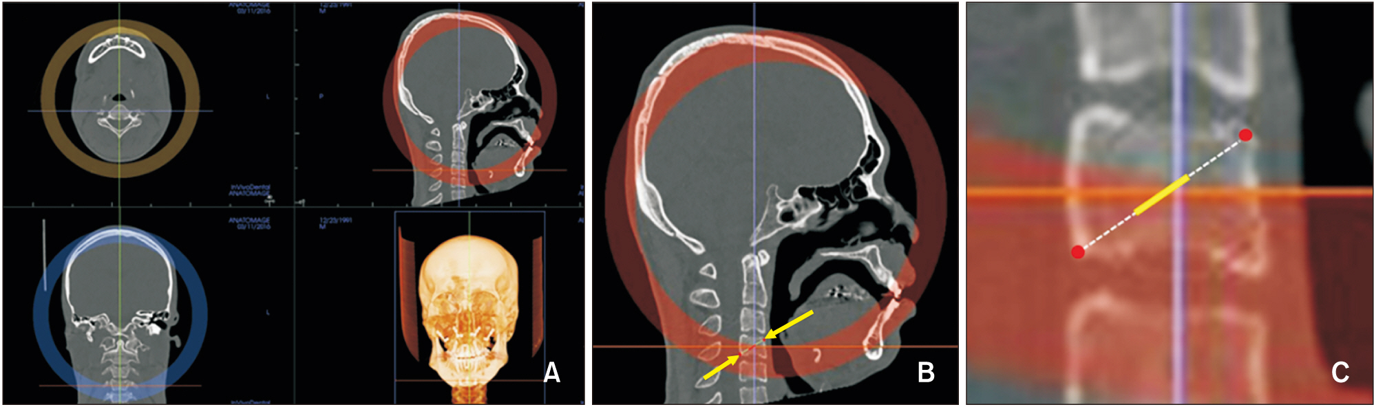
Figure 7 Bone density measurements for the fourth cervical vertebrae (C4). A, Orientation of computed tomography images for C4. B, Measurement sites for C4 (yellow arrow). C, Magnified view of C4. Cortical bone densities were measured at the most anterosuperior and posteroinferior red points and cancellous bone density was measured in the middle 5 mm of the line (yellow) connecting the two red points.
Reference
-
1. Köle H. 1959; Surgical operations on the alveolar ridge to correct occlusal abnormalities. Oral Surg Oral Med Oral Pathol. 12:515–29. DOI: 10.1016/0030-4220(59)90153-7. PMID: 13644913.
Article2. Frost HM. 1981; The regional accelerated phenomenon. Orthop Clin North Am. 12:725–6.3. Frost HM. 1983; The regional acceleratory phenomenon: a review. Henry Ford Hosp Med J. 31:3–9. PMID: 6345475.4. Frost HM. 1989; The biology of fracture healing. An overview for clinicians. Part I. Clin Orthop Relat Res. (248):283–93. DOI: 10.1097/00003086-198911000-00045. PMID: 2680202.5. Melsen B. 2001; Tissue reaction to orthodontic tooth movement--a new paradigm. Eur J Orthod. 23:671–81. DOI: 10.1093/ejo/23.6.671. PMID: 11890063.
Article6. Mueller M, Schilling T, Minne HW, Ziegler R. 1991; A systemic acceleratory phenomenon (SAP) accompanies the regional acceleratory phenomenon (RAP) during healing of a bone defect in the rat. J Bone Miner Res. 6:401–10. DOI: 10.1002/jbmr.5650060412. PMID: 1858523.
Article7. Liou EJ, Chen PH, Wang YC, Yu CC, Huang CS, Chen YR. 2011; Surgery-first accelerated orthognathic surgery: postoperative rapid orthodontic tooth movement. J Oral Maxillofac Surg. 69:781–5. DOI: 10.1016/j.joms.2010.10.035. PMID: 21353934.
Article8. Verna C, Zaffe D, Siciliani G. 1999; Histomorphometric study of bone reactions during orthodontic tooth movement in rats. Bone. 24:371–9. DOI: 10.1016/S8756-3282(99)00009-5. PMID: 10221549.
Article9. Melsen B. 1999; Biological reaction of alveolar bone to orthodontic tooth movement. Angle Orthod. 69:151–8. DOI: 10.1043/0003-3219(1999)069<0151:BROABT>2.3.CO;2. PMID: 10227556.10. White SC, Rudolph DJ. 1999; Alterations of the trabecular pattern of the jaws in patients with osteoporosis. Oral Surg Oral Med Oral Pathol Oral Radiol Endod. 88:628–35. DOI: 10.1016/S1079-2104(99)70097-1. PMID: 10556761.
Article11. Peiró-Guijarro MA, Guijarro-Martínez R, Hernández-Alfaro F. 2016; Surgery first in orthognathic surgery: a systematic review of the literature. Am J Orthod Dentofacial Orthop. 149:448–62. DOI: 10.1016/j.ajodo.2015.09.022. PMID: 27021449.
Article12. Kang HJ, Jeong SW, Jo BH, Kim YD, Kim SS. 2012; Observation of trabecular changes of the mandible after orthognathic surgery using fractal analysis. J Korean Assoc Oral Maxillofac Surg. 38:96–100. DOI: 10.5125/jkaoms.2012.38.2.96.
Article13. Shahlaie M, Gantes B, Schulz E, Riggs M, Crigger M. 2003; Bone density assessments of dental implant sites: 1. Quantitative computed tomography. Int J Oral Maxillofac Implants. 18:224–31. PMID: 12705300.14. Parfitt AM. 2002; Misconceptions (2): turnover is always higher in cancellous than in cortical bone. Bone. 30:807–9. DOI: 10.1016/S8756-3282(02)00735-4. PMID: 12052445.
Article15. Funk JF, Krummrey G, Perka C, Raschke MJ, Bail HJ. 2009; Distraction osteogenesis enhances remodeling of remote bones of the skeleton: a pilot study. Clin Orthop Relat Res. 467:3199–205. DOI: 10.1007/s11999-009-0902-y. PMID: 19475465. PMCID: PMC2772934.
Article16. Roberts WE. Graber TM, Vanarsdall RL, Vig KWL, editors. 2005. Bone physiology, metabolism and biomechanics in orthodontic practice. Orthodontics: current principles and techniques. 4th ed. Mosby-Year Book;St Louis: p. 221–92.17. Deguchi T, Takano-Yamamoto T, Yabuuchi T, Ando R, Roberts WE, Garetto LP. 2008; Histomorphometric evaluation of alveolar bone turnover between the maxilla and the mandible during experimental tooth movement in dogs. Am J Orthod Dentofacial Orthop. 133:889–97. DOI: 10.1016/j.ajodo.2006.12.013. PMID: 18538254.
Article18. Huja SS, Fernandez SA, Hill KJ, Li Y. 2006; Remodeling dynamics in the alveolar process in skeletally mature dogs. Anat Rec A Discov Mol Cell Evol Biol. 288:1243–9. DOI: 10.1002/ar.a.20396. PMID: 17075846. PMCID: PMC2612758.
Article19. Kotze MJ, Bütow KW, Olorunju SA, Kotze HF. 2014; A comparison of mandibular and maxillary alveolar osteogenesis over six weeks: a radiological examination. Head Face Med. 10:50. DOI: 10.1186/1746-160X-10-50. PMID: 25429901. PMCID: PMC4289173.
Article20. McBride MD, Campbell PM, Opperman LA, Dechow PC, Buschang PH. 2014; How does the amount of surgical insult affect bone around moving teeth? Am J Orthod Dentofacial Orthop. 145(4 Suppl):S92–9. DOI: 10.1016/j.ajodo.2013.10.020. PMID: 24680029.
Article21. Kausal S, Agrawal A, Misal AN, Toshniwal NG. 2015; Accelerted orthodontic tooth movement: a new paradigm in orthodontics. Int J Oral Health Med Res. 2:94–6.22. Schilling T, Müller M, Minne HW, Ziegler R. 1998; Influence of inflammation-mediated osteopenia on the regional acceleratory phenomenon and the systemic acceleratory phenomenon during healing of a bone defect in the rat. Calcif Tissue Int. 63:160–6. DOI: 10.1007/s002239900508. PMID: 9685523.
Article23. Sebaoun JD, Kantarci A, Turner JW, Carvalho RS, Van Dyke TE, Ferguson DJ. 2008; Modeling of trabecular bone and lamina dura following selective alveolar decortication in rats. J Periodontol. 79:1679–88. DOI: 10.1902/jop.2008.080024. PMID: 18771369. PMCID: PMC2563959.
Article24. Zhang Y, Zhou Z, Wu C, Zhao D, Wang C, Cheng X, et al. 2016; Population-stratified analysis of bone mineral density distribution in cervical and lumbar vertebrae of Chinese from quantitative computed tomography. Korean J Radiol. 17:581–9. DOI: 10.3348/kjr.2016.17.5.581. PMID: 27587947. PMCID: PMC5007385.
Article
- Full Text Links
-
- Actions
-
Cited
- CITED
-
- Close
- Share
-
- Similar articles
-
- Assessment of the increased calcification of the jaw bone with CT-Scan after dental implant placement
- Study for hounsfield units in computed tomogram with jaw lesion
- Evaluation of alveolar bone density by intraoral periapical radiography
- Can Dental Cone Beam Computed Tomography Assess Bone Mineral Density?
- A clinical pilot study of jawbone mineral density measured by the newly developed dual-energy cone-beam computed tomography method compared to calibrated multislice computed tomography
