2019 Novel Coronavirus (COVID-19) Pneumonia: Serial Computed Tomography Findings
- Affiliations
-
- 1Department of Radiology, Jiangxi Provincial People's Hospital, Nanchang, China. 26171381@qq.com
- 2Department of Medical Cosmetology, Jiangxi Provincial People's Hospital, Nanchang, China.
- 3Department of Radiology, Jiangxi Chest Hospital, Nanchang, China.
- 4Institute of Clinical Medicine, Jiangxi Provincial People's Hospital, Nanchang, China.
- KMID: 2471816
- DOI: http://doi.org/10.3348/kjr.2020.0112
Abstract
- From December 2019, Coronavirus disease 2019 (COVID-19) pneumonia (formerly known as the 2019 novel Coronavirus [2019-nCoV]) broke out in Wuhan, China. In this study, we present serial CT findings in a 40-year-old female patient with COVID-19 pneumonia who presented with the symptoms of fever, chest tightness, and fatigue. She was diagnosed with COVID-19 infection confirmed by real-time reverse-transcriptase-polymerase chain reaction. CT showed rapidly progressing peripheral consolidations and ground-glass opacities in both lungs. After treatment, the lesions were shown to be almost absorbed leaving the fibrous lesions.
Keyword
MeSH Terms
Figure
-
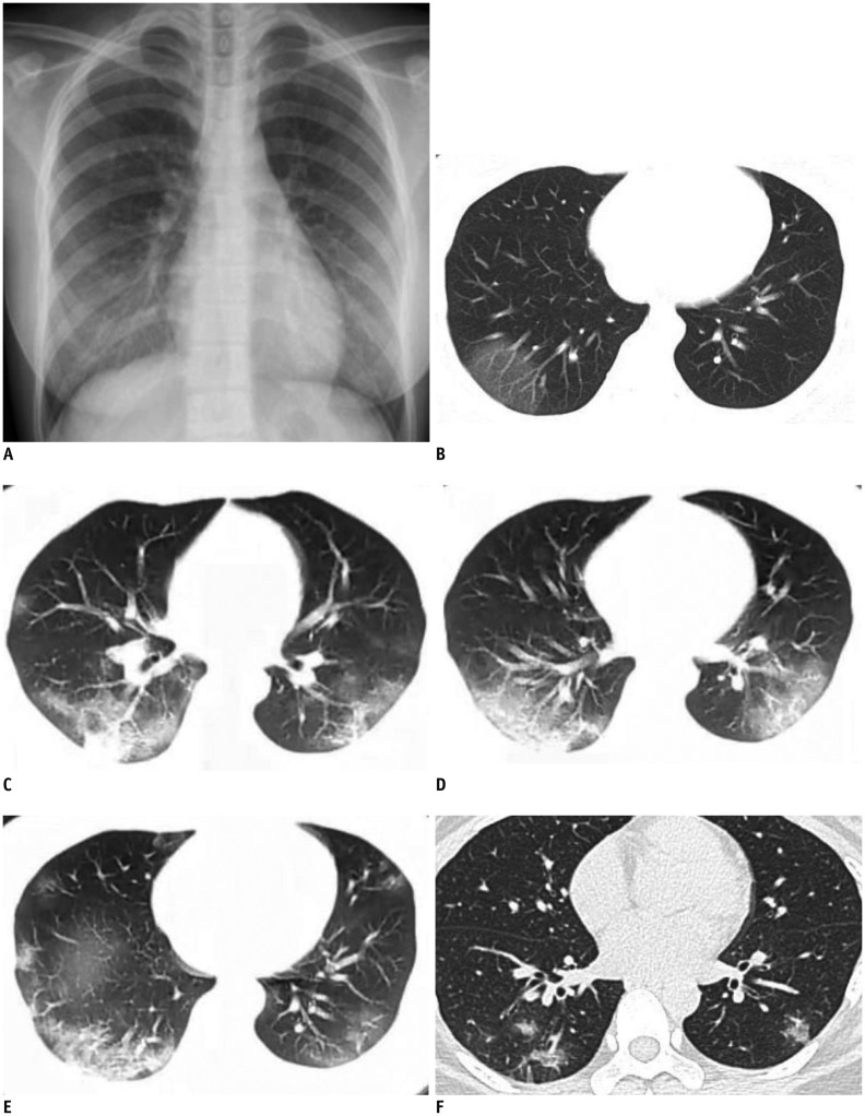
Fig. 1 40-year-old female patient with Coronavirus disease 2019 pneumonia.A, B. Initial posteroanterior chest radiograph and chest CT scan were performed on day of admission (3 days after onset of fever). Chest radiograph (A) shows no thoracic abnormalities. Axial CT scan (B) shows GGOs in subpleural area of right lower lobe. Left lung is normal. C–E. Follow-up chest CT scans taken 6 days after onset of fever show increased density of GGOs in right lower lobe, which then progressed into consolidations with perilobular thickening. Multifocal peripheral patchy areas of nodular consolidations and nodular GGO lesions are newly developed in subpleural areas of both lower lobes. F. Progressive resolution of parenchymal lesions is seen in follow-up high-resolution CT scan obtained on day 12. Patchy consolidations and GGOs in both lungs were almost absorbed leaving a few fibrous lesions that may represent residual organizing pneumonia. Repeat real-time reverse-transcriptase-polymerase chain reaction was negative and patient was discharged.CT = computer tomography, GGO = ground-glass opacity
Reference
-
1. Rubin EJ, Baden LR, Morrissey S, Campion EW. Medical journals and the 2019-nCoV outbreak. N Engl J Med. 2020; 1. 27. [Epub]. DOI: 10.1056/NEJMe2001329.
Article2. The Lancet. Emerging understandings of 2019-nCoV. Lancet. 2020; 395:311. PMID: 31986259.3. Carlos WG, Dela Cruz CS, Cao B, Pasnick S, Jamil S. Novel Wuhan (2019-nCoV) Coronavirus. Am J Respir Crit Care Med. 2020; 201:P7–P8. PMID: 32004066.
Article4. Lei J, Li J, Li X, Qi X. CT imaging of the 2019 Novel Coronavirus (2019-nCoV) pneumonia. Radiology. 2020; 1. 31. [Epub]. DOI: 10.1148/radiol.2020200236.5. Zhu N, Zhang D, Wang W, Li X, Yang B, Song J, et al. China Novel Coronavirus Investigating and Research Team. A novel coronavirus from patients with pneumonia in China, 2019. N Engl J Med. 2020; 1. 24. [Epub]. DOI: 10.1056/NEJMoa2001017.
Article6. Lu H. Drug treatment options for the 2019-new Coronavirus (2019-nCoV). Biosci Trends. 2020; 1. 28. [Epub]. DOI: 10.5582/bst.2020.01020.
Article7. Corman VM, Landt O, Kaiser M, Molenkamp R, Meijer A, Chu DK, et al. Detection of 2019 novel Coronavirus (2019-nCoV) by real-time RT-PCR. Euro Surveill. 2020; 25:2000045.
Article
- Full Text Links
-
- Actions
-
Cited
- CITED
-
- Close
- Share
-
- Similar articles
-
- Clinical and Radiologic Findings of COVID-19 Pneumonia: South Korean Experience from Three Cases
- Mediastinal Emphysema, Giant Bulla, and PneumothoraxDeveloped during the Course of COVID-19 Pneumonia
- Pulmonary Contusion Similar to COVID-19 Pneumonia
- Small Solitary Ground-Glass Nodule on CT as an InitialManifestation of Coronavirus Disease 2019 (COVID-19)Pneumonia
- Novel Coronavirus Pneumonia Outbreak in 2019: Computed Tomographic Findings in Two Cases
